Review
. 2017 Aug 4;121(4):451-468.
doi: 10.1161/CIRCRESAHA.117.311170.

Brain-Heart Interaction: Cardiac Complications After Stroke

Affiliations
Review

Brain-Heart Interaction: Cardiac Complications After Stroke

Zhili Chen et al. Circ Res. .

Abstract

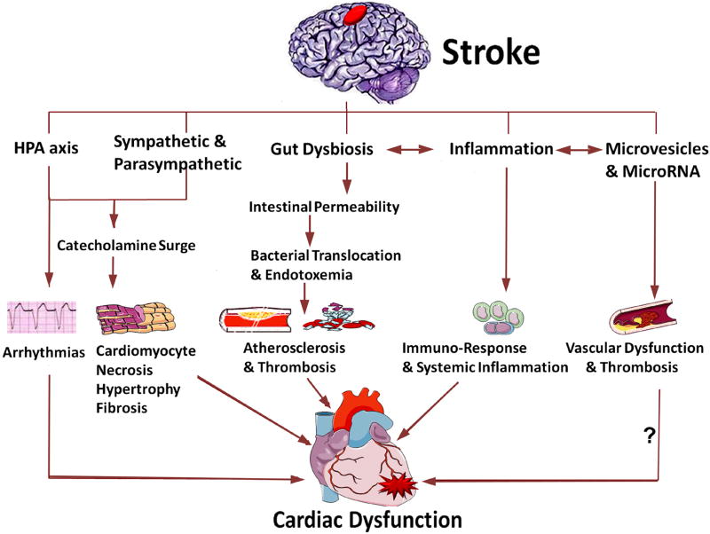
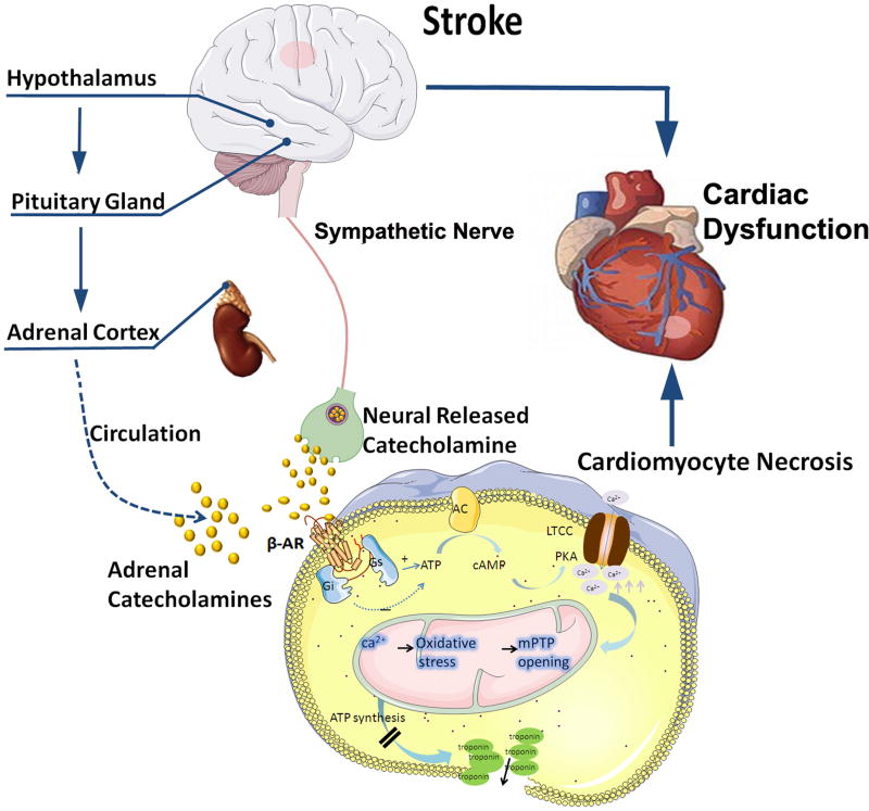
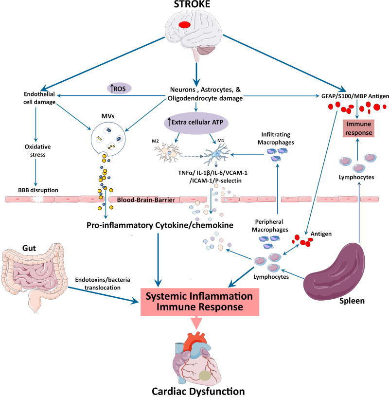
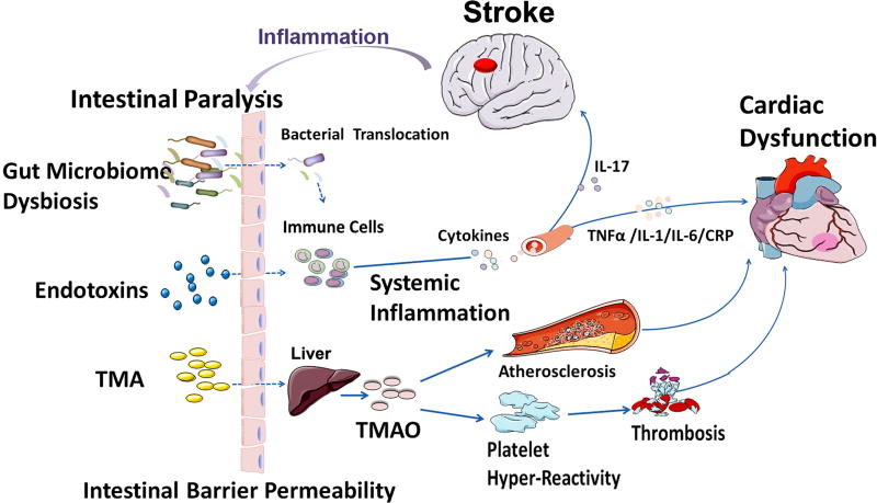
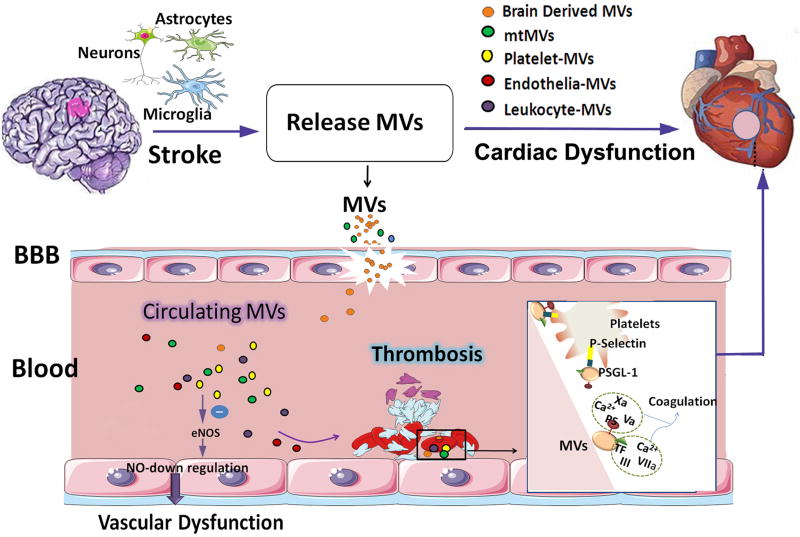
Neurocardiology is an emerging specialty that addresses the interaction between the brain and the heart, that is, the effects of cardiac injury on the brain and the effects of brain injury on the heart. This review article focuses on cardiac dysfunction in the setting of stroke such as ischemic stroke, brain hemorrhage, and subarachnoid hemorrhage. The majority of post-stroke deaths are attributed to neurological damage, and cardiovascular complications are the second leading cause of post-stroke mortality. Accumulating clinical and experimental evidence suggests a causal relationship between brain damage and heart dysfunction. Thus, it is important to determine whether cardiac dysfunction is triggered by stroke, is an unrelated complication, or is the underlying cause of stroke. Stroke-induced cardiac damage may lead to fatality or potentially lifelong cardiac problems (such as heart failure), or to mild and recoverable damage such as neurogenic stress cardiomyopathy and Takotsubo cardiomyopathy. The role of location and lateralization of brain lesions after stroke in brain-heart interaction; clinical biomarkers and manifestations of cardiac complications; and underlying mechanisms of brain-heart interaction after stroke, such as the hypothalamic-pituitary-adrenal axis; catecholamine surge; sympathetic and parasympathetic regulation; microvesicles; microRNAs; gut microbiome, immunoresponse, and systemic inflammation, are discussed.

Keywords: brain injury; brain ischemia; heart failure; inflammation; stroke.


Figures

Figure 1
Figure 1. Summary of mechanisms for brain heart interaction after stroke
Cardiac dysfunction after stroke may be caused by several mechanisms, including activation of the HPA axis, sympathetic and parasympathetic regulation, catecholamine surge, gut microbiome dysbiosis, immune responses and inflammation as well as the release of microvesicles and microRNA (? indicates that future studies are warranted).
Figure 2
Figure 2. The HPA axis, catecholamine surge and sympathetic and parasympathetic regulation mediate cardiac dysfunction after stroke
Catecholamines act on β receptors, β receptors couple to the stimulatory G protein (Gs) and activate adenylyl cyclase (AC), and increase cytosolic cAMP. cAMP binds to the regulatory subunits of protein kinase A (PKA), which phosphorylates sarcolemmal L-type Ca2+ channels (LTCC) and sarcoplasmic phospholamban, which over loads mitochondria. Mitochondrial Ca2+ overload triggers oxidative stress, and the subsequent opening of their inner membrane permeability transition pore with ensuing osmotic swelling and loss of ATP synthesis, leads to myocardial cell death.
Figure 3
Figure 3. Role of immune response and inflammation in mediating cardiac dysfunction after stroke
Local inflammatory responses in the brain, endothelial cell damage and increased oxidative stress lead to BBB disruption after stroke. Release of proinflammatory cytokine and chemokines can be stimulated by damaged endothelial cells and astrocytes, activated resident microglia and infiltrating macrophages that assume M1 phenotype, as well as the spleen. Systemic inflammatory responses are mainly driven by a number of cytokines, chemokines, stress hormones, as well as parasympathetic and sympathetic regulation. Gut microbiome dysbiosis after stroke, can also lead to bacterial and endotoxin translocation to the blood, leading to systemic inflammation. Damage associated molecular patterns (DAMPs) released by dying brain cells after ischemic stroke release brain derived antigens which can pass the ruptured BBB and enter the systemic circulation. In the presence of systemic inflammation, the antigen specific autoimmune response leads to the secretion of proinflammatory cytokines.
Figure 4
Figure 4. The brain-gut axis and gut-heart axis may mediate cardiac damage after stroke
Stroke increases intestinal barrier permeability, enabling bacterial and endotoxin translocation to bloodstream inducing inflammatory responses and proinflammatory cytokine production. Cytokine production can trigger inflammation, fibrosis and microvascular and myocardial dysfunction. TMAO is the hepatic oxidation product of the microbial metabolite TMA. TMAO promotes the development of hyper responsive platelet phenotype and enhances elevating the risk for heart failure and/or heart attack.
Figure 5
Figure 5. The role of microvesicles released by damaged astrocytes, neurons and microglia after stroke in mediating cardiac damage warrants future investigation
Stroke induced elevation of circulating platelets and microvesicles (MVs) are associated with endothelial dysfunction and coagulation leading to thrombosis. MVs induce endothelial dysfunction by decreasing nitric oxide (NO) synthesis as the result of endothelial nitric oxide synthase (eNOS) function inhibition and an increase in caveolin-1. The outer leaflet of the MVs membrane contains two pro-coagulants: phosphatidylserine and tissue factor. MVs can bind to coagulation factors and promote their activation. In addition, MVs harbor tissue factor, which can initiate extrinsic coagulation pathways and promote the assembly of clotting enzymes leading to thrombin generation. At sites of vascular injury, P-selectin exposure by activated endothelial cells or platelets leads to the rapid recruitment of MVs bearing the P-selectin glycoprotein ligand-1 (PSGL-1). The interactions between PSGL-1 and platelet P-selectin are necessary to concentrate tissue factor activity at the thrombus.

References

    1. Ay H, Koroshetz WJ, Benner T, Vangel MG, Melinosky C, Arsava EM, Ayata C, Zhu M, Schwamm LH, Sorensen AG. Neuroanatomic correlates of stroke-related myocardial injury. Neurology. 2006;66:1325–1329. - PubMed
    1. Oppenheimer SM. Neurogenic cardiac effects of cerebrovascular disease. Current opinion in neurology. 1994;7:20–24. - PubMed
    1. Tokgozoglu SL, Batur MK, Topcuoglu MA, Saribas O, Kes S, Oto A. Effects of stroke localization on cardiac autonomic balance and sudden death. Stroke; a journal of cerebral circulation. 1999;30:1307–1311. - PubMed
    1. Byer E, Ashman R, Toth LA. Electrocardiograms with large, upright t waves and long q-t intervals. American heart journal. 1947;33:796–806. - PubMed
    1. Samuels MA. The brain-heart connection. Circulation. 2007;116:77–84. - PubMed