. 2017 Oct;38(10):2008-2014.
doi: 10.3174/ajnr.A5311. Epub 2017 Aug 3.

Localizing the L5 Vertebra Using Nerve Morphology on MRI: An Accurate and Reliable Technique

Affiliations

Localizing the L5 Vertebra Using Nerve Morphology on MRI: An Accurate and Reliable Technique

M E Peckham et al. AJNR Am J Neuroradiol. 2017 Oct.

Abstract

Background and purpose: Multiple methods have been used to determine the lumbar vertebral level on MR imaging, particularly when full spine imaging is unavailable. Because postmortem studies show 95% accuracy of numbering the lumbar vertebral bodies by counting the lumbar nerve roots, attention to lumbar nerve morphology on axial MR imaging can provide numbering clues. We sought to determine whether the L5 vertebra could be accurately localized by using nerve morphology on MR imaging.

Materials and methods: One hundred eight cases with full spine MR imaging were numbered from the C2 vertebral body to the sacrum with note of thoracolumbar and lumbosacral transitional states. The origin level of the L5 nerve and iliolumbar ligament were documented in all cases. The reference standard of numbering by full spine imaging was compared with the nerve morphology numbering method. Five blinded raters evaluated all lumbar MRIs with nerve morphology technique twice. Prevalence and bias-adjusted κ were used to measure interrater and intrarater reliability.

Results: The L5 nerve arose from the 24th presacral vertebra (L5) in 106/108 cases. The percentage of perfect agreement with the reference standard was 98.1% (95% CI, 93.5%-99.8%), which was preserved in transitional and numeric variation states. The iliolumbar ligament localization method showed 83.3% (95% CI, 74.9%-89.8%) perfect agreement with the reference standard. Inter- and intrarater reliability when using the nerve morphology method was strong.

Conclusions: The exiting L5 nerve can allow accurate localization of the corresponding vertebrae, which is essential for preprocedure planning in cases where full spine imaging is not available. This neuroanatomic method displays higher agreement with the reference standard compared with previously described methods, with strong inter- and intrarater reliability.


Figures

Fig 1.
Fig 1.
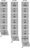
Graphic demonstrating our method for vertebral body numbering. When counting down from C2, patients with only 4 lumbar-type vertebral bodies (sacralized L5) have 23 PSV (A), patients with 5 lumbar-type vertebral bodies have 24 PSV (B), and patients with 6 lumbar-type vertebral bodies (lumbarized S1) have 25 PSV (C).
Fig 2.
Fig 2.
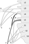
Schematic demonstrating the divisions of the lumbosacral plexus. The L4 nerve divides soon after exiting the neural foramen into peroneal (black) (A) and tibial (B) components, with the peroneal component joining the lateral fibers of L5 (gray) (C). The L4 nerve also contributes to both the femoral (D) and obturator (E) nerves. L5 is the only lumbar nerve that does not have a proximal division. Branches of L4–S2 make up the common peroneal nerve (F), and branches of L4–S3 make up the tibial nerve (G), which together comprise components of the sciatic nerve (not illustrated). The MRI morphology of the L4 peroneal component and L5 nerve are of special importance for localization; thus, they are shaded in this figure.
Fig 3.
Fig 3.
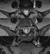
Consecutive cranial to caudal axial T2-weighted MR images demonstrate L4 and L5 nerve root anatomy. The L4 nerve root splits proximally into tibial and peroneal branches (solid arrows). The peroneal branch extends caudally and joins with the L5 nerve root (dashed arrow) along its anterolateral aspect at the level of the lateral sacrum.
Fig 4.
Fig 4.
Axial T2-weighted MR images at the level of the sacrum with corresponding graphics demonstrating how the caliber of the nerve roots along the sacrum can be used to identify the number of lumbar vertebral segments. In patients with 4 lumbar segments, the L4 nerve root is seen splitting over the lateral sacrum (A, arrows). In patients with 5 lumbar segments, the peroneal branch of L4 joins the L5 nerve root, which is twice the caliber of L4 (B, arrows). In patients with more than 5 lumbar segments, 2 nerves of similar caliber will be seen along the lateral sacral wing, representing L5 laterally and S1 medially (C, arrows).

Similar articles

Cited by

References

    1. Paik NC, Lim CS, Jang HS. Numeric and morphological verification of lumbosacral segments in 8280 consecutive patients. Spine 2013;38:E573–78 10.1097/BRS.0b013e31828b7195 - DOI - PubMed
    1. Hughes RJ, Saifuddin A. Imaging of lumbosacral transitional vertebrae. Clin Radiol 2004;59:984–91 10.1016/j.crad.2004.02.019 - DOI - PubMed
    1. Mahato NK. Relationship of sacral articular surfaces and gender with occurrence of lumbosacral transitional vertebrae. Spine J 2011;11:961–65 10.1016/j.spinee.2011.08.007 - DOI - PubMed
    1. Konin GP, Walz DM. Lumbosacral transitional vertebrae: classification, imaging findings, and clinical relevance. AJNR Am J Neuroradiol 2010;31:1778–86 10.3174/ajnr.A2036 - DOI - PMC - PubMed
    1. Apazidis A, Ricart PA, Diefenbach CM, et al. . The prevalence of transitional vertebrae in the lumbar spine. Spine J 2011;11:858–62 10.1016/j.spinee.2011.08.005 - DOI - PubMed