. 2017 Oct;37(10):1828-1839.
doi: 10.1161/ATVBAHA.117.309868. Epub 2017 Aug 3.

First Selective 12-LOX Inhibitor, ML355, Impairs Thrombus Formation and Vessel Occlusion In Vivo With Minimal Effects on Hemostasis

Affiliations

First Selective 12-LOX Inhibitor, ML355, Impairs Thrombus Formation and Vessel Occlusion In Vivo With Minimal Effects on Hemostasis

Reheman Adili et al. Arterioscler Thromb Vasc Biol. 2017 Oct.

Abstract

Objective: Adequate platelet reactivity is required for maintaining hemostasis. However, excessive platelet reactivity can also lead to the formation of occlusive thrombi. Platelet 12(S)-lipoxygenase (12-LOX), an oxygenase highly expressed in the platelet, has been demonstrated to regulate platelet function and thrombosis ex vivo, supporting a key role for 12-LOX in the regulation of in vivo thrombosis. However, the ability to pharmacologically target 12-LOX in vivo has not been established to date. Here, we studied the effect of the first highly selective 12-LOX inhibitor, ML355, on in vivo thrombosis and hemostasis.

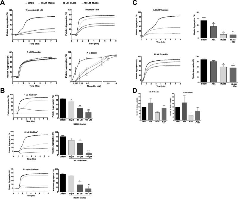
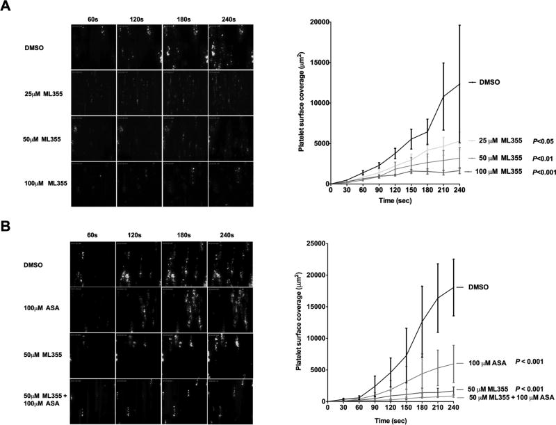
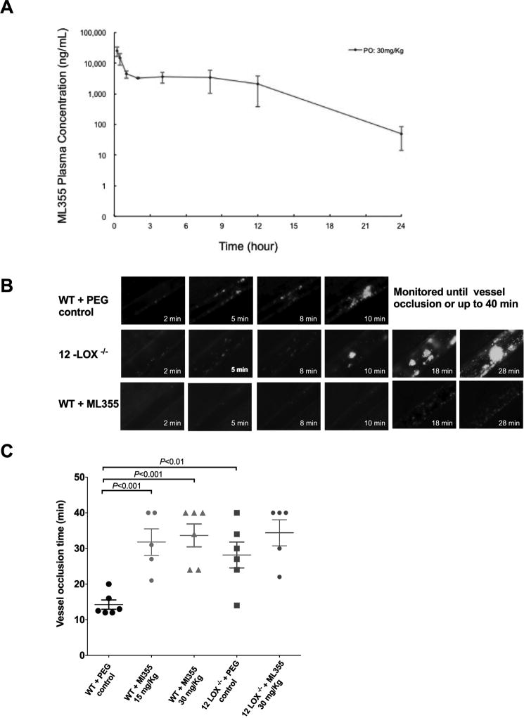
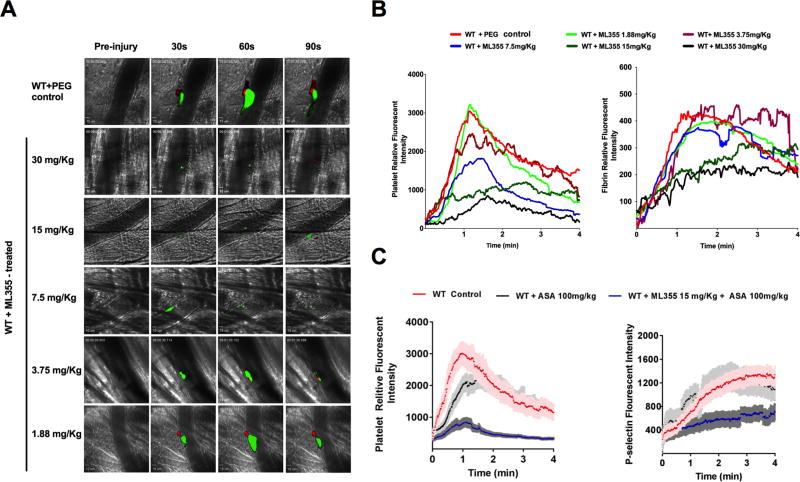
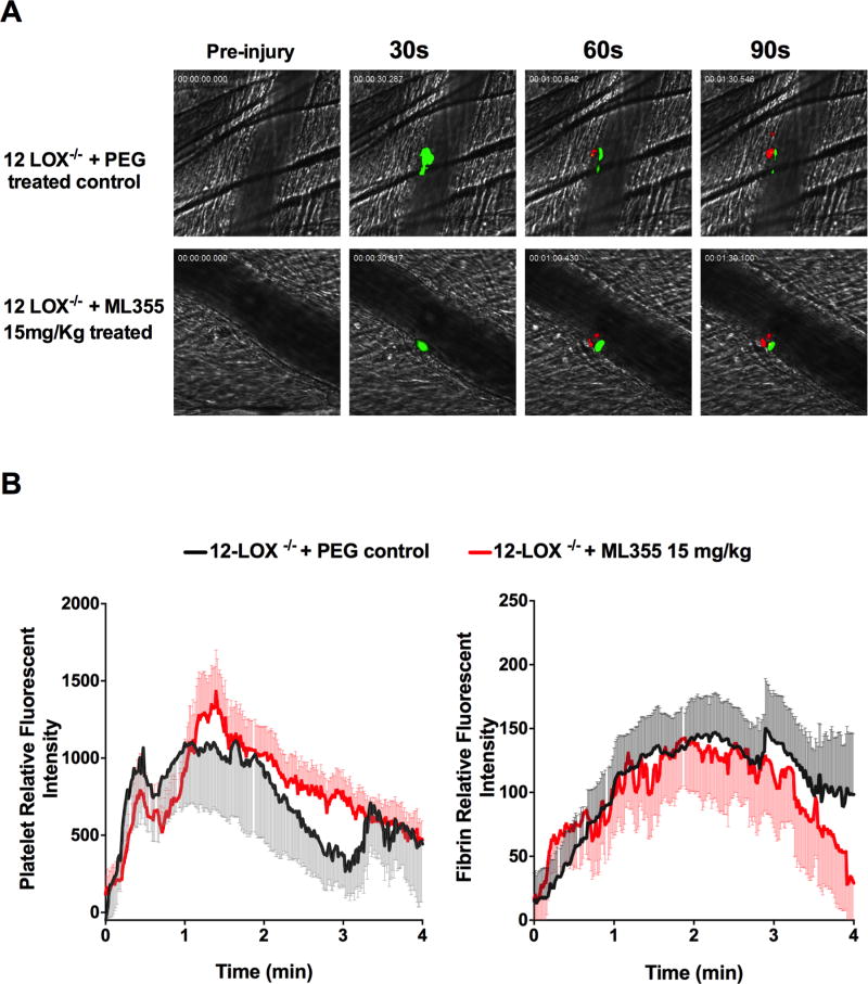
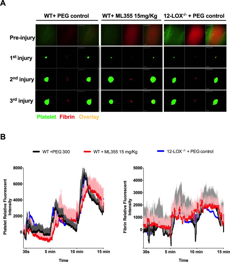
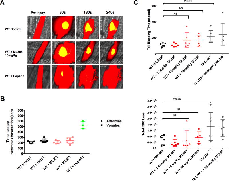
Approach and results: ML355 dose-dependently inhibited human platelet aggregation and 12-LOX oxylipin production, as confirmed by mass spectrometry. Interestingly, the antiplatelet effects of ML355 were reversed after exposure to high concentrations of thrombin in vitro. Ex vivo flow chamber assays confirmed that human platelet adhesion and thrombus formation at arterial shear over collagen were attenuated in whole blood treated with ML355 comparable to aspirin. Oral administration of ML355 in mice showed reasonable plasma drug levels by pharmacokinetic assessment. ML355 treatment impaired thrombus growth and vessel occlusion in FeCl3-induced mesenteric and laser-induced cremaster arteriole thrombosis models in mice. Importantly, hemostatic plug formation and bleeding after treatment with ML355 was minimal in mice in response to laser ablation on the saphenous vein or in a cremaster microvasculature laser-induced rupture model.

Conclusions: Our data strongly support 12-LOX as a key determinant of platelet reactivity in vivo, and inhibition of platelet 12-LOX with ML355 may represent a new class of antiplatelet therapy.

Keywords: collagen; hemostasis; lipoxygenase; platelet reactivity; thrombosis.


Figures

Figure 1
Figure 1. ML355 inhibits human platelet aggregation induced by various agonists
ML355 dose-dependently inhibits human platelet aggregation induced by various platelet agonists. Washed human platelets were incubated with 25–100 µM of ML355 or DMSO for 5 min and platelet aggregation was measured following stimulation with various platelet agonists. A) Thrombin-induced platelet aggregation (n=7 independent experiments). B) Platelet aggregation induced by PAR1-AP (1 µM), PAR4-AP (50 µM) and collagen (0.5 µg/mL). N=4 independent experiments. *, P <0.05; **, P < 0.01). C) ML355 inhibits human platelet aggregation in the presence of COX-1 inhibitor ASA. Washed human platelets were incubated with DMSO, 25 µM ML355, 100 µM ASA, or ML355 + ASA prior to aggregation assays. ML355 showed stronger inhibition when compared with ASA alone. D) ML355 treatment inhibited 12-HETE production in thrombin-stimulated human platelets. Human platelets (3×10⁸ platelets/mL) treated with DMSO, 25 µM ML355, 100 µM ASA or ML355 + ASA prior to activation by 0.25 nM thrombin (left panel) and 0.5 nM thrombin (right panel). 12-HETE production (ng/1×106 platelets) was determined by LC/MS/MS.
Figure 2
Figure 2. ML355 potently inhibits platelet function ex vivo under arterial shear
A) ML355 attenuates platelet adhesion, aggregation, and thrombus formation under arterial flow conditions. Human whole blood was pre-incubated with 25–100 µM of ML355 or DMSO for 5 min prior to being perfused over a collagen-coated surface at arterial shear rate (1800/s) for 4 min. Representative pictures of platelet adhesion and aggregation at the end of each minute of perfusion (upper panel). Dynamics of platelet surface coverage in each 30-sec interval (lower panel). N=8. P value determined by ANOVA analysis compared to DMSO condition. B) ML355 inhibits platelet adhesion and aggregation in the presence of COX-1 inhibitor ASA. Human whole blood was pre-incubated with DMSO, 25 µM ML355, 100 µM ASA, or ML355 + ASA prior to undergoing perfusion as described above.
Figure 3
Figure 3. ML355 treatment inhibited the formation of an occlusive thrombus in mesenteric artery in vivo
A) Concentrations of ML355 in mice plasma over time following 30mg/kg via oral gavage (PO). Values represent mean and standard deviation; N=3 per time point. Pharmacological inhibition of 12-LOX by ML355 impairs thrombus formation and vessel occlusion in FeCI3-induced mesenteric arteriole thrombosis model. B) Representative images of platelet adhesion, aggregation, and thrombus formation after application of FeCI3 on mesenteric arterioles in WT (upper), 12-LOX−/− (middle) treated with PEG control, and WT mice after orally administered with ML355 (30 mg/kg twice a day for 2 days; bottom). Time after FeCI3 injury is indicated. C) Vessel occlusion time in mesenteric arterioles in WT mice (red) with PEG control or treated with ML355 (15 mg/Kg 30 mg/kg) and 12-LOX −/− mice (grey) with or without ML355 treatments (30mg/Kg) (5 to 6 mice each group). Thrombus formation was recorded up to complete vessel occlusion or stopped at 40 min if occlusion did not occur following the topical application of FeCI3.
Figure 4
Figure 4. 12-LOX inhibition impairs thrombus formation in laser-induced cremaster arteriole thrombosis models
A) Representative images of platelet accumulation (green) and fibrin formation (red) in growing thrombi in cremaster arterioles in WT control treated with PEG (control, upper panel) and WT treated with ML355 (30 to 1.88 mg/kg, twice a day for 2 days, lower panels). B) Dynamics of platelet accumulation and fibrin formation in thrombi analyzed by change in fluorescent intensity. The kinetic curves represent the mean fluorescence intensity of platelet (left) and fibrin (right). Platelet recruitment was significantly inhibited with all of the doses of ML355 treatments above 1.88 mg/kg (P<0.001) while inhibition of fibrin was significant at only higher doses of ML355 (15, 30 mg/kg, (P<0.001) when compared to PEG-treated control (8–10 thrombi in each mouse, 3 mice in each group. Standard error (SEM) was not shown in the figures for clarity. C) ASA (100 mg/Kg) treatment impaired platelet recruitment without significantly effect P-selectin positive platelets within thrombi. However, 15 mg/kg ML355 and ASA dual treatments significantly inhibited platelet recruitment and platelet surface P-selectin expression in thrombi (P<0.001).
Figure 5
Figure 5. ML355 inhibition of thrombus formation requires platelet 12-LOX
A) Representative images of laser-induced cremaster arteriole thrombosis in 12-LOX−/− mice treated with PEG (control, upper panel) or treated with ML355 (15mg/kg, lower panel) twice a day for two days. Platelet accumulation is shown in green and fibrin formation is shown in red. Time after injury is indicated above. B) Dynamics of fluorescent intensity of platelet (right) and fibrin (left) in residual thrombi in 12-LOX−/− mice treated with PEG (control, shown in black) or treated with ML355 (15mg/kg, shown in red). The kinetic curves represent the mean fluorescence intensity of platelets and fibrin and the shaded regions are representative of the standard error (SEM) (8–10 thrombi in each mouse and 3 mice in each group, P>0.05).
Figure 6
Figure 6. ML355 treatment did not impair hemostatic plug formation in laser ablation saphenous vein hemostasis model
A) Representative images of platelet accumulation (green) and fibrin formation (red) within hemostatic plug forming at the injured site of repeated vascular injury on saphenous vein of WT mice treated with PEG (control) or with ML355 (15 mg/kg,) and 12 LOX−/− mice treated with PEG control twice a day for 2 days. B) Quantitative analysis of platelet accumulation and fibrin formation after laser ablation at 30 sec and repeated at 5 min and 10 min time points. The kinetic curves represent the mean platelet (left) and fibrin (right) fluorescence intensity and the shaded regions are representative of the standard error (SEM). ML355 treatment in WT mice attenuated platelet adhesion and accumulation (P<0.001) when compared to WT mice or 12 LOX−/− mice treated with PEG N= 6 (2 independent injuries in each mouse, 3 mice in each group).
Figure 7
Figure 7. ML355 treatment did not significantly increase bleeding as assessed by plasma extravasation following laser-induced rupture of cremaster microvasculature and tail bleeding assays
A) Fluorescein isothiocyanate-dextran (10,000 MW) was infused prior to laser injury in order to visualize the blood flow (plasma shown in red) in cremaster arteriole of WT mice treated with PEG control (upper panel), WT mice treated with ML355 (15 mg/kg, twice a day for 2 days; middle panel) and WT intravenously injected with 25U heparin 10 min prior to microscopy. The cremaster muscle arteriole wall was exposed to a high intensity laser pulse to puncture a hole, which resulted in plasma extravasation and subsequent formation of a platelet hemostatic plug (shown in yellow) leading to cessation of extravasation of plasma. B) The time required for the cessation of plasma dextran extravasation from arterioles (shown in circle), venules (shown in square) in PEG-treated WT control mice (shown in black), and ML355-treated WT mice (shown in red). Heparin-treated WT mice control shown in green. Data from 1–2 independent injuries per mouse, 3 mice in each group. P>0.05. C) Tail bleeding time and total red blood cell loss was assessed in WT treated with PEG control (Black), WT mice treated with ML355 (3.5 mg/kg,15 mg/kg and 30 mg/kg twice a day for 2 days; red) and 12 LOX−/− mice with or without ML355 (30 mg/kg) treatment (grey). Compared to PEG 300 treated control mice, ML355 treatment mice did not significantly increase tail-bleeding time all tested doses (P>0.05, upper panel) and total blood loss (lower panel, P>0.05) while 12 LOX−/− mice has increased tail bleeding time (P<0.01) and blood loss (P<0.05).

References

    1. Ruggeri ZM. Platelets in atherothrombosis. Nature medicine. 2002;8:1227–1234. - PubMed
    1. Go AS, Mozaffarian D, Roger VL, et al. American Heart Association Statistics C, Stroke Statistics S. Executive summary: Heart disease and stroke statistics--2014 update: A report from the american heart association. Circulation. 2014;129:399–410. - PubMed
    1. Steg PG, Dorman SH, Amarenco P. Atherothrombosis and the role of antiplatelet therapy. J Thromb Haemost. 2011;9(Suppl 1):325–332. - PubMed
    1. Ikei KN, Yeung J, Apopa PL, Ceja J, Vesci J, Holman TR, Holinstat M. Investigations of human platelet-type 12-lipoxygenase: Role of lipoxygenase products in platelet activation. J Lipid Res. 2012;53:2546–2559. - PMC - PubMed
    1. Steele VE, Holmes CA, Hawk ET, Kopelovich L, Lubet RA, Crowell JA, Sigman CC, Kelloff GJ. Lipoxygenase inhibitors as potential cancer chemopreventives. Cancer Epidemiol Biomarkers Prev. 1999;8:467–483. - PubMed

Publication types

MeSH terms