. 2018 Jan 30;137(5):488-503.
doi: 10.1161/CIRCULATIONAHA.117.028533. Epub 2017 Aug 3.

Chronic Rejection of Cardiac Allografts Is Associated With Increased Lymphatic Flow and Cellular Trafficking

Affiliations

Chronic Rejection of Cardiac Allografts Is Associated With Increased Lymphatic Flow and Cellular Trafficking

Lindsey A Edwards et al. Circulation. .

Abstract

Background: Cardiac transplantation is an excellent treatment for end-stage heart disease. However, rejection of the donor graft, in particular, by chronic rejection leading to cardiac allograft vasculopathy, remains a major cause of graft loss. The lymphatic system plays a crucial role in the alloimmune response, facilitating trafficking of antigen-presenting cells to draining lymph nodes. The encounter of antigen-presenting cells with T lymphocytes in secondary lymphoid organs is essential for the initiation of alloimmunity. Donor lymphatic vessels are not anastomosed to that of the recipient during transplantation. The pathophysiology of lymphatic disruption is unknown, and whether this disruption enhances or hinders the alloimmune responses is unclear. Although histological analysis of lymphatic vessels in donor grafts can yield information on the structure of the lymphatics, the function following cardiac transplantation is poorly understood.

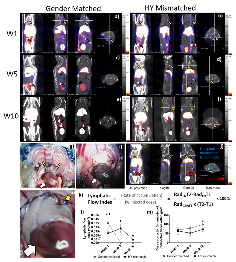
Methods: Using single-photon emission computed tomography/computed tomography lymphoscintigraphy, we quantified the lymphatic flow index following heterotrophic cardiac transplantation in a murine model of chronic rejection.

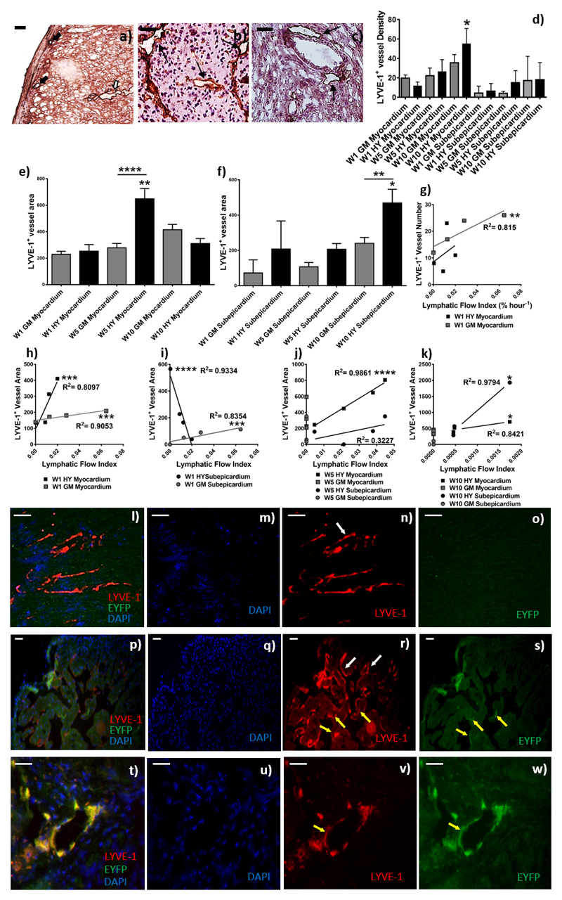
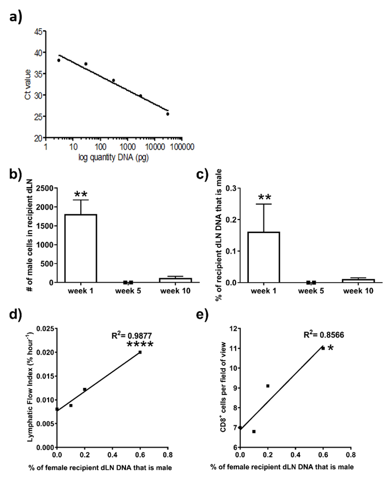
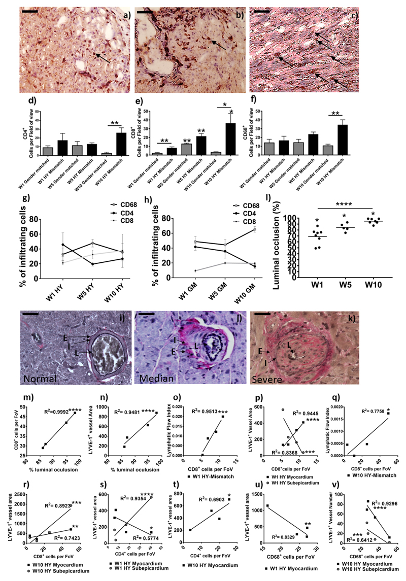
Results: Ten weeks following transplantation of a minor antigen (HY) sex-mismatched heart graft, the lymphatic flow index was significantly increased in comparison with sex-matched controls. Furthermore, the enhanced lymphatic flow index correlated with an increase in donor cells in the mediastinal draining lymph nodes; increased lymphatic vessel area; and graft infiltration of CD4+, CD8+ T cells, and CD68+ macrophages.

Conclusions: Chronic rejection results in increased lymphatic flow from the donor graft to draining lymph nodes, which may be a factor in promoting cellular trafficking, alloimmunity, and cardiac allograft vasculopathy.

Keywords: immune system; inflammation; rejection; transplantation.


Conflict of interest statement

Disclosures

The authors have no conflicts of interest to disclose

Figures

Figure 1
Figure 1. Quantification of the Lymphatic Flow Index from the cardiac grafts to the mediastinal lymph nodes.
SPECT/CT images were taken after injection of nanocoll into the apex of transplanted hearts. Representative images are shown (n = 4-5 per group) in four planes the anterior-posterior view, sagittal, coronal and transverse taken at the same point of the “crosshairs” (white). One (a-b), five (c-d) or ten (e-f) weeks after either gender-matched (a, c & e) or HY-mismatched (b, d & f) abdominal heart transplantation, the animal was scanned 30 min and four hours after nanocoll injection. Images are shown at four-hours post-injection. Representative examples of direct visualisation of lymph drainage from donor heart grafts after transplantation using Evan's Blue injection (n=3 in each group). Recipient of HY-mismatched donor graft showing mdLN, above the native heart (white arrows), staining blue with Evan's Blue dye (g & h, yellow arrows). Recipient of gender-matched donor graft showing mediastinal not staining blue (i). A sphere is drawn around each “region of interest” (j; blue & red sphere) and the radioactivity quantified in the mdLN (j; Yellow arrow; RadLN) and the transplanted donor cardiac graft (j; Red arrow; Radgraft). Decay corrected values are used to quantify the lymphatic flow index with T1 being the time of the first scan and T2 being the time of the second scan (k). Shown here is the mean ± SEM, of four to five animals at each time point, of the lymphatic flow index (l) or the percentage loss of injected radiation from the graft (m); with gender-matched and HY-mismatched grafts at weeks, one, five and ten (W1**p = 0.0080, W5*p = 0.0286 & W10*p = 0.0156).
Figure 2
Figure 2. Quantification of lymphatic density in cardiac donor grafts and determination of the origin of LYVE-1+ lymphatic vessels; Lymphatic flow index correlates with lymphatic vessel area, but not density.
After SPECT/CT scanning donor grafts were harvested and used for immunofluorescence and immunohistochemistry. Images are representative examples of four independent experiments; scale bars = 50 μm. Immunohistochemical staining for LYVE-1 was conducted in order to quantify the density of subepicardial (a, the black arrows) and myocardial (a, the white arrows) lymphatic vessels. Shown is a representative of gender-matched (b) and HY-mismatched grafts (c) at ten-weeks post-transplant. The black arrows show LYVE-1+ vessels (magnification 200x). LYVE-1+ vessels density (d, *p=0.038) and myocardial (e, ****p <0.0001) and subepicardial (f, *p = 0.0091) area (μm2) was determined by two independent observers in at least 10 medium power fields, with area determined with Image J software. Data are shown as a mean of the two independent observations and four independent experiments ± SEM; of gender-matched (GM) and HY-mismatched (HY) grafts at weeks, one, five and ten. The lymphatic flow index from each individual mouse was correlated with the corresponding LYVE-1+vessel number (g, *p=0.0021) and area at week one (h, Myocardium, HY ***p=0.0008, GM ***p=0.0003; i, subepicardium, HY ****p = 0.0008, GM ***p=0.0003), week five (j, ****p <0.0001) and week ten (k, *p=0.0222). To determine if vessels were of the donor or recipient origin, immunofluorescence was performed with anti-LYVE-1 and anti-YFP. Donor lymphatic vessels are EYFP- and LYVE-1+(l-s; white arrows). However, the transplant recipients express EYFP and thus recipient lymphatic vessels are EYFP+ and LYVE-1+(t-w; yellow arrows). Shown are a representative of lymphatic vessel staining in gender-matched grafts (l-o) and HY-mismatched grafts (p-s) ten-weeks post-transplant. A representative high power image showing a recipient lymphatic vessel within a donor organ is shown (t-w).
Figure 3
Figure 3. QPCR for donor cells in draining lymph nodes of transplant recipients.
After SPECT/CT imaging, the mdLN were harvested. Genomic DNA was isolated and the Y-chromosome specific gene, Zfy1, was amplified using quantitative Real-time PCR. The presence of male donor-derived DNA in the female recipient lymph nodes at week one, five and ten post-transplant was calculated against a standard curve (a) with a known percentage of male DNA, which corresponds to a known number of cells. Allowing for quantification of the number of male donor-derived cells (b, **p = 0.0019) as a percentage (c, **p = 0.0019) of the total number of cells within the female lymph node. Shown is the mean ±SD of seven mice. The percentage of male cells present in the draining mediastinal lymph nodes at week one post-transplant from each individual mouse was correlated with the corresponding lymphatic flow index (d, ****p <0.0001) and the number of CD8+ cells (e, *p = 0.0081) within the donor graft at week one; the number of CD4+ cells; CD68+ cells; the number of vessels and the area of vessels within the donor graft at week one.
Figure 4
Figure 4. Determination of graft vasculopathy and inflammatory cellular infiltration within the transplanted donor grafts.
After SPECT/CT scanning donor grafts were harvested, immunohistochemical staining for CD4 (a) CD8 (b), and CD68 (c) were conducted to determine cell numbers (black arrows show an example); shown is a representative of HY-mismatched grafts at ten weeks post-transplant. Images are representative examples of 4 independent experiments; scale bars = 50 μm. Positive CD4 (d, **p = 0.0079) and CD8 (e, W5GM**p =0.0019, W5HY**p = 0.0066, W10HY**p = 0.0429, W1GM vs. HY**p = 0.0077, W10GM vs. HY*p = 0.0233) cells were counted in 20 random high-power fields (magnification 400x) by two independent observers. For CD68+ macrophage staining (f, W10**p = 0.0079), a 63-point grid was overlaid onto a 400x magnification of macrophage staining. The percentages of grid points positive for CD68 staining were counted by two independent observers. Data are shown as the mean number of cells, per field of view (FoV), of the two independent observations and four independent experiments ± SEM; of a gender-matched and HY-mismatched grafts at weeks, one, five and ten. Within the infiltrating cells, the percentage of CD68+, CD4+ and CD8+ cells was determined in the HY-mismatched (g) and gender-matched grafts (h). Immunohistochemical staining for Elastic Van Gieson was performed to determine graft vasculopathy. Shown is a representative of a gender-matched (i) and HY-mismatched grafts (j & k). Histologically the allograft vasculopathy ranged from median (j) to severe (k) in the HY mismatched graft. Characterised by inflammatory cell invasion into the layers of the internal to external elastic; shown is the lumen of the vessel (L), the internal elastic (I) and the external elastic (E). Measurement of the internal elastic (I) and the external elastic (E) can be used to calculate the percentage of luminal occlusion (l, W1*p=0.0121, W5*p=0.0357 & W10*p=0.0238; ****p<0.0001). Shown is the percentage luminal occlusion in four independent experiments with mean at one, five and ten weeks. The percentage luminal occlusion was correlated with CD8+ cell number (m, ****p<0.0001) and the mean lymphatic vessel area (n, ****p<0.0001). CD8+ cell number was correlated with the LFI (o, W1****p<0.0001 & q, W10*p=0.0218) and the lymphatic vessel area (p, myocardial W1****p<0.0001, subepicardial W1***p=0.0008 & r, myocardial W10**p=0.006, subepicardial W10***p=0.0004). CD4+ cell number was correlated with the lymphatic vessel area (s, myocardial W1*p=0.0287 & subepicardial W1****p<0.0001 & t, W10*p=0.0206). CD68+ cell number was correlated with the lymphatic vessel area (u, W1**p=0.0016) and density (v, myocardial W10****p<0.0001 & subepicardial W10***p=0.0008).

Comment in

References

    1. Taylor R, Lannon J, Wong E, Collett D. Annual Report on Cardiothoracic Transplantation. Statistics & Clinical Studies, NHS Blood & Transplant. 2015:1–131. http://www.odt.nhs.uk/pdf/organ_specific_report_cardiothoracic_2015.pdf.
    1. Sayegh MH, Carpenter CB. Transplantation 50 years later--progress, challenges, and promises. N Engl J Med. 2004;23:2761–6. - PubMed
    1. Lund LH, Edwards LB, Kucheryavaya AY, Benden C, Christie JD, Dipchand AI, Dobbels F, Goldfarb SB, Levvey BJ, Meiser B, Yusen RD, et al. The Registry of the International Society for Heart and Lung Transplantation: Thirty-first Official Adult Heart Transplant Report 2014; Focus Theme: Retransplantation. The Journal of Heart and Lung Transplantation. 2014;33:996–1008. - PubMed
    1. Nykanen AI, Sandelin H, Krebs R, Keranen MA, Tuuminen R, Karpanen T, Wu Y, Pytowski B, Koskinen PK, Yla-Herttuala S, Alitalo K, et al. Targeting lymphatic vessel activation and CCL21 production by vascular endothelial growth factor receptor-3 inhibition has novel immunomodulatory and antiarteriosclerotic effects in cardiac allografts. Circulation. 2010;121:1413–22. - PubMed
    1. Hasegawa T, Visovatti SH, Hyman MC, Hayasaki T, Pinsky DJ. Heterotopic vascularized murine cardiac transplantation to study graft arteriopathy. Nat Protoc. 2007;2:471–80. - PubMed

Publication types

MeSH terms