. 2018 Feb;176(2):1262-1285.
doi: 10.1104/pp.17.00478. Epub 2017 Aug 3.

OsbZIP48, a HY5 Transcription Factor Ortholog, Exerts Pleiotropic Effects in Light-Regulated Development

Affiliations

OsbZIP48, a HY5 Transcription Factor Ortholog, Exerts Pleiotropic Effects in Light-Regulated Development

Naini Burman et al. Plant Physiol. 2018 Feb.

Abstract

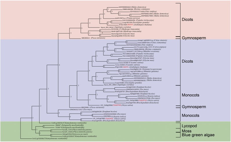
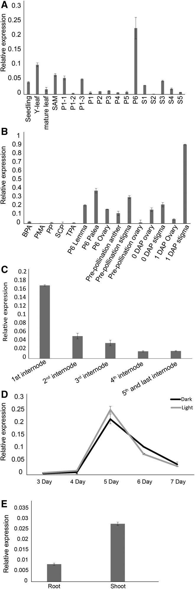
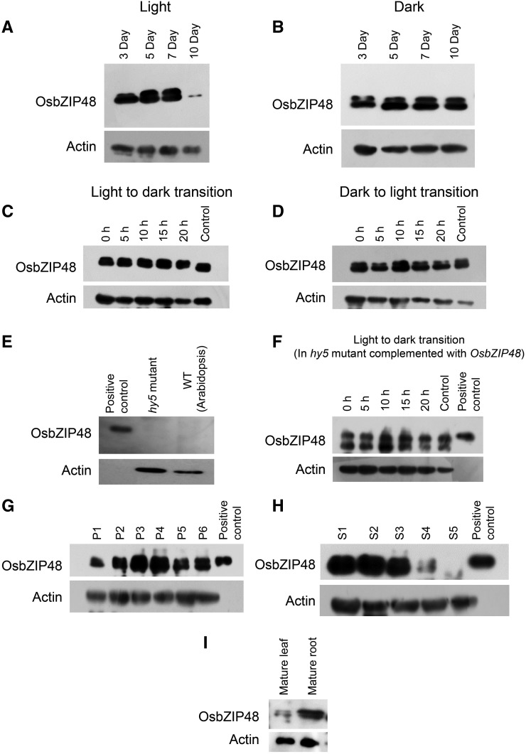
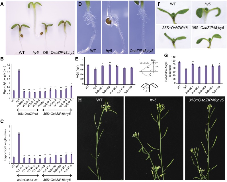
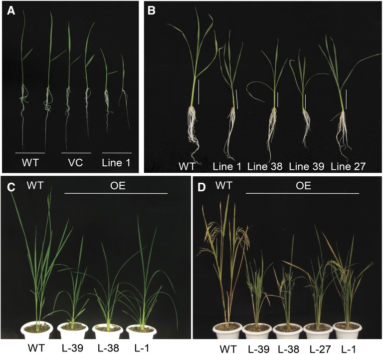
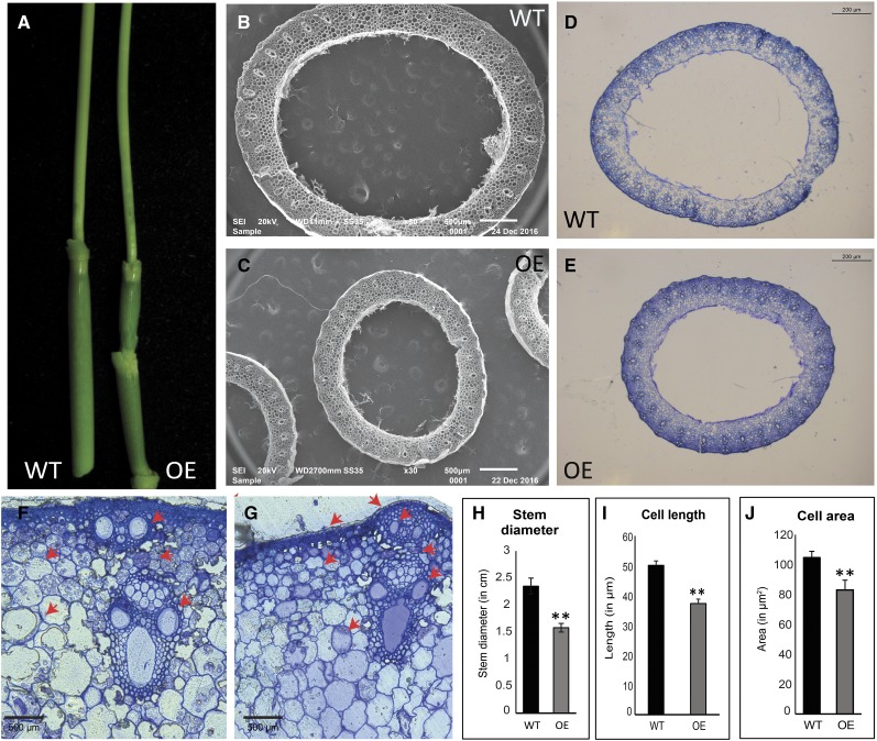
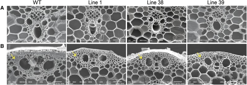
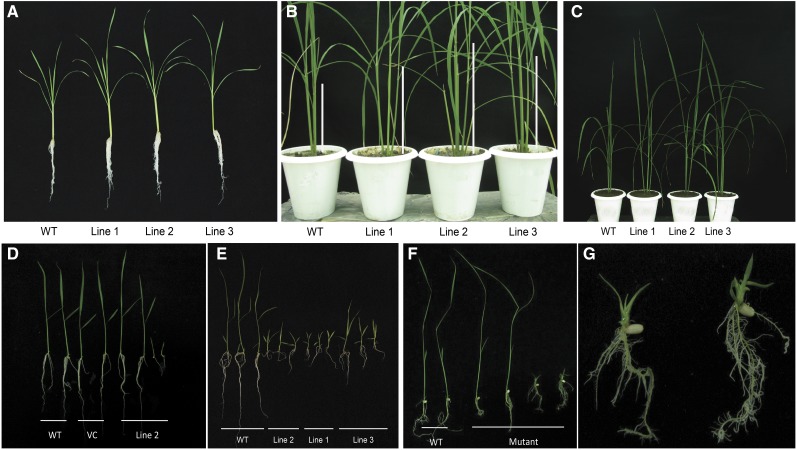
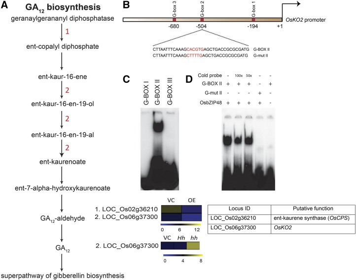
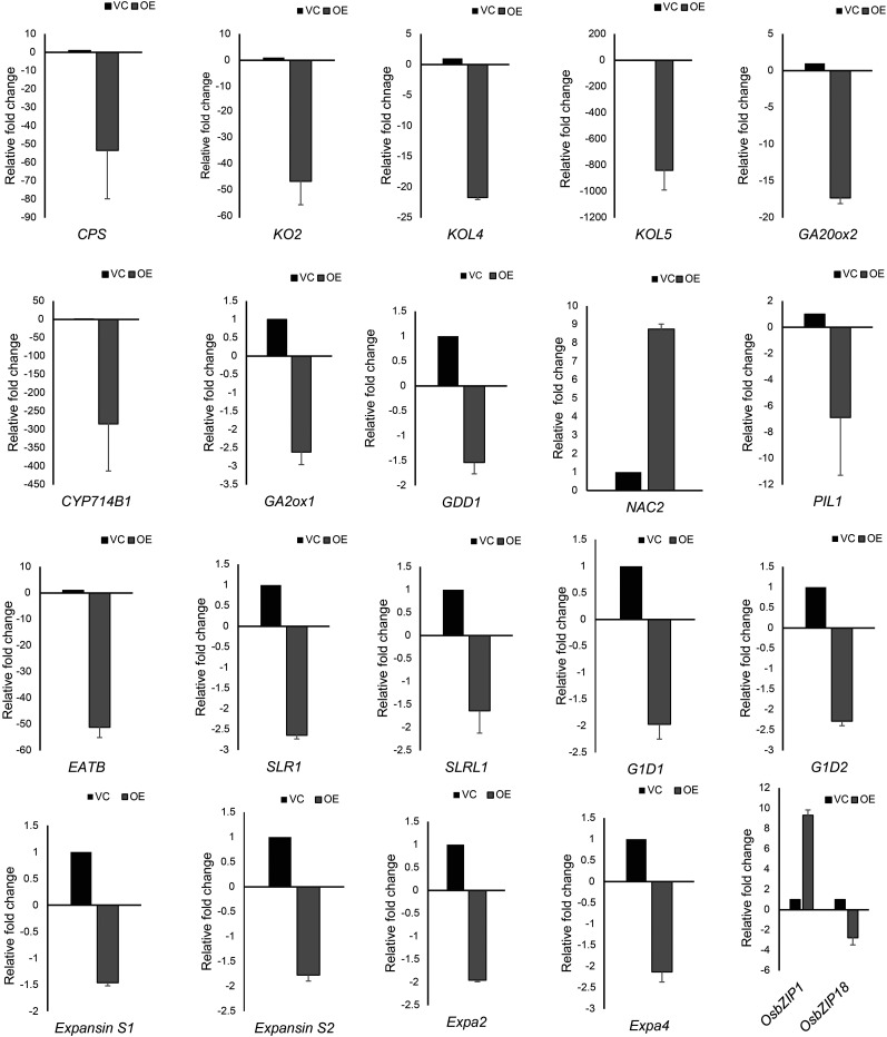
Plants have evolved an intricate network of sensory photoreceptors and signaling components to regulate their development. Among the light signaling components identified to date, HY5, a basic leucine zipper (bZIP) transcription factor, has been investigated extensively. However, most of the work on HY5 has been carried out in Arabidopsis (Arabidopsis thaliana), a dicot. In this study, based on homology search and phylogenetic analysis, we identified three homologs of AtHY5 in monocots; however, AtHYH (HY5 homolog) homologs are absent in the monocots analyzed. Out of the three homologs identified in rice (Oryza sativa), we have functionally characterized OsbZIP48OsbZIP48 was able to complement the Athy5 mutant. OsbZIP48 protein levels are developmentally regulated in rice. Moreover, the OsbZIP48 protein does not degrade in dark-grown rice and Athy5 seedlings complemented with OsbZIP48, which is in striking contrast to AtHY5. In comparison with AtHY5, which does not cause any change in hypocotyl length when overexpressed in Arabidopsis, the overexpression of full-length OsbZIP48 in rice transgenics reduced the plant height considerably. Microarray analysis revealed that OsKO2, which encodes ent-kaurene oxidase 2 of the gibberellin biosynthesis pathway, is down-regulated in OsbZIP48OE and up-regulated in OsbZIP48KD transgenics as compared with the wild type. Electrophoretic mobility shift assay showed that OsbZIP48 binds directly to the OsKO2 promoter. The RNA interference lines and the T-DNA insertional mutant of OsbZIP48 showed seedling-lethal phenotypes despite the fact that roots were more proliferative during early stages of development in the T-DNA insertional mutant. These data provide credible evidence that OsbZIP48 performs more diverse functions in a monocot system like rice in comparison with its Arabidopsis ortholog, HY5.


Figures

Figure 1.
Figure 1.
Phylogenetic tree of HY5 and HYH homologous proteins from across species (asterisks represent manually reannotated proteins, and daggers represent incomplete proteins even after manual reannotation but having the bZIP domain). The consensus tree was generated after merging the individual trees generated by phyml, neighbor joining, and the maximum parsimony approach. The numbering at the nodes represents the number of trees (generated by three different methods) that have the same topology as the consensus tree.
Figure 2.
Figure 2.
Expression profile of OsbZIP48 in various tissues and at different stages of development. A, Expression of OsbZIP48 in vegetative (seedling, young [Y] leaf, mature leaf, and shoot apical meristem [SAM]), panicle, and seed stages of development in rice variety IR64 as analyzed by real-time PCR. (Panicle stages are as follows: P1-1, 0.5–2 mm; P1-2, 2–5 mm; P1-3, 5–10 mm; P1, 0–3 cm; P2, 3–5 cm; P3, 5–10 cm; P4, 10–15 cm; P5, 15–22 cm; and P6, 22–30 cm. Seed stages are as follows: S1, 0–2 DAP; S2, 3–4 DAP; S3, 5–10 DAP; S4, 11–20 DAP; and S5, 21–29 DAP.) B, Real-time PCR analysis of OsbZIP48 using different organs of the inflorescence: PMA, premeiotic anther; SCP, single-cell pollen; and TPA, trinucleate pollen anther. C, Real-time PCR analysis to check the expression of OsbZIP48 in different internodes of the mature rice stem. D, Expression analysis of OsbZIP48 using real-time PCR in 3- to 7-d-old light- and dark-grown rice seedlings. E, Expression analysis of OsbZIP48 root and shoot of 5-d-old light-grown seedlings using real-time PCR. Data shown are means ± se. The expression data presented are relative to UBIQUITIN5.
Figure 3.
Figure 3.
Western blots showing OsbZIP48 protein expression levels in different tissues of rice and the Arabidopsis hy5 mutant complemented with OsbZIP48. A, OsbZIP48 protein levels in 3-, 5-, 7-, and 10-d-old light-grown rice seedlings (100 μmol m−2 s−1). B, OsbZIP48 protein levels in 3-, 5-, 7-, and 10-d old dark-grown rice seedlings. C, OsbZIP48 protein levels in seedlings grown in continuous light for 4 d and then transferred to dark for 5, 10, 15, and 20 h; the control is 5-d-old seedlings grown in continuous light. D, OsbZIP48 protein levels in seedlings grown in continuous dark for 4 d and then transferred to the light for 5, 10, 15, and 20 h; 5-d-old seedlings grown in continuous dark were used as the control. E, Western blot using OsbZIP48 antibodies shows no cross-reactivity with Athy5 mutant protein extracts. F, OsbZIP48 protein levels in Arabidopsis hy5 mutant seedlings complemented with OsbZIP48, grown in continuous light for 4 d, and then transferred to the dark for 5, 10, 15, and 20 h; control represents 5-d-old seedlings grown in continuous light. G, Changes in OsbZIP48 protein levels during various stages of panicle development in rice. H, OsbZIP48 protein levels during seed development (S1–S5) stages in rice. I, OsbZIP48 protein levels in mature leaf and root in rice. The positive control in E to H is bacterially expressed 6× His-tagged OsbZIP48 protein.
Figure 4.
Figure 4.
OsbZIP48 is localized in the nucleus, forms a homodimer, and lacks transactivation activity. A, Particle bombardment of the YFP-OsbZIP48 construct in onion cells. The first column shows photographs taken in dark field, and the second column shows merged photographs of dark field and bright field captured using a Leica microscope. The first row (YFP control vector) shows localization of only YFP protein, the second row (OsbZIP48-YFP) shows localization of OsbZIP48 tagged to YFP protein, and the third row (DAPI) shows DAPI-stained nucleus. B, Prebleach and postbleach images showing bleaching of YFP-OsbZIP48 for FRET analysis. C, Histogram showing FRET efficiency of CFP-OsbZIP48 and YFP-OsbZIP48 interaction as compared with the controls. Data shown are means ± se; n = 10. D, BiFC analysis using onion peel cells showing the homodimerization of nEYFPC1-OsbZIP48 and cEYFPC1-OsbZIP48 in the nucleus. E, Transactivation assay of OsbZIP48 in yeast cells. OsbZIP48 lacks transactivation activity, as the yeast cells containing the OsbZIP48-pGBKT construct were unable to grow on SD-HW medium (synthetic defined medium without histidine and tryptophan amino acids).
Figure 5.
Figure 5.
Phenotypic analyses of Arabidopsis hy5 mutant seedlings/plants overexpressing OsbZIP48. A, Phenotypes of 3-d-old white light-grown wild-type (WT), hy5, OsbZIP48OE, and OsbZIP48;hy5 seedlings. B and C, Hypocotyl lengths of 3- and 6-d-old white light (200 μmol m−2 s−1)-grown wild-type, hy5, OsbZIP48OE, and OsbZIP48;hy5 seedlings. D and E, VGI of roots of 3-d-old wild-type, hy5, and OsbZIP48;hy5 seedlings. F and G, Cotyledon opening angle in response to white light. H, Altered gravitropic set angle in siliques of Arabidopsis hy5 mutant plants. Data presented are means ± se, n = 15 plants in each case. Statistically significant differences (*, P < 0.05 and **, P < 0.005) were identified by Dunnett’s test using the wild type as a control for overexpression transgenics and the hy5 mutant as a control for OsbZIP48;hy5 transgenics in B and C and the hy5 mutant as a control in E and G.
Figure 6.
Figure 6.
Phenotypes of OsbZIP48OE rice transgenics at different developmental stages. A, Photograph of 10-d-old seedlings of the wild type (WT), pB4NU vector control (VC), and OsbZIP48OE transgenics grown in white light (75 μmol m−2 s−1). B, Photograph of 30-d-old seedlings grown in white light (75 μmol m−2 s−1). C, Photograph of plants at the vegetative phase of life. D, Photograph of plants grown in a greenhouse at the reproductive stage.
Figure 7.
Figure 7.
Phenotypic comparison of the stems of wild-type (WT) and OsbZIP48OE rice transgenic plants. A, Photograph showing the difference in the stem diameter of mature green plants. B and C, Scanning electron microscopic images showing differences in the diameter of the stems of wild-type and OsbZIP48OE transgenic plants, respectively. D and E, Methylene Blue-stained transverse sections of wild-type and OsbZIP48OE transgenic stems showing differences in the diameter of the stems taken at 2.5× magnification. F and G, Methylene Blue-stained transverse section of wild-type and OsbZIP48OE transgenic stems, with red arrows showing differences in the size of the vascular bundle, parenchyma cells, and the secondary cell wall thickening of the sclerenchyma cells. H to J, Histograms showing differences in stem diameter, cell length, and cell area of the wild type and overexpression transgenics, respectively. Cortical cells were used to measure cell length and cell area. Data presented are means ± se, n = 10 in each case. Statistically significant differences (*, P < 0.05 and **, P < 0.005) were identified by Student’s t test.
Figure 8.
Figure 8.
Scanning electron microscopic images of second last internodes of wild-type (WT) and OsbZIP48OE transgenic plants at different magnifications showing the size of vascular bundles and secondary cell wall thickenings. In B, the yellow arrows show the thickness of secondary cell wall thickenings in wild-type and OsbZIP48OE transgenic plants.
Figure 9.
Figure 9.
Phenotypes of OsbZIP48KD lines and the T-DNA insertion mutant of OsbZIP48. A, T1 OsbZIP48KD lines showing profuse root growth at the seedling stage as compared with the wild type (WT). B and C, T1 OsbZIP48KD plants at the vegetative stage showing increased height at two different magnifications; note the elongated stem of OsbZIP48KD plants in B. D, T2 OsbZIP48KD transgenics showing two types of plants; seedlings of the 10-d-old wild type, pANDA vector control (VC), and T2 OsbZIP48KD transgenics grown in white light (75 μmol m−2 s−1). E, T2 OsbZIP48KD 10-d-old transgenics showing a lethal phenotype in multiple lines. F, Photograph of 15-d-old wild-type and OsbZIP48 mutant lines showing their phenotypes. G, T-DNA insertional mutant plants showing profuse rooting (at higher magnification).
Figure 10.
Figure 10.
A, Schematic representation of the GA biosynthesis pathway showing altered gene expression in OsbZIP48OE and OsbZIP48KD transgenics. The color bar at the base represents log2 expression values, with blue representing low-level expression, black representing medium-level expression, and yellow signifying high-level expression. The numbers in red in the pathway correspond to the serial number of the locus identifier in the heat map (i.e. the gene with the serial number performs in the step where it is mentioned in the pathway). VC, Vector control. B, Schematic representation of the OsKO2 1-kb promoter showing the location of three G-box motifs. In the diagram, the sequence of the probe is given with the G-box sequence (CACGTG) highlighted in red. The G-mut II probe sequence shows the mutated G-box sequence and is highlighted in red. These probes were labeled with 32P for electrophoretic mobility shift assay (EMSA). C, OsbZIP48 binds in vitro to G-box II in the OsKO2 promoter in EMSA. D, EMSA gel showing OsbZIP48 in vitro binding to G-box II in the OsKO2 promoter with proper controls. For the assay, the radiolabeled probes were incubated with OsbZIP48 protein. Cold (unlabeled) probe (100× or 50×), G-box II probe, and G-mut II probe were used as indicated.
Figure 11.
Figure 11.
Real-time PCR-based expression analysis of genes known to be involved in regulating plant height in OsbZIP48OE seedlings. The last histogram shows the transcript levels of OsbZIP1 and OsbZIP18 in the OsbZIP48OE line. Data shown are means ± se. VC, Vector control.

References

    1. Alabadí D, Gallego-Bartolomé J, Orlando L, García-Cárcel L, Rubio V, Martínez C, Frigerio M, Iglesias-Pedraz JM, Espinosa A, Deng XW, et al. (2008) Gibberellins modulate light signaling pathways to prevent Arabidopsis seedling de-etiolation in darkness. Plant J 53: 324–335 - PubMed
    1. Ang LH, Chattopadhyay S, Wei N, Oyama T, Okada K, Batschauer A, Deng XW (1998) Molecular interaction between COP1 and HY5 defines a regulatory switch for light control of Arabidopsis development. Mol Cell 1: 213–222 - PubMed
    1. Bandyopadhyay A, Blakeslee JJ, Lee OR, Mravec J, Sauer M, Titapiwatanakun B, Makam SN, Bouchard R, Geisler M, Martinoia E, et al. (2007) Interactions of PIN and PGP auxin transport mechanisms. Biochem Soc Trans 35: 137–141 - PubMed
    1. Bennett MJ, Marchant A, Green HG, May ST, Ward SP, Millner PA, Walker AR, Schulz B, Feldmann KA (1996) Arabidopsis AUX1 gene: a permease-like regulator of root gravitropism. Science 273: 948–950 - PubMed
    1. Bradford MM. (1976) A rapid and sensitive method for the quantitation of microgram quantities of protein utilizing the principle of protein-dye binding. Anal Biochem 72: 248–254 - PubMed

Publication types

MeSH terms