. 2017 Oct 1;77(19):5236-5247.
doi: 10.1158/0008-5472.CAN-16-3089. Epub 2017 Aug 3.

Loss of Igf2 Gene Imprinting in Murine Prostate Promotes Widespread Neoplastic Growth

Affiliations

Loss of Igf2 Gene Imprinting in Murine Prostate Promotes Widespread Neoplastic Growth

Nathan A Damaschke et al. Cancer Res. .

Abstract

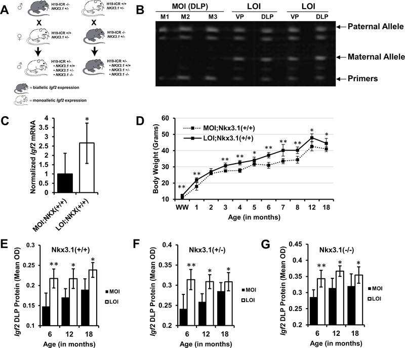
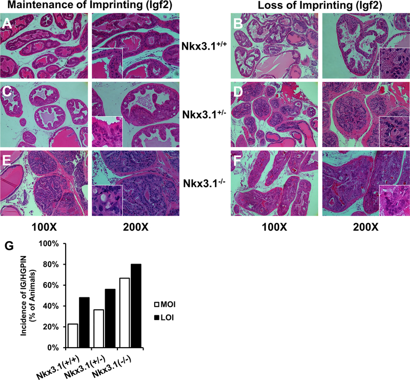
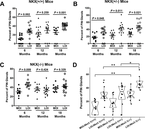
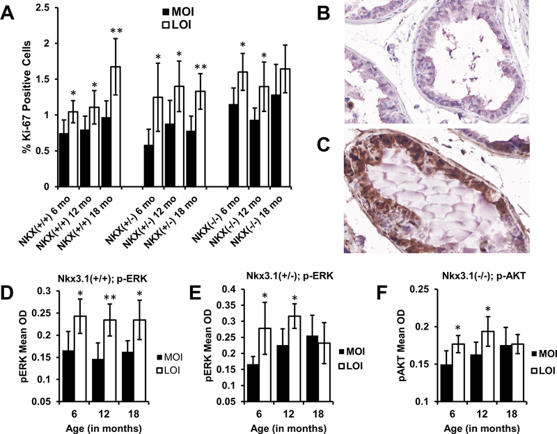
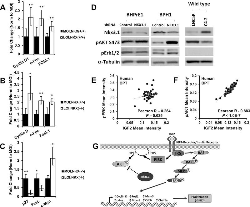
Loss of imprinting (LOI) is an epigenetic event that relaxes an allele-specific restriction on gene expression. One gene that experiences LOI is the paracrine insulin-like growth factor IGF2, which occurs commonly in human prostate tissues during aging and tumorigenesis. However, the relationship between IGF2 LOI and prostate tumorigenesis has not been established functionally. In this study, we created a mouse model with CTCF-binding site mutations at the Igf2-H19 imprint control region that abolishes CTCF insulator activity, resulting in biallelic Igf2 expression that mimics increased levels seen with aging-induced LOI. We found that Igf2 LOI increased the prevalence and severity of prostatic intraepithelial neoplasia (PIN), a premalignant lesion. Engineering Nkx3.1 deficiency into our model increased the frequency of PIN lesions in an additive fashion. Prostates harboring LOI displayed increased MAPK signaling and epithelial proliferation. In human prostate tissue arrays, we documented a positive correlation in benign tissues of IGF2 levels with phospho-ERK and phospho-AKT levels. Overall, our results establish that Igf2 LOI is sufficient on its own to increase rates of neoplastic development in the prostate by upregulating critical cancer-associated signaling pathways. Cancer Res; 77(19); 5236-47. ©2017 AACR.


Figures

Figure 1.
Figure 1.. Biallelic expression of Igf2 in 142* mice.
A, Breeding schemes for generation of maintenance (monoallelic Igf2) and loss of imprinting (biallelic Igf2) mice. Maternal transmission of 142* mutation (H19-ICR +/−) results in biallelic expression in offspring by abolishing CTCF insulator activity at the normally silenced maternal allele. Paternal transmission leads to the typical monoallelic expression. B, Ventral prostate (VP) and Dorsolateral prostate (DLP) tissues were analyzed for maintenance of imprinting (MOI) and loss of imprinting (LOI) in mice and maternal and paternal allelic expression was quantitated using FluPE as described in methods. MOI mice maintain expression from a single allele, while LOI mice exhibit biallelic expression. C, Igf2 mRNA expression measured by qPCR in mouse prostates (*P<0.05, mean+SD; n = 6 each group). D, Body weights of experimental animals were recorded throughout their lifetime. LOI animals expressing biallelic Igf2 were persistently heavier throughout their life (mean+95% CI; **P<0.01, *P<0.05). E, F, G, Igf2 protein expression was quantitatively analyzed on tissue microarrays constructed from mouse prostate tissues. Igf2 protein expression was significantly elevated in LOI animals with (E) Nkx3.1+/+ (P=0.001, P=0.015, P=0.012; 6, 12, 18 months), (F) Nkx3.1+/− (P=0.002, P=0.02, P=0.04; 6, 12, 18 months) and (G) Nkx3.1−/− (P<0.001, P=0.029, P=0.043; 6, 12, 18 months), backgrounds across all ages (*P<0.05, columns represent Mean+SD; n > 8 for each group).
Figure 2.
Figure 2.. Histopathology of PIN lesions and demonstration of increased prevalence of PIN in LOI mice.
H&E stained FFPE mouse prostate tissues matched by Nkx3.1 status (horizontally) and Igf2 imprint status (vertically) at 12 mo. PIN grading was performed as described by Park et al. (25) and low (PIN 1 and 2) versus intermediate/high grade (PIN 3 and 4) lesions were based on the most severe lesion within the prostate. (A) In normal MOI;Nkx3.1+/+ prostate tissues, glands are lined with columnar epithelial cells and exhibit uniform staining of cytoplasmic and nucleic structures (400X Inset). Low grade (LG) PIN lesions (B,C) were seen in all genotypes surveyed, marked by nuclear pleomorphism and mild disorganization of cell layers projecting into the lumen. More severe PIN lesions (D, E, F) contain predominantly cribriform patterns, with increasing severity seen with Nkx3.1 deletion. G, Incidence of IG/HGPIN in 6 and 12 month prostate tissues. LOI prostates display increased prevalence and severity of PIN lesions. Multiple adjacent glands are affected with similar grade PIN in LOI animals.
Figure 3.
Figure 3.. Igf2 LOI results in an increased multifocality within PIN affected prostate glands.
H&E stained sections of mouse prostate tissue were quantified for the number of normal and PIN appearing glands. Proportions of PIN affected glands were compared in (A) Nkx3.1+/+, (B) Nkx3.1+/−, and (C) Nkx3.1−/− mice. A, LOI;Nkx3.1+/+ animals present significantly greater PIN affected glands at 6 and 18 months, at 12 months similar differences were apparent but non-significant. B, LOI;Nkx3.1+/− animals present significantly greater PIN affected glands at all ages (6, 12, 18 mo). C, Nkx3.1−/− animals exhibit a significantly increased proportion of PIN affected glands at 6 months, but non-significant differences at 12 and 18 months. D, Frequency of PIN lesions increase with Nkx3.1 mutation and in the presence of LOI in an additive fashion in 6mo animals (*P<0.05, **P<0.01).
Figure 4.
Figure 4.. Igf2 LOI animals display increased proliferation and phosphorylated ERK expression.
Tissue microarrays constructed from mouse prostate tissues were IHC stained for Ki-67 and Igf2 downstream signaling markers. A, Igf2 LOI resulted in significantly higher rates of prostate epithelial cell proliferation (Ki-67) ((**P<0.01, *P<0.05; columns represent mean+95% CI). Protein expression levels were quantified by Vectra analysis and average levels of p-ERK staining analyzed by age and genotype. Representative p-ERK staining (brown) in (B) MOI and (C) LOI prostate tissues from 12 mo Nkx3.1+/+ mice. D, LOI;Nkx3.1+/+ prostate tissues showed significantly elevated levels of p-ERK staining at 6, 12, and 18 months (P=0.013, P=0.004, P=0.012 respectively). E, Levels of p-ERK were increased at 6 (P=0.028) and 12 months (P=0.016) in LOI;Nkx3.1+/− mice compared to their age matched counterparts. F, Expression of p-AKT significantly increased in LOI;Nkx3.1−/− prostate tissues at 6 (P=0.038) and 12 (P=0.02) months. Figures 4D, 4E, 4F, columns represent mean+95% CI; **P<0.01; *P<0.05.
Figure 5.
Figure 5.. Transcription of p-ERK and p-AKT effector genes are altered in Igf2 LOI prostates, IGF2 correlates with p-ERK and p-AKT in human prostate tissues.
Gene transcription was analyzed in MOI and LOI mouse prostate tissues (Minimum n=5 each group). A, Increased mRNA expression of p-ERK target genes Cyclin D1 (P=0.003), c-Fos (P=0.003), and Fosl1 (P=0.009) was demonstrated in LOI;Nkx3.1+/+ prostates. B, CyclinD1 (P=0.029), c-Fos (P=0.004), and Fosl1 (P=0.017) were significantly increased in LOI;Nkx3.1+/− prostates. C, Altered mRNA expression of p-AKT target genes p27 (P=0.0081), Fasl (P=0.033), and c-Myc (P=0.044) in LOI;Nkx3.1−/− prostate tissues D, Western blot of NKX3.1 knockdown. An siRNA for NKX3.1 (and non-silencing control) was transfected into the BHPrE1, a nontumorigenic human epithelial cell line, and BPH1 cells and protein lysates analyzed. Knockdown of NXX3.1 results in a pAKT increase in BHPrE1 and BPH1 cells and pERK downregulation in BHPrE1. An inverse correlation between NKX3.1 expression and pAKT is noted in LNCaP and its derivative C4–2. E, Quantitative IHC in human prostate tissues demonstrates a significant correlation between IGF2 and p-ERK expression in benign prostate tissues (BPT). F, IGF2 and p-AKT protein expression strongly correlate in human benign prostate tissues. Correlation coefficient and P-value calculated using Pearson’s product moment correlation (BPT, n=48). G, In LOI affected prostates containing at least one Nkx3.1 allele, proliferation is predominantly mediated via MAPK/ERK signaling (right). In contrast, Nkx3.1 negatively regulates AKT signaling in Nkx3.1+/n animals, and Nkx3.1−/− prostates exhibit increased p-AKT.

Comment in

References

    1. Siegel RL, Miller KD, Jemal A. Cancer statistics, 2015. CA Cancer J Clin 2015;65:5–29 - PubMed
    1. Damaschke NA, Yang B, Bhusari S, Svaren JP, Jarrard DF. Epigenetic susceptibility factors for prostate cancer with aging. Prostate 2013;73:1721–30 - PMC - PubMed
    1. Guileyardo JM, Johnson WD, Welsh RA, Akazaki K, Correa P. Prevalence of latent prostate carcinoma in two U.S. populations. J Natl Cancer Inst 1980;65:311–6 - PubMed
    1. Fu VX, Dobosy JR, Desotelle JA, Almassi N, Ewald JA, Srinivasan R, et al. Aging and Cancer-Related Loss of Insulin-like Growth Factor 2 Imprinting in the Mouse and Human Prostate. Cancer Res 2008;68:6797–802 - PMC - PubMed
    1. Jarrard DF, Bussemakers MJ, Bova GS, Isaacs WB. Regional loss of imprinting of the insulin-like growth factor II gene occurs in human prostate tissues. Clin Cancer Res 1995;1:1471–8 - PubMed

Publication types

MeSH terms