. 2017 Aug 4;8(1):189.
doi: 10.1038/s41467-017-00205-3.

HIPP neurons in the dentate gyrus mediate the cholinergic modulation of background context memory salience

Affiliations

HIPP neurons in the dentate gyrus mediate the cholinergic modulation of background context memory salience

Syed Ahsan Raza et al. Nat Commun. .

Abstract

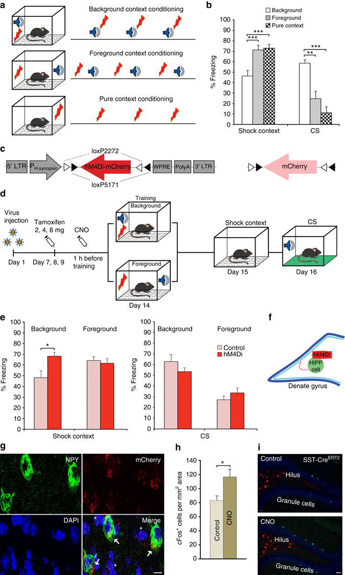
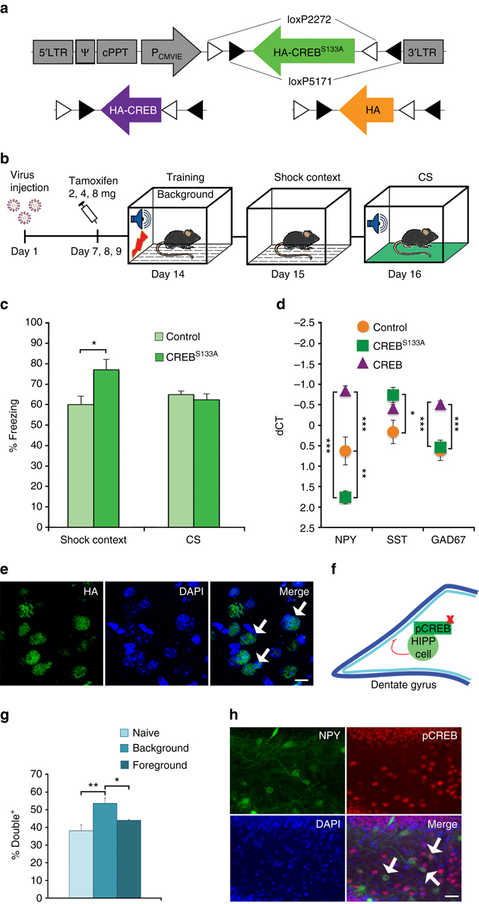
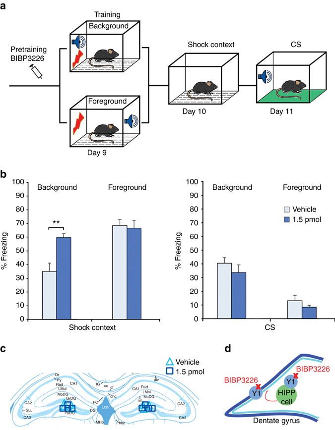
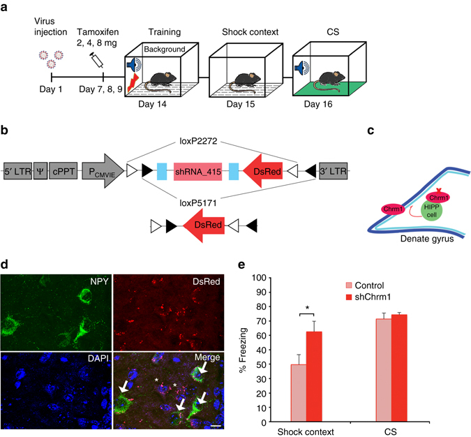
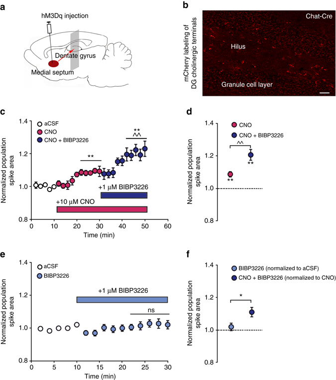
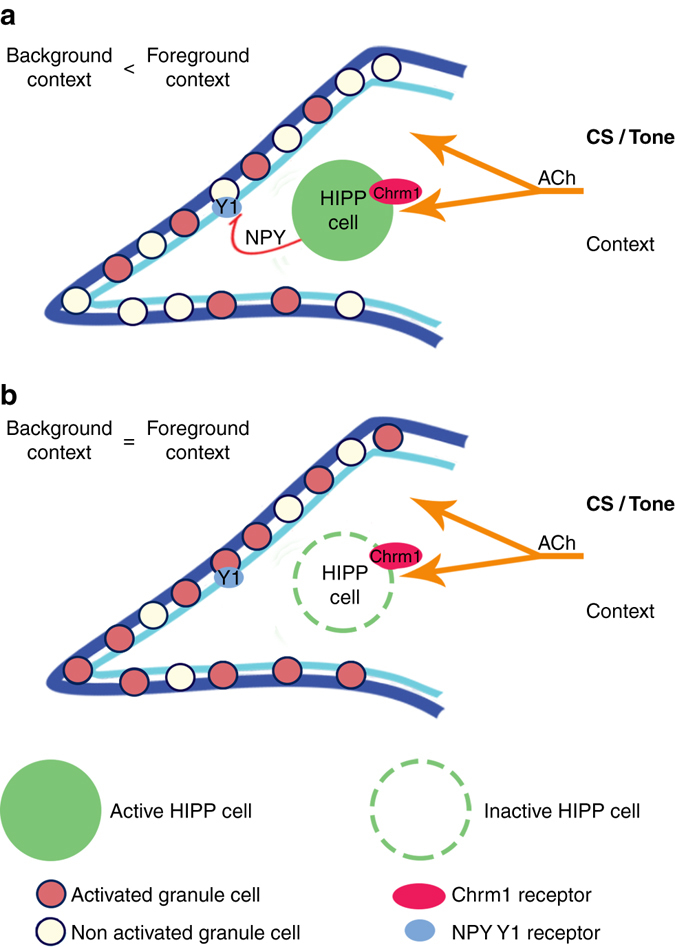
Cholinergic neuromodulation in the hippocampus controls the salience of background context memory acquired in the presence of elemental stimuli predicting an aversive reinforcement. With pharmacogenetic inhibition we here demonstrate that hilar perforant path-associated (HIPP) cells of the dentate gyrus mediate the devaluation of background context memory during Pavlovian fear conditioning. The salience adjustment is sensitive to reduction of hilar neuropeptide Y (NPY) expression via dominant negative CREB expression in HIPP cells and to acute blockage of NPY-Y1 receptors in the dentate gyrus during conditioning. We show that NPY transmission and HIPP cell activity contribute to inhibitory effects of acetylcholine in the dentate gyrus and that M1 muscarinic receptors mediate the cholinergic activation of HIPP cells as well as their control of background context salience. Our data provide evidence for a peptidergic local circuit in the dentate gyrus that mediates the cholinergic encoding of background context salience during fear memory acquisition.Intra-hippocampal circuits are essential for associating a background context with behaviorally salient stimuli and involve cholinergic modulation at SST+ interneurons. Here the authors show that the salience of the background context memory is modulated through muscarinic activation of NPY+ hilar perforant path associated interneurons and NPY signaling in the dentate gyrus.


Conflict of interest statement

The authors declare no competing financial interests.

Figures

Fig. 1
Fig. 1
Pharmacogenetic inactivation of HIPP cells increases background context conditioning. a Schematic of the context conditioning paradigms. b Contextual and cued fear memory of C57Bl/6 mice trained according to those protocols. The contextual fear response is significantly lower in the background context group (n = 8) than in both foreground context (n = 8) and pure context (n = 8) groups in agreement with previous reports , , . At the same time, freezing to the CS is higher in the background context group, as expected. c Conditional hM4Di-mCherry and mCherry-control vectors. d Schematic of viral transduction and behavioral testing paradigms, depicting the virus injection, the activation of CRE recombination with increasing doses of tamoxifen and the activation of hM4Di with clozapine-N-oxide (CNO). Background and foreground context conditioning were applied to different groups and contextual and auditory memory was tested in both 24 h and 48 h later, respectively. e (Left) Memory to the background context is increased when HIPP cells are silenced during background context conditioning in SST-CreERT2 mice (n = 8; controls n = 10), whereas no effect is observed upon HIPP cells silencing during foreground conditioning (n = 8; controls n = 7). (Right) As expected the CS response is high in the background context group compared to the foreground context group. Viral intervention has no effect on cued memory, as the response to auditory stimuli remains unaltered in both training paradigms. f A schematic of the proposed circuitry. g Representative microscopic images show the adenoviral expression of mCherry-tagged hM4Di receptors in NPY+ cells (arrows) and NPY cells of the hilus (asterisks). For an overview of the viral expression please see Supplementary Fig. 1d. Scale bar, 10 μm. h The number of cFos+ granule cells is increased after background context conditioning if hM4Di targeted HIPP cells are silenced with CNO (n = 6 each). i Representative images depicting cFos labeling and mCherry-tagged hM4Di in the DG following background context conditioning. Scale bar, 50 μm. Data are means + s.e.m. Statistical analysis was done with Fisher’s LSD following one-way ANOVA in a and with Student’s unpaired t-test in e, h. *P < 0.05; **P < 0.01; ***P< 0.001
Fig. 2
Fig. 2
CREB activation in HIPP cells during background context conditioning. a Lentiviral vector constructs for the conditional expression of control (HA), native CREB and dominant negative CREBS133A. b Schematic of the viral transduction and behavioral testing paradigm. c CREBS133A (n = 8) increases fear memory to the background context, while the fear response to the CS remains unaltered (controls n = 10). d The overexpression of native CREB in SST-CreERT2 mice increases, whereas dominant negative CREBS133A reduces NPY mRNA levels in the hilus (n = 6 each). GAD67 mRNA is also increased by CREB overexpression. SST mRNA by contrast is increased in the CREBS133A group with a non-significant trend for native CREB, indicative of a phosphorylation-independent constitutive expression regulation. Note the inverted scaling of the ordinate, the lower values indicate higher expression. e A representative microscopic image shows the lentiviral expression in the hilus. Green, HA; blue, DAPI. Scale bar, 10 μm. For an overview, please see Supplementary Fig. 4c. f A schematic of the proposed circuit and the site of CREB manipulation. g The proportion of pCREBS133/GFP double positive cells in the hilus of NPY-GFP transgenic mice is selectively increased 1 h after background context conditioning (n = 6 each). h Representative microscopic images show phosphorylated CREB expression in the hilus and granule cell layer of the DG. Arrows, pCREBS133/GFP double-labeled cells. Scale bar, 25 μm. Data are means + s.e.m. Statistical analysis was done with Student’s unpaired t-test in c, and by Fisher’s LSD following one-way ANOVA in d and g. *P < 0.05; **P < 0.01; ***P < 0.001
Fig. 3
Fig. 3
NPY in the dorsal DG controls background context fear memory. a A schematic of the behavioral paradigms. b Blocking Y1 receptors with BIBP3226 (1.5 pmol per hemisphere; n = 9) during training increases background context conditioning compared to vehicle (n = 11). BIBP3226 treatment does not induce any change, when used in foreground context conditioning (vehicle n = 7; 1.5 pmol BIBP3226 n = 8). No effect is observed on auditory fear memory, as the background context group maintains unchanged cue responding after injection, while the foreground context group continues to show only low levels of freezing. c Localization of guide cannulas in the dorsal DG. d A schematic of the proposed local circuit and site of drug activity. Data are means + s.e.m. Statistical analysis was done with Student’s unpaired t-test. **P < 0.01
Fig. 4
Fig. 4
Chrm1 expression and function in HIPP cells. a Hilar GFP+ cells of NPY-GFP mice (n = 6–8) are enriched with mRNA for NPY and M1 muscarinic receptors (Chrm1) but not M2 receptor (Chrm2). Values are expressed relative to the housekeeping gene GAPDH. b A 3D-reconstruction of a GFP+ neuron targeted by cholinergic fibers (arrows) at the soma. Scale bar, 10 μm. c A GFP+ neuron (green) in the dorsal hilus of an NPY-GFP mouse with somatic immunolabeling of M1 receptors (red). Scale bar, 5 μm. d (Left) Representative current-clamp recording from GFP+ interneurons. Application of 10 µM oxotremorine M (oxo) induces a pirenzepine-sensitive transient depolarization paralleled by a significant increase in input membrane resistance and an increase in spike activity. (Right) Quantification of spike activity. Oxo induces a transient membrane depolarization from resting membrane potential in 7 out of 7 recorded neurons, paralleled by a significant increase in input membrane resistance and mean spike frequency elicited by positive current injections. These effects are abolished in the presence of the M1 receptor antagonist pirenzepine (10 μM; n = 5). e In field recordings, either Y1 receptor blockage (left) (n = 6 slices with BIBP3226 and n = 6 slices without BIBP3226) or pharmacogenetic HIPP cell inactivation (right) (n = 8 slices with BIBP3226 and n = 10 slices without BIBP3226) counteracts the depression of population spikes induced by oxo bath application. Example traces to the right of the graph illustrate the difference in response to perforant path stimulation. Values are means ± s.e.m. Statistical analysis was done with Fisher’s LSD following one-way ANOVA in a, Wilcoxon signed rank test in d and Student’s unpaired t-test in e. *P < 0.05; **P < 0.01; ***P < 0.001
Fig. 5
Fig. 5
Chrm1 knock down in HIPP cells increases background context memory. a A schematic of the behavioral paradigm. b Lentiviral vector constructs for the conditional knockdown of Chrm1 and control vector. c Schematic of the proposed circuit. d Representative microscopic images show lentiviral expression of the DsRed marker in NPY+ cells (arrows) and NPY cells of the hilus (asterisks). Scale bar, 10 μm. For an overview refer to Supplementary Fig. 9f. e The knockdown of Chrm1 in SST-CreERT2 mice (n = 8) significantly increases the memory to background context (controls n = 7), while the conditioned response to the CS remains unaffected. Data are means + s.e.m. Statistical analysis was done with Student’s unpaired t-test. *P < 0.05
Fig. 6
Fig. 6
Facilitatory effect of endogenous ACh-release on the DG population spikes is augmented by Y1 receptor blockage. a Schematic of conditional hM3Dq viral injection into the medial septum of Chat-Cre driver mice. b A representative microscopic image shows the expression of the virally expressed mCherry tag in cholinergic terminals throughout the dentate gyrus. Scale bar, 25 μm. c The hippocampal cholinergic terminals expressing hM3Dq are activated by perfusion of hippocampal slices with CNO. Total of 10 µM CNO results in a moderate increase of the DG population spike area, which is further augmented by 1 µM BIBP3226 (n = 8 slices). For statistical analysis the last 10 min of each condition are averaged and compared, to ensure full availability or the bath-applied drugs. d Summary graph illustrating the change in average population spike area after CNO and CNO + BIBP3226 application relative to aCSF. e By contrast, BIBP3226 application in the absence of CNO does not affect the population spike area (n = 16 slices). f Comparison of normalized BIBP3226 effects on population spike area shows a significant change only after CNO application (n = 16 slices without CNO and n = 8 slices with CNO). Values are means ± s.e.m. Statistical analysis was done with Fisher’s LSD following one-way repeated measures ANOVA (^^P < 0.01: CNO vs. CNO + BIBP3226; **P < 0.01; CNO or CNO + BIBP3226 vs. aCSF condition) in c and d, paired t-test in e and Student’s unpaired t-test in f (*P < 0.05)
Fig. 7
Fig. 7
A local circuit model for the cholinergic modulation of background context salience in the dentate gyrus. a HIPP cells act as relay stations in the septo-hippocampal pathway that adjust context memory strength during fear learning. Acetylcholine released from medial septal afferences during memory acquisition activates both granule cells and HIPP cells in the dentate gyrus. The cholinergic stimulation of HIPP cells via muscarinic M1 receptors (Chrm1) triggers their NPY release, which in turn via Y1 receptors attenuates the excitation of granule cells. This feed forward inhibition is effective when conditioning occurs to an elemental cue (context in background) and restricts the number of granule cells that are recruited to the fear memory engram, resulting in reduced context salience. b Upon experimental silencing of HIPP cells and blockage of NPY-mediated inhibition the cue-induced devaluation of the background context fails and an increased number of granule cells are recruited to the context memory trace. Experience-dependent changes in HIPP cell function, e.g., via CREB-mediated transcription can adjust the strength of this local circuit and are critical for the function of the DG in adaptively encoding context salience

References

    1. Rescorla, R. A. & Wagner, A. R. in Classical Conditioning II (eds Black A. H. & Prokasy W. F.) 64–99 (Appleton-Century-Crofts, 1972).
    1. Phelps EA. Human emotion and memory: interaction of the amygdala and hippocampal complex. Curr. Opin. Neurobiol. 2004;14:198–202. doi: 10.1016/j.conb.2004.03.015. - DOI - PubMed
    1. Calandreau L, Jaffard R, Desmedt A. Dissociated roles for the lateral and medial septum in elemental and contextual fear conditioning. Learn. Mem. 2007;14:422–429. doi: 10.1101/lm.531407. - DOI - PMC - PubMed
    1. Calandreau L, Desmedt A, Decorte L, Jaffard R. A different recruitment of the lateral and basolateral amygdala promotes contextual or elemental conditioned association in Pavlovian fear conditioning. Learn. Mem. 2005;12:383–388. doi: 10.1101/lm.92305. - DOI - PMC - PubMed
    1. Calandreau L, Desgranges B, Jaffard R, Desmedt A. Switching from contextual to tone fear conditioning and vice versa: the key role of the glutamatergic hippocampal-lateral septal neurotransmission. Learn. Mem. 2010;17:440–443. doi: 10.1101/lm.1859810. - DOI - PubMed

Publication types

MeSH terms