Review
. 2017 Jul 20:7:324.
doi: 10.3389/fcimb.2017.00324. eCollection 2017.

Cytokines and Chemokines in Cerebral Malaria Pathogenesis

Affiliations
Review

Cytokines and Chemokines in Cerebral Malaria Pathogenesis

Josefine Dunst et al. Front Cell Infect Microbiol. .

Abstract

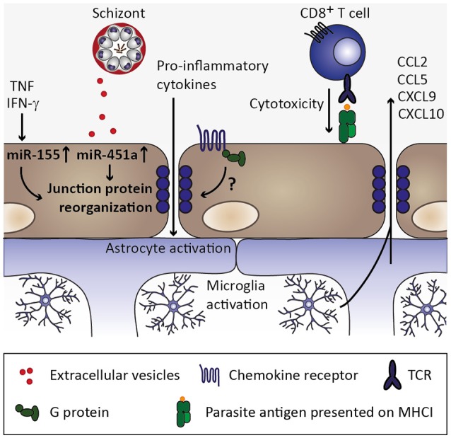
Cerebral malaria is among the major causes of malaria-associated mortality and effective adjunctive therapeutic strategies are currently lacking. Central pathophysiological processes involved in the development of cerebral malaria include an imbalance of pro- and anti-inflammatory responses to Plasmodium infection, endothelial cell activation, and loss of blood-brain barrier integrity. However, the sequence of events, which initiates these pathophysiological processes as well as the contribution of their complex interplay to the development of cerebral malaria remain incompletely understood. Several cytokines and chemokines have repeatedly been associated with cerebral malaria severity. Increased levels of these inflammatory mediators could account for the sequestration of leukocytes in the cerebral microvasculature present during cerebral malaria, thereby contributing to an amplification of local inflammation and promoting cerebral malaria pathogenesis. Herein, we highlight the current knowledge on the contribution of cytokines and chemokines to the pathogenesis of cerebral malaria with particular emphasis on their roles in endothelial activation and leukocyte recruitment, as well as their implication in the progression to blood-brain barrier permeability and neuroinflammation, in both human cerebral malaria and in the murine experimental cerebral malaria model. A better molecular understanding of these processes could provide the basis for evidence-based development of adjunct therapies and the definition of diagnostic markers of disease progression.

Keywords: Plasmodium; blood-brain barrier; cerebral malaria; chemokines; cytokines; endothelial activation; malaria; neuroinflammation.


Figures

Figure 1
Figure 1
Overview of a potential inflammatory cascade culminating in cerebral malaria pathology. Five consecutive events shape the outcome of a Plasmodium infection and contribute to cerebral malaria. During Plasmodium infection of the mammalian host, two consecutive parasite replication phases in the liver and red blood cells lead to distinct innate responses, which modulate downstream parasite/host cell interactions. Upon parasite accumulation in the microvasculature, endothelial cells become activated, leading to enhanced chemokine secretion, which in turn enhances leukocyte recruitment. Acute pathology is caused by permeabilization of the endothelial barrier. See Figures 2–6 for the central roles of chemokines and cytokines in the individual events.
Figure 2
Figure 2
Innate immune response to Plasmodium liver stage infection. Plasmodium infection of hepatocytes activates interferon regulatory factors (IRF), which induce transcription of type I interferons (IFN) IFNα and IFNβ. Secretion of type I IFNs activates IFNα/β receptor IFNAR in an autocrine or paracrine manner. IFNAR signaling results in transcription of IFN-stimulated genes (ISGs), which includes chemokines, such as CXCL9 and CXCL10. Upon secretion from hepatocytes, these chemokines might recruit cells expressing the corresponding chemokine receptor CXCR3, including natural killer (NK), T, and NKT cells. Upon activation by type I IFN at the site of infection, these cell types could contribute to limiting Plasmodium liver stage expansion by IFN-γ secretion. Based on Liehl et al. (2014) and Miller et al. (2014).
Figure 3
Figure 3
Innate immune response to Plasmodium blood stage infection in the spleen. Macrophages as well as dendritic cells (DC) remove infected erythrocytes from the circulation by phagocytosis. In macrophages, uptake of infected erythrocytes might not lead to secretion of pro-inflammatory cytokines due to phagosomal acidification (Wu et al., 2015). Upon rupture of infected erythrocytes, pathogen-associated molecular patterns (PAMPs) and danger-associated molecular patterns (DAMPs) are released, including microvesicles, hemozoin, and glycosylphosphatidylinositols (GPI). These potential PAMPs and DAMPs might be recognized by DC through pattern recognition receptors, resulting in the secretion of interleukin 12 (IL-12), tumor necrosis factor (TNF), and IL-6 (Wu et al., 2015). DC-derived IL-12 might activate natural killer (NK) cells, which in turn secrete interferon γ (IFN-γ) and could thereby activate macrophages (Stevenson and Riley, 2004).
Figure 4
Figure 4
Endothelial activation and chemokine secretion. A characteristic feature of Plasmodium infection is endothelial activation, which is likely induced by elevated serum tumor necrosis factor (TNF) levels. Binding of TNF to its receptor (TNFR1) induces transcription of adhesion molecules, including ICAM-1 and VCAM-1, as well as chemokines (Pober and Sessa, 2007). Endothelial activation might be directly induced by infected erythrocytes, possibly through activation of pattern recognition receptors (PRR), resulting in elevated expression of ICAM-1 and chemokine secretion (Viebig et al., ; Tripathi et al., , ; Chakravorty et al., 2007).
Figure 5
Figure 5
Chemokine-mediated leukocyte recruitment and progression of local inflammation. As a consequence of endothelial activation by pro-inflammatory cytokines, such as TNF, and infected erythrocytes, chemokines are secreted from endothelial cells, which initiate recruitment of leukocytes expressing the respective chemokine receptors, including macrophages, monocytes, neutrophils, and T cell. These cell types were also found to sequester in the microvasculature during human CM as well as murine ECM (Hunt and Grau, ; Renia et al., 2012). Upon arrival at the site of the inflammatory insult, these leukocyte subsets may in turn secrete cytokines as well as chemokines, thereby further promoting endothelial activation and leukocyte recruitment. Thus, a feed-forward loop is initiated which exacerbates local inflammation in the brain.
Figure 6
Figure 6
Endothelial permeability and neuroinflammation. Through continued inflammatory insults toward endothelial cells by, for instance, circulating tumor necrosis factor (TNF) and interferon γ (IFN-γ), miR-155 might be upregulated in endothelial cells. Along with uptake of miR-451a from P. falciparum-infected erythrocyte-derived extracellular vesicles, reorganization of tight junction proteins such as zonula occludens 1 (ZO-1) is induced and could contribute to endothelial permeability during cerebral malaria (Lopez-Ramirez et al., ; Mantel et al., 2016). Additionally, chemokine receptors might induce redistribution of tight junction proteins in a G protein-dependent manner (Stamatovic et al., ; Song and Pachter, ; Yao and Tsirka, 2011), while the contribution of chemokine-induced opening of tight junctions is less clear in the context of cerebral malaria. CD8+ T cell-mediated cytotoxity toward endothelial cells through recognition of parasite antigen presented on MHC class I molecules on endothelial cells likely contributes substantially to blood-brain barrier permeability during cerebral malaria (Howland et al., 2015a,b). Consequently, pro-inflammatory cytokines enter the brain parenchyma and could thereby activate astrocytes and microglia, which in turn could secrete chemokines (Capuccini et al., 2016) and thus promote leukocyte recruitment and local inflammation.

References

    1. Adams Y., Kuhnrae P., Higgins M. K., Ghumra A., Rowe J. A. (2014). Rosetting Plasmodium falciparum-infected erythrocytes bind to human brain microvascular endothelial cells in vitro, demonstrating a dual adhesion phenotype mediated by distinct P. falciparum erythrocyte membrane protein 1 domains. Infect. Immun. 82, 949–959. 10.1128/IAI.01233-13 - DOI - PMC - PubMed
    1. Anstey N. M., Russell B., Yeo T. W., Price R. N. (2009). The pathophysiology of vivax malaria. Trends Parasitol. 25, 220–227. 10.1016/j.pt.2009.02.003 - DOI - PubMed
    1. Armah H. B., Wilson N. O., Sarfo B. Y., Powell M. D., Bond V. C., Anderson W., et al. . (2007). Cerebrospinal fluid and serum biomarkers of cerebral malaria mortality in Ghanaian children. Malar. J. 6:147. 10.1186/1475-2875-6-147 - DOI - PMC - PubMed
    1. Bakmiwewa S. M., Weiser S., Grey M., Heng B., Guillemin G. J., Ball H. J., et al. . (2016). Synergistic induction of CXCL10 by interferon-gamma and lymphotoxin-alpha in astrocytes: possible role in cerebral malaria. Cytokine 78, 79–86. 10.1016/j.cyto.2015.11.024 - DOI - PubMed
    1. Barker K. R., Lu Z., Kim H., Zheng Y., Chen J., Conroy A. L., et al. . (2017). miR-155 modifies inflammation, endothelial activation and blood-brain barrier dysfunction in cerebral malaria. Mol. Med. 23, 24–33. 10.2119/molmed.2016.00139 - DOI - PMC - PubMed

Publication types