. 2018 Aug;596(15):3271-3283.
doi: 10.1113/JP274726. Epub 2017 Sep 2.

Hypercapnia-induced active expiration increases in sleep and enhances ventilation in unanaesthetized rats

Affiliations

Hypercapnia-induced active expiration increases in sleep and enhances ventilation in unanaesthetized rats

Isabela P Leirão et al. J Physiol. 2018 Aug.

Abstract

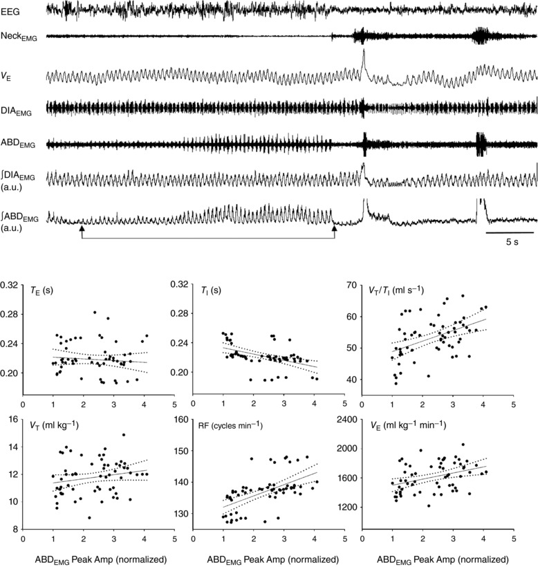
Key points: Expiratory muscles (abdominal and thoracic) can be recruited when respiratory drive increases under conditions of increased respiratory demand such as hypercapnia. Studying hypercapnia-induced active expiration in unanaesthetized rats importantly contributes to the understanding of how the control system is integrated in vivo in freely moving animals. In unanaesthetized rats, hypercapnia-induced active expiration was not always recruited either in wakefulness or in sleep, suggesting that additional factors influence the recruitment of active expiration. The pattern of abdominal muscle recruitment varied in a state-dependent manner with active expiration being more predominant in the sleep state than in quiet wakefulness. Pulmonary ventilation was enhanced in periods with active expiration compared to periods without it.

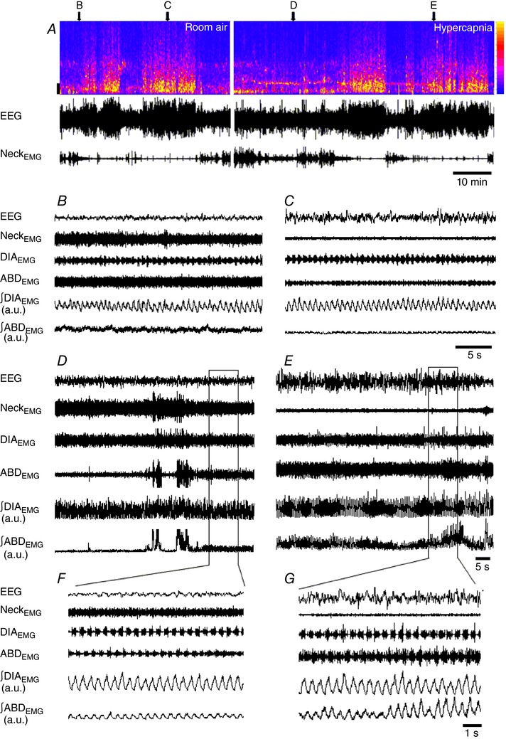
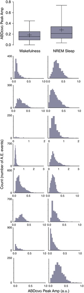
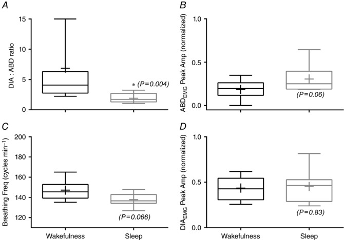
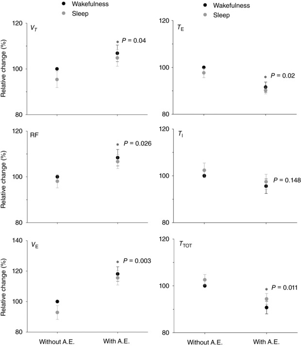
Abstract: Expiration is passive at rest but becomes active through recruitment of abdominal muscles under increased respiratory drive. Hypercapnia-induced active expiration has not been well explored in unanaesthetized rats. We hypothesized that (i) CO2 -evoked active expiration is recruited in a state-dependent manner, i.e. differently in sleep or wakefulness, and (ii) recruitment of active expiration enhances ventilation, hence having an important functional role in meeting metabolic demand. To test these hypotheses, Wistar rats (280-330 g) were implanted with electrodes for EEG and electromyography EMG of the neck, diaphragm (DIA) and abdominal (ABD) muscles. Active expiratory events were considered as rhythmic ABDEMG activity interposed to DIAEMG . Animals were exposed to room air followed by hypercapnia (7% CO2 ) with EEG, EMG and ventilation ( V̇E ) recorded throughout the experimental protocol. No active expiration was observed during room air exposure. During hypercapnia, CO2 -evoked active expiration was predominantly recruited during non-rapid eye movement sleep. Its increased occurrence during sleep was evidenced by the decreased DIA-to-ADB ratio (1:1 ratio means that each DIA event is followed by an ABD event, indicating a high occurrence of ABD activity). Moreover, V̇E was also enhanced (P < 0.05) in periods with active expiration. V̇E had a positive correlation (P < 0.05) with the peak amplitude of ABDEMG activity. The data demonstrate strongly that hypercapnia-induced active expiration increases during sleep and provides an important functional role to support V̇E in conditions of increased respiratory demand.

Keywords: EEG; EMG; breathing control; expiratory activity; sleep; wakefulness.


Figures

Figure 1
Figure 1. State‐dependent activity of abdominal and diaphragm activity during room air and hypercapnia exposure
A, EEG spectrogram, EEG and NeckEMG raw recordings (top, middle and bottom, respectively) of a representative animal during a typical experimental protocol in room air (left) and hypercapnia exposure (right). In the EEG spectrogram, the colour intensity, as indicated by the colour calibration bar to the right side, corresponds to the amount of signal power at a given time and frequency. On the left side, frequency calibration bar: 2 Hz. Note that the animal cycles between wake and sleep in both conditions: room air and hypercapnia. Above the spectrogram, the letters and arrows indicate for each of the respective panels where their traces were withdrawn and expanded. The expanded EEG and EMGs [raw and integrated abdominal (∫ABDEMG) and diaphragm (∫DIAEMG) signals] during room air are shown in periods of wakefulness (B) and sleep (C), and during hypercapnia in wakefulness (D) and sleep (E). The horizontal rectangles in D and E depict the expanded trace shown in F and G, respectively. Note the absence of active expiration during room air (B and C) and its recruitment during hypercapnia (DG). [Color figure can be viewed at http://wileyonlinelibrary.com]
Figure 2
Figure 2. Distribution of active expiration (A.E.) events (∫ABDEMG Peak Amp) during hypercapnia in wakefulness and sleep
Top panel shows the group data as box plots in which the centre line shows the median; box limits indicate the 25th and 75th percentiles; whiskers extend to 5th and 95th percentiles; and crosses represent sample means. Each histogram (row) represents the distribution for one individual animal in both vigilance states: wakefulness (left column) and sleep (right column). Note that one animal (bottom row) did not present active expiration in wakefulness, but only in sleep. [Color figure can be viewed at http://wileyonlinelibrary.com]
Figure 3
Figure 3. Respiratory muscle EMG and breathing frequency during hypercapnia in different vigilance states: wakefulness and sleep
Box plots: centre line shows the median; box limits indicate the 25th and 75th percentiles; whiskers extend to 5th and 95th percentiles; and crosses represent sample means. The panels show the DIA‐to‐ADB ratio (A), ∫ABDEMG Peak Amp (B), breathing frequency (C) and ∫DIAEMG Peak Amp (D) (n = 8). The P values are indicated for the sleep data set compared to wakefulness. *Difference (P < 0.05) of sleep state compared to wakefulness state.
Figure 4
Figure 4. Comparison of ventilatory variables during hypercapnia in periods with or without recruitment of active expiration
V T and T E top panels, RF and T I middle panels, and VE and T TOT bottom panels. (*) indicates difference (P < 0.05) comparing the ventilatory variables with and without active expiration (A.E.) (n = 7). The P values are indicated.
Figure 5
Figure 5. Functional impact of active expiration with increasing ABDEMG activity on ventilatory variables during hypercapnia
Top traces depict EEG, NeckEMG, barometric V E recording and raw and integrated DIAEMG and ABDEMG. Two arrows below the ∫ABDEMG trace show the interval time with increasing ABDEMG activity from which correlation analyses between active expiration (∫ABDEMG Peak Amp) and ventilation were performed. A breath‐by‐breath analysis was run in 60 events. Scatter plots show the correlation between increasing ∫ABDEMG Peak Amp and T E, T I and V T/T I (top row) and V T, RF and V E (bottom row).

Comment in

  • Sleep awakens active expiration.
    O'Halloran KD. O'Halloran KD. J Physiol. 2018 Aug;596(15):2947-2948. doi: 10.1113/JP275056. Epub 2017 Sep 2. J Physiol. 2018. PMID: 28833135 Free PMC article. No abstract available.

Similar articles

Cited by

References

    1. Abdala AP, Rybak IA, Smith JC & Paton JFR (2009). Abdominal expiratory activity in the rat brainstem–spinal cord in situ: patterns, origins and implications for respiratory rhythm generation. J Physiol 587, 3539–3559. - PMC - PubMed
    1. Abe T, Kusuhara N, Yoshimura N, Tomita T & Easton PA (1996). Differential respiratory activity of four abdominal muscles in humans. J Appl Physiol 80, 1379–1389. - PubMed
    1. Andrews CG & Pagliardini S (2015). Expiratory activation of abdominal muscle is associated with improved respiratory stability and an increase in minute ventilation in REM epochs of adult rats. J Appl Physiol 119, 968–974. - PMC - PubMed
    1. Badr MS, Skatrud JB, Dempsey JA & Begle RL (1990). Effect of mechanical loading on expiratory and inspiratory muscle activity during NREM sleep. J Appl Physiol 68, 1195–1202. - PubMed
    1. Bartlett D & Tenney SM (1970). Control of breathing in experimental anemia. Respir Physiol 10, 384–395. - PubMed

Publication types

LinkOut - more resources