Assessing Tumor-Infiltrating Lymphocytes in Solid Tumors: A Practical Review for Pathologists and Proposal for a Standardized Method from the International Immuno-Oncology Biomarkers Working Group: Part 2: TILs in Melanoma, Gastrointestinal Tract Carcinomas, Non-Small Cell Lung Carcinoma and Mesothelioma, Endometrial and Ovarian Carcinomas, Squamous Cell Carcinoma of the Head and Neck, Genitourinary Carcinomas, and Primary Brain Tumors
- PMID: 28777143
- PMCID: PMC5638696
- DOI: 10.1097/PAP.0000000000000161
Assessing Tumor-Infiltrating Lymphocytes in Solid Tumors: A Practical Review for Pathologists and Proposal for a Standardized Method from the International Immuno-Oncology Biomarkers Working Group: Part 2: TILs in Melanoma, Gastrointestinal Tract Carcinomas, Non-Small Cell Lung Carcinoma and Mesothelioma, Endometrial and Ovarian Carcinomas, Squamous Cell Carcinoma of the Head and Neck, Genitourinary Carcinomas, and Primary Brain Tumors
Abstract
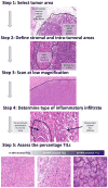
Assessment of the immune response to tumors is growing in importance as the prognostic implications of this response are increasingly recognized, and as immunotherapies are evaluated and implemented in different tumor types. However, many different approaches can be used to assess and describe the immune response, which limits efforts at implementation as a routine clinical biomarker. In part 1 of this review, we have proposed a standardized methodology to assess tumor-infiltrating lymphocytes (TILs) in solid tumors, based on the International Immuno-Oncology Biomarkers Working Group guidelines for invasive breast carcinoma. In part 2 of this review, we discuss the available evidence for the prognostic and predictive value of TILs in common solid tumors, including carcinomas of the lung, gastrointestinal tract, genitourinary system, gynecologic system, and head and neck, as well as primary brain tumors, mesothelioma and melanoma. The particularities and different emphases in TIL assessment in different tumor types are discussed. The standardized methodology we propose can be adapted to different tumor types and may be used as a standard against which other approaches can be compared. Standardization of TIL assessment will help clinicians, researchers and pathologists to conclusively evaluate the utility of this simple biomarker in the current era of immunotherapy.
Conflict of interest statement
The authors have no conflicts of interest to disclose.
Figures


References
-
- Denkert C, Wienert S, Poterie A, et al. Standardized evaluation of tumor-infiltrating lymphocytes in breast cancer: results of the ring studies of the international immuno-oncology biomarker working group. Mod Pathol. 2016;29:1155–1164. - PubMed
-
- McGovern VJ, Mihm MC, Jr, Bailly C, et al. The Classification of Malignant Melanoma and its Histologic Reporting. Cancer. 1973;32:1446–1457. - PubMed
-
- Elder DE, DuPont G, Van Horn M, et al. The Role of Lymph Node Dissection for Clinical Stage I Malignant Melanoma of Intermediate Thickness (1.51–3. 99 mm) Cancer. 1985;56:413–418. - PubMed
-
- Clark WH, Jr, Elder DE, DuPont G, et al. Model Predicting Survival in Stage I Melanoma Based on Tumor Progression. J Natl Cancer Inst. 1989;81:1893–1904. - PubMed
Publication types
MeSH terms
Substances
Grants and funding
LinkOut - more resources
Full Text Sources
Other Literature Sources
Medical

