

Co-Infection of Mosquitoes with Chikungunya and Dengue Viruses Reveals Modulation of the Replication of Both Viruses in Midguts and Salivary Glands of Aedes aegypti Mosquitoes
- PMID: 28777313
- PMCID: PMC5578098
- DOI: 10.3390/ijms18081708
Co-Infection of Mosquitoes with Chikungunya and Dengue Viruses Reveals Modulation of the Replication of Both Viruses in Midguts and Salivary Glands of Aedes aegypti Mosquitoes
Abstract
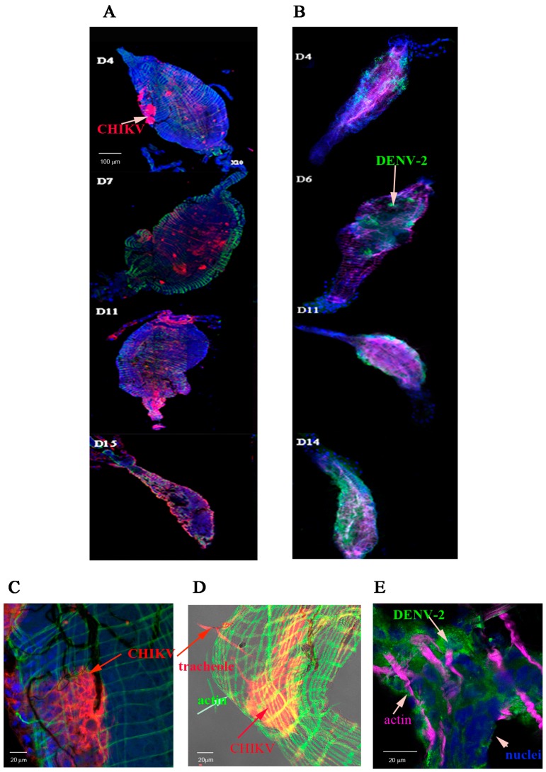
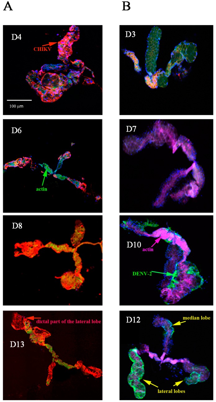
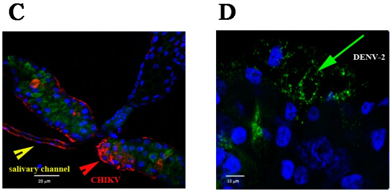
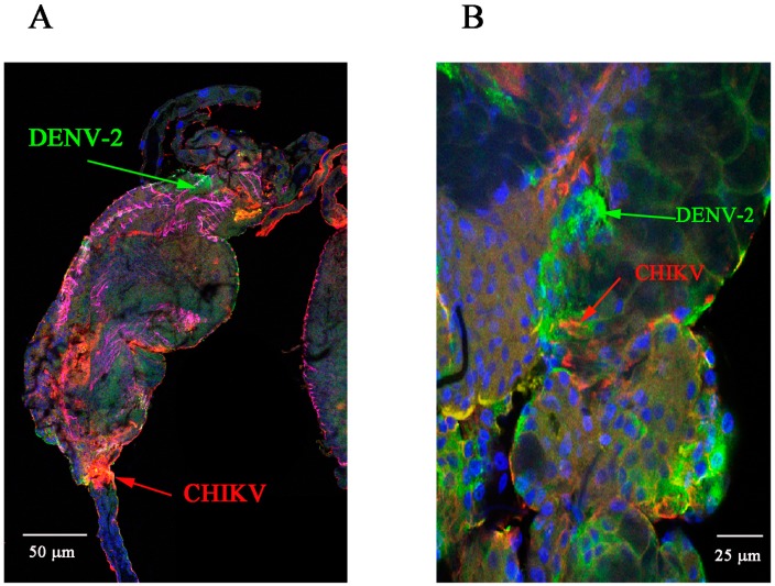
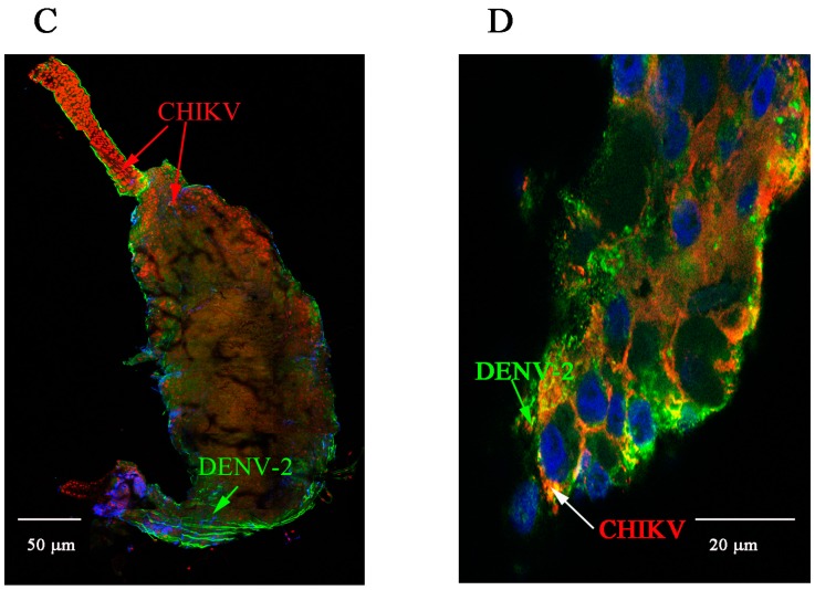
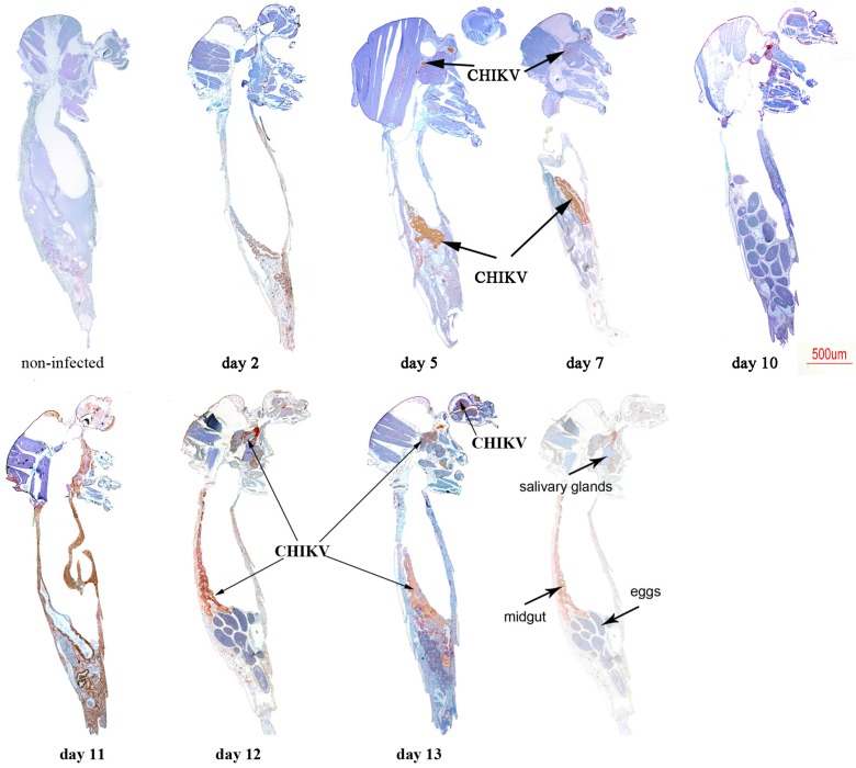
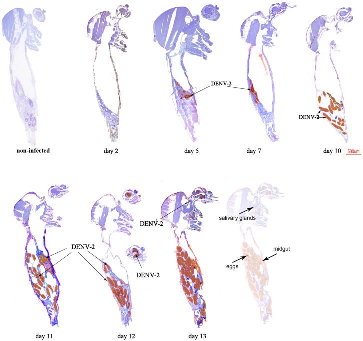
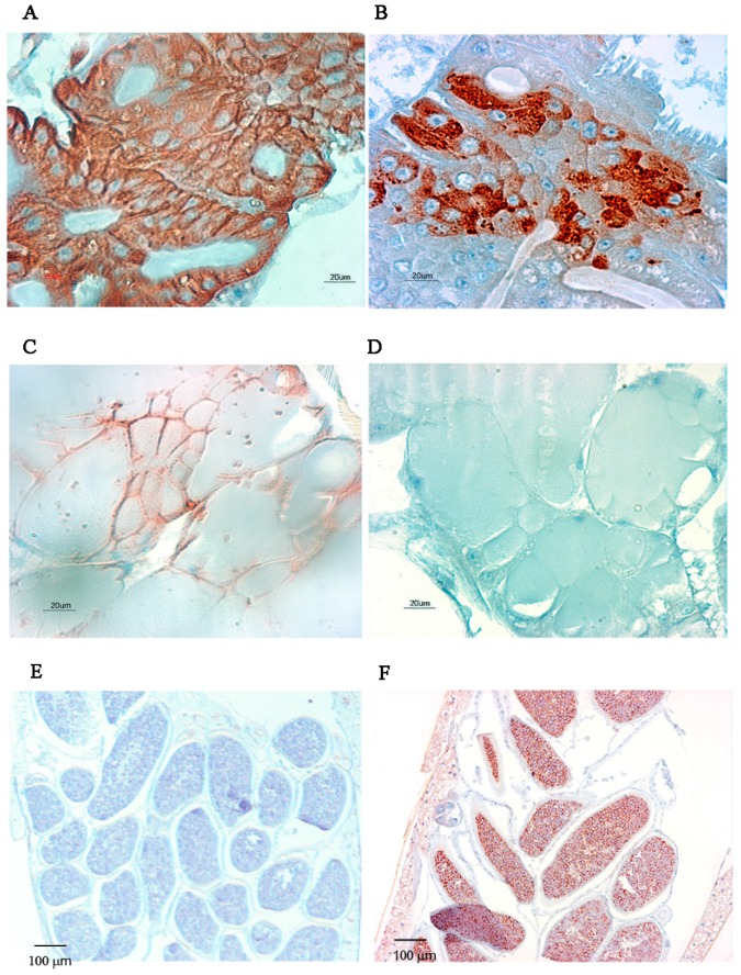
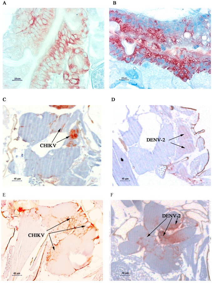
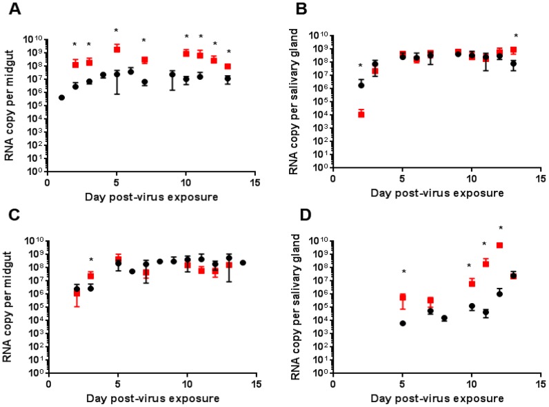
Arthropod-borne virus (arbovirus) infections cause several emerging and resurgent infectious diseases in humans and animals. Chikungunya-affected areas often overlap with dengue-endemic areas. Concurrent dengue virus (DENV) and chikungunya virus (CHIKV) infections have been detected in travelers returning from regions of endemicity. CHIKV and DENV co-infected Aedes albopictus have also been collected in the vicinity of co-infected human cases, emphasizing the need to study co-infections in mosquitoes. We thus aimed to study the pathogen-pathogen interaction involved in these co-infections in DENV/CHIKV co-infected Aedes aegypti mosquitoes. In mono-infections, we detected CHIKV antigens as early as 4 days post-virus exposure in both the midgut (MG) and salivary gland (SG), whereas we detected DENV serotype 2 (DENV-2) antigens from day 5 post-virus exposure in MG and day 10 post-virus exposure in SG. Identical infection rates were observed for singly and co-infected mosquitoes, and facilitation of the replication of both viruses at various times post-viral exposure. We observed a higher replication for DENV-2 in SG of co-infected mosquitoes. We showed that mixed CHIKV and DENV infection facilitated viral replication in Ae. aegypti. The outcome of these mixed infections must be further studied to increase our understanding of pathogen-pathogen interactions in host cells.
Keywords: Aedes aegypti; arbovirus; chikungunya; co-infection; dengue.
Conflict of interest statement
The authors declare no conflict of interest.
Figures
Similar articles
-
Transcriptome analysis of Aedes aegypti in response to mono-infections and co-infections of dengue virus-2 and chikungunya virus.Biochem Biophys Res Commun. 2017 Oct 28;492(4):617-623. doi: 10.1016/j.bbrc.2017.01.162. Epub 2017 Feb 1. Biochem Biophys Res Commun. 2017. PMID: 28161634
-
wMel limits zika and chikungunya virus infection in a Singapore Wolbachia-introgressed Ae. aegypti strain, wMel-Sg.PLoS Negl Trop Dis. 2017 May 19;11(5):e0005496. doi: 10.1371/journal.pntd.0005496. eCollection 2017 May. PLoS Negl Trop Dis. 2017. PMID: 28542240 Free PMC article.
-
Experimental Zika virus infection in Aedes aegypti: Susceptibility, transmission & co-infection with dengue & chikungunya viruses.Indian J Med Res. 2018 Jan;147(1):88-96. doi: 10.4103/ijmr.IJMR_1142_17. Indian J Med Res. 2018. PMID: 29749366 Free PMC article.
-
The worldwide seroprevalence of DENV, CHIKV and ZIKV infection: A systematic review and meta-analysis.PLoS Negl Trop Dis. 2021 Apr 28;15(4):e0009337. doi: 10.1371/journal.pntd.0009337. eCollection 2021 Apr. PLoS Negl Trop Dis. 2021. PMID: 33909610 Free PMC article.
-
Dengue Chikungunya co-infection: A live-in relationship??Biochem Biophys Res Commun. 2017 Oct 28;492(4):608-616. doi: 10.1016/j.bbrc.2017.02.008. Epub 2017 Feb 9. Biochem Biophys Res Commun. 2017. PMID: 28189673 Review.
Cited by
-
Unlike Zika, Chikungunya virus interferes in the viability of Aedes aegypti eggs, regardless of females' age.Sci Rep. 2020 Aug 12;10(1):13642. doi: 10.1038/s41598-020-70367-6. Sci Rep. 2020. PMID: 32788625 Free PMC article.
-
Biased virus transmission following sequential coinfection of Aedes aegypti with dengue and Zika viruses.PLoS Negl Trop Dis. 2024 Apr 1;18(4):e0012053. doi: 10.1371/journal.pntd.0012053. eCollection 2024 Apr. PLoS Negl Trop Dis. 2024. PMID: 38557981 Free PMC article.
-
Performing Immunohistochemistry in Mosquito Salivary Glands.Cold Spring Harb Protoc. 2022 Oct 3;2022(10):Pdb.top107699. doi: 10.1101/pdb.top107699. Cold Spring Harb Protoc. 2022. PMID: 35960615 Free PMC article.
-
Arbovirus coinfection and co-transmission: A neglected public health concern?PLoS Biol. 2019 Jan 22;17(1):e3000130. doi: 10.1371/journal.pbio.3000130. eCollection 2019 Jan. PLoS Biol. 2019. PMID: 30668574 Free PMC article.
-
Comodulation of Dengue and Chikungunya Virus Infection During a Coinfection Scenario in Human Cell Lines.Front Cell Infect Microbiol. 2022 Apr 28;12:821061. doi: 10.3389/fcimb.2022.821061. eCollection 2022. Front Cell Infect Microbiol. 2022. PMID: 35573775 Free PMC article.
References
-
- Woodring J.L., Higgs S., Beaty B.J. Natural cycles of vector borne pathogens. In: William M., editor. Book Biology of Disease Vectors. 2nd ed. Volume 2. Elsevier Academic Press; Burlington, MA, USA: 1996. pp. 167–185.
MeSH terms
Substances
LinkOut - more resources
Full Text Sources
Other Literature Sources
Medical
