. 2017 Dec;11(12):2754-2766.
doi: 10.1038/ismej.2017.125. Epub 2017 Aug 4.

Sulfide production and oxidation by heterotrophic bacteria under aerobic conditions

Affiliations

Sulfide production and oxidation by heterotrophic bacteria under aerobic conditions

Yongzhen Xia et al. ISME J. 2017 Dec.

Abstract

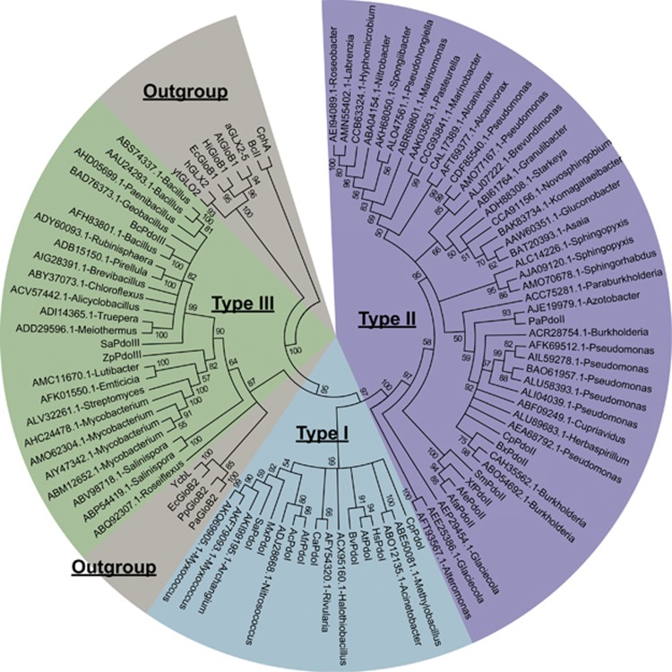
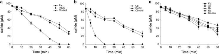
Sulfide (H2S, HS- and S2-) oxidation to sulfite and thiosulfate by heterotrophic bacteria, using sulfide:quinone oxidoreductase (SQR) and persulfide dioxygenase (PDO), has recently been reported as a possible detoxification mechanism for sulfide at high levels. Bioinformatic analysis revealed that the sqr and pdo genes were common in sequenced bacterial genomes, implying the sulfide oxidation may have other physiological functions. SQRs have previously been classified into six types. Here we grouped PDOs into three types and showed that some heterotrophic bacteria produced and released H2S from organic sulfur into the headspace during aerobic growth, and others, for example, Pseudomonas aeruginosa PAO1, with sqr and pdo did not release H2S. When the sqr and pdo genes were deleted, the mutants also released H2S. Both sulfide-oxidizing and non-oxidizing heterotrophic bacteria were readily isolated from various environmental samples. The sqr and pdo genes were also common in the published marine metagenomic and metatranscriptomic data, indicating that the genes are present and expressed. Thus, heterotrophic bacteria actively produce and consume sulfide when growing on organic compounds under aerobic conditions. Given their abundance on Earth, their contribution to the sulfur cycle should not be overlooked.


Conflict of interest statement

The authors declare no conflict of interest.

Figures

Figure 1
Figure 1
The phylogenetic tree of representative PDOs whose genes are physically linked to sqr in bacterial genomes. 69 representative PDOs were used for phylogenetic tree construction with reference sequences. PDOs belong to the metallo-beta-lactamase superfamily, and several related proteins, such as glyoxalase II (GloB) proteins, were also included as references. The representative proteins were labeled with their GenBank accession numbers and bacterial genera. These sequences were aligned by using ClustalW, and the tree was built by using MEGA6. Reference proteins with accession number were given below. Type I PDOs: SaPdoI (YP_003957083.1), CpPdoI (YP_297536.1), MxPdoI (YP_633997.1), HsPdoI (NP_055112.2), CaPdoI (YP_007162862.1), BvPdoI (ZP_00420127.1), AtPdoI (NP_974018.3), AcPdoI (AEK59246.1). Type II PDOs: XfPdoII (NP_298058.1), SmPdoII (NP_435818.1), CpPdoII (YP_297791.1), PaPdoII (NP_251605.1), BxPdoII (YP_554628.1), AfaPdoII (AAK89929.1), AfePdoII (ZP_11421028.1), AfrPdoII (YP_002424776.1). Type III PDOs: ZpPdoIII (ADF52140.1), SaPdoIII (WP_000465474.1), BcPdoIII (EEK49737.1). Glyoxalase II and related proteins: aGLX2-5 (NP_850166.1), AtGloB1 (NP_356997.2), BcII (AAA22276.1), CphA (CAA40386.1), EcGloB1 (NP_414748.1), EcGloB2 (NP_415447.1), hGLX2 (CAA62483.1), HiGloB1 (ADO96205.1), PaGloB2 (NP_249523.1), PpGloB2 (ABQ76961.1), YcbL (CAD05397.1) and ytGLO2 (CAA71335.1).
Figure 2
Figure 2
Sulfide oxidation by recombinant E. coli BL21(DE3) cells. Cells were suspended in 100 mm Tris buffer (pH 8) with 50 μm DTPA at OD600nm of 2. Sulfide (500 μm) was added to initiate the reaction. (a) E. coli BL21(DE3) (Ec) with cloned Zppdo-Pasqr2, Ec(Zppdo-Pasqr2); (b) Ec(Bcpdo-Pasqr2); (c) Ec(Sapdo-Pasqr2); (d) Ec(Pasqr2) and Ec(pMCS5). Sulfide, ● thiosulfate, ▪ and sulfane sulfur, formula image 
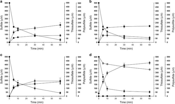
In Figure 2d, control Ec(pMCS5): sulfide, ○ thiosulfate, □ and sulfane sulfur, formula image Sulfide oxidation, polysulfide production and thiosulfate production by E. coli with cloned Zppdo, Bcpdo or Sapdo were essentially the same as Ec(pMCS5) (Data not shown).
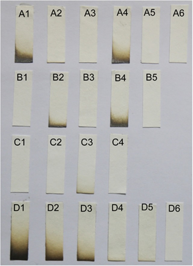
Figure 3
Figure 3
Testing H2S Production by bacteria in LB. Bacteria were inoculated in 2 ml of LB in 15-ml tubes with the lead-acetate filter paper fixed at the top of the headspace. (Row A) A1, E. coli BL21(DE3) (Ec); A2, Ec(Pasqr1); A3, Ec(Pasqr2); A4, Ec(Papdo); A5, Ec(Papdo-Pasqr2); A6, Ec(Cppdo2-Cpsqr). (Row B) B1, P. aeruginosa PAO1 (Pa); B2, PaΔsqrsqr2; B3, PaΔpdo; B4, PaΔsqrsqrpdo (Pa3K), B5: Pa3K(Papdo-Pasqr2). (Row C) C1, C. pinatubonensis JMP134 (Cp); C2, CpΔsqr; C3, CpΔpdosqr (Cp2K); C4, Cp2K(Cppdo2-Cpsqr). (Row D) NaHS standards in LB: D1, 100 μm; D2, 50 μm; D3, 25 μm; D4, 10 μm; D5, 5 μm; D6, 0 μm. E. coli and P. aeruginosa were incubated at 37 °C, and C. pinatubonensis was incubated at 30 °C for 18 h.
Figure 4
Figure 4
Resting bacterial cells oxidize spiked sulfide. Cells were suspended in 50 mm Tris buffer, pH 8.0, with 50 μm DTPA at OD600nm of 1, and NaHS was added to initiate the reaction. Controls were done in the same buffer without bacterial cells. All data are average of three samples with standard deviation (error bar). (a) C. pinatubonensis JMP134 (Cp) and C. pinatubonensis 2 K (Cp2K); (b) P. aeruginosa PAO1 (Pa) and P. aeruginosa 3 K (Pa3K); (c) B. subtilis 168 (Bs), A. tumefaciens C58 (At), K. pneumonia DSM30104 (Kp) and E. coli BL21(DE3) (Ec).
Figure 5
Figure 5
Sulfide oxidation by mixed bacterial cells with SQR or PDO. (a) Combination of C. vitaeruminis DSM20294 (Cv) (with sqr) and S. meliloti 1021 (Sm) (with pdo). (b) Combination of E. coli BL21(DE3) (Ec) with Cpsqr [Ec(Cpsqr)] or Cppdo2 [(Ec(Cppdo2)]. Cv and Sm were induced with sulfide before harvesting, and the recombinant E. coli cells were induced with IPTG before harvesting. Cells were suspended in 50 mm Tris buffer, pH 8.0, with 50 μm DTPA at OD600nm of 2 for individual strains or 4 for mixed strains and 1 mm NaHS was added to initiate the reaction. Controls were done with individual bacterial strains. Sulfide oxidation and the production of polysulfide, sulfite and thiosulfate were detected. All data are average of three samples with standard deviation (error bar).

Similar articles

Cited by

References

    1. Alcolombri U, Ben-Dor S, Feldmesser E, Levin Y, Tawfik DS, Vardi A. (2015). MARINE SULFUR CYCLE. Identification of the algal dimethyl sulfide-releasing enzyme: a missing link in the marine sulfur cycle. Science 348: 1466–1469. - PubMed
    1. Amin S, Hmelo L, van Tol H, Durham B, Carlson L, Heal K et al. (2015). Interaction and signalling between a cosmopolitan phytoplankton and associated bacteria. Nature 522: 98–101. - PubMed
    1. Anandham R, Indiragandhi P, Madhaiyan M, Ryu KY, Jee HJ, Sa TM. (2008). Chemolithoautotrophic oxidation of thiosulfate and phylogenetic distribution of sulfur oxidation gene (soxB) in rhizobacteria isolated from crop plants. Res Microbiol 159: 579–589. - PubMed
    1. Boden R, Borodina E, Wood AP, Kelly DP, Murrell JC, Schafer H. (2011). Purification and characterization of dimethylsulfide monooxygenase from Hyphomicrobium sulfonivorans. J Bacteriol 193: 1250–1258. - PMC - PubMed
    1. Clarke PH. (1953). Hydrogen sulphide production by bacteria. J Gen Microbiol 8: 397–407. - PubMed

LinkOut - more resources