. 2017 Aug 1;58(10):3940-3949.
doi: 10.1167/iovs.16-20912.

Effect of Methotrexate on an In Vitro Patient-Derived Model of Proliferative Vitreoretinopathy

Affiliations

Effect of Methotrexate on an In Vitro Patient-Derived Model of Proliferative Vitreoretinopathy

Dhanesh Amarnani et al. Invest Ophthalmol Vis Sci. .

Abstract

Purpose: The purpose of this study was to develop a method for isolating, culturing, and characterizing cells from patient-derived membranes in proliferative vitreoretinopathy (PVR) to be used for drug testing.

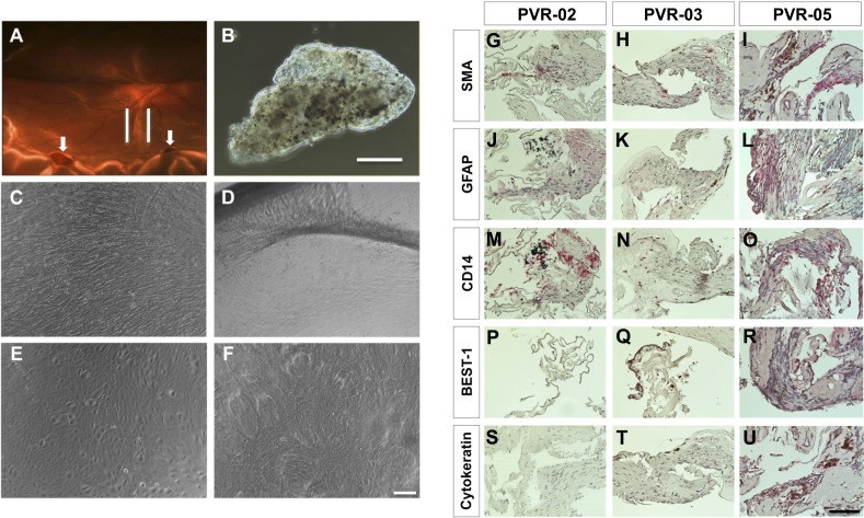
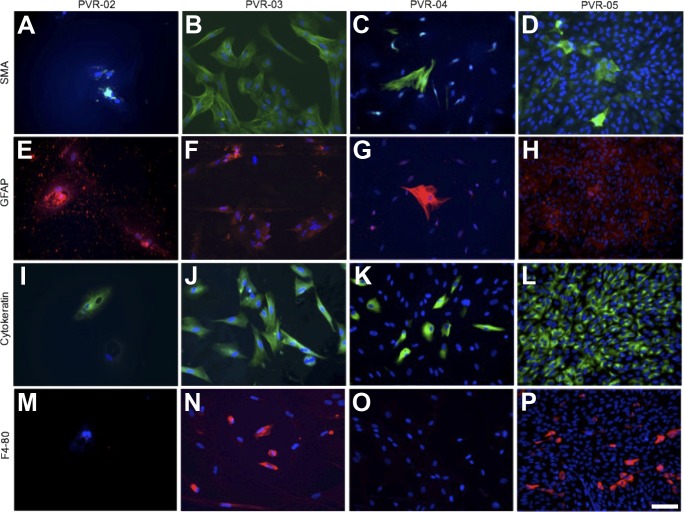
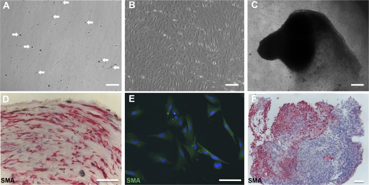
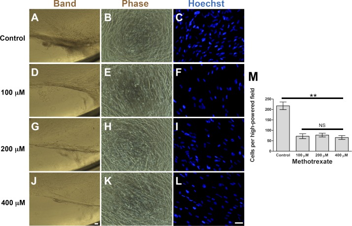
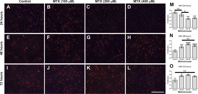
Methods: PVR membranes were obtained from six patients with grade C PVR. Membrane fragments were analyzed by gross evaluation, fixed for immunohistologic studies to establish cell identity, or digested with collagenase II to obtain single cell suspensions for culture. PVR-derived primary cultures were used to examine the effects of methotrexate (MTX) on proliferation, migration, and cell death.

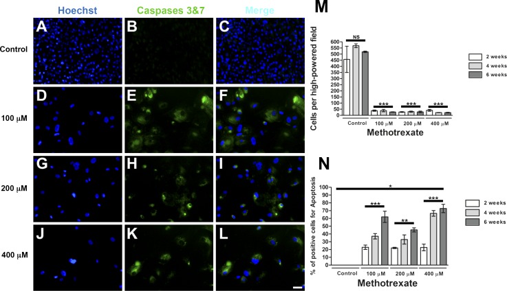
Results: Gross analysis of PVR membranes showed presence of pigmented cells, indicative of retinal pigment epithelial cells. Immunohistochemistry identified cells expressing α-smooth muscle actin, glial fibrillary acidic protein, Bestrophin-1, and F4/80, suggesting the presence of multiple cell types in PVR. Robust PVR primary cultures (C-PVR) were successfully obtained from human membranes, and these cells retained the expression of cell identity markers in culture. C-PVR cultures formed membranes and band-like structures in culture reminiscent of the human condition. MTX significantly reduced the proliferation and band formation of C-PVR, whereas it had no significant effect on cell migration. MTX also induced regulated cell death within C-PVR as assessed by increased expression of caspase-3/7.

Conclusions: PVR cells obtained from human membranes can be successfully isolated, cultured, and profiled in vitro. Using these primary cultures, we identified MTX as capable of significantly reducing growth and inducing cell death of PVR cells in vitro.


Figures

Figure 1
Figure 1
Culture of human PVR membranes and histopathology of PVR membranes. (A) Fundus photograph of the left eye of a patient, case 6 (PVR-06) with recurrent retinal detachment; note presence of a gas bubble within the eye from previous retinal surgery. There is a band of pigmented PVR along the inferior arcade (outlined by white lines). Also note inferior retinal holes in area of detached retina (white arrows). (B) Phase contrast view of the PVR membrane after surgical excision from PVR-06. Scale bar denotes 500 μm. (C) C-PVR cells from case 3 (PVR-03) after 1 week in culture. (D) After 4 weeks in culture, note the formation of bands between the membrane and the rim of the culture dish. (E) C-PVR cells from case 5 (PVR-05) were similarly confluent after 1 week. (F) After 4 weeks in culture, cells grew on top of each other with loss of cell contact inhibition. Scale bar denotes 100 μm. (G–U) Light micrographs of PVR membranes from three different cases (PVR-02, PVR-03, and PVR-05) using primary antibodies (all in red) against SMA (G–I), GFAP (J–L), CD14 (M–O), BEST-1 (P–R), cytokeratin (S–U), and counterstained with hematoxylin (blue). Scale bar denotes 100 μm.
Figure 2
Figure 2
Characterization of cultured cells by immunofluorescence. Immunofluorescence of C-PVR cells from four different cases using primary antibodies against (A–D) SMA all in green, (E–H) GFAP all in red, (I–L) cytokeratin (intermediate filaments in epithelial cells) all in green, and (M–P) F4/80 (immune cells) all in red and counterstained with DAPI (blue). Scale bar denotes 100 μm.
Figure 3
Figure 3
Expression of cell identity molecular markers in PVR membranes is retained in C-PVR primary cultures. (A) Specific populations of C-PVR cells contained pigmented granules (white arrows), characteristic of RPE cells. These granules were released from the cells after 48 to 72 hours. Scale bar denotes 500 μm. (B) C-PVR cells grew as a confluent monolayer in growth medium containing EBM-2 supplemented with SingleQuots Kit (Lonza) and 12% FBS. Scale bar denotes 100 μm. (C) A transformation from a monolayer to invasive cell clusters was triggered by switching to a differentiation culture medium. The phenotypic change was reversible by switching back to the growth medium. Scale bar denotes 500 μm. (D) PVR membrane from clinical case 3 was processed for immunohistochemistry using the antibody against SMA in red and counterstained with hematoxylin (blue). Scale bar denotes 100 μm. (E) Immunofluorescence of the C-PVR primary cultures showing positive cells for SMA in green, suggesting that this molecular marker is retained in C-PVR primary cultures. Nuclei are counterstained with DAPI. Scale bar denotes 100 μm. (F) Cell cluster shown in (C) was harvested and processed for immunocytochemistry using antibody against SMA. Scale bar denotes 100 μm.
Figure 4
Figure 4
Effect of MTX on cell density and band formation of C-PVR. Phase contrast and Hoechst-stained immunofluorescence images of C-PVR cells from PVR-03 after 6 weeks of culture without MTX (A–C) and with 100 (D–F), 200 (G–I), and 400 μM (J–L) MTX. (M) Graph shows quantification of cell density after treatment with different concentrations of MTX or control. Error bars represent SEM. Scale bar denotes 100 μm.
Figure 5
Figure 5
Effect of MTX on proliferation of C-PVR cells. Immunofluorescence images of C-PVR cells from after 24, 48, and 72 hours without (A, E, I) and with 100 (B, F, J), 200 (C, G, K), and 400 mM (D, H, L) MTX. Quantification of proliferation by Ki67 immunofluorescence (red) at 24, 48, and 72 hours (M–O, respectively). Error bars represent SEM. Scale bar denotes 100 μm.
Figure 6
Figure 6
Effect of MTX on C-PVR survival. Hoechst-stained immunofluorescence images of C-PVR cells from PVR-05 after 6 weeks of culture without MTX (A–C) and with 100 (D–F), 200 (G–I), and 400 μM (J–L) MTX. (M) Quantification of cell density showing a significant reduction in cell density after 2, 4, and 6 weeks under the treatment with MTX. (N) Quantification of apoptosis showing a significant increase in the presence of Caspases 3 and 7 after different time points (2, 4, and 6 weeks) with MTX. Error bars represent SEM. Scale bar denotes 100 μm.

References

    1. Weichel ED, Colyer MH. . Combat ocular trauma and systemic injury. Curr Opin Ophthalmol. 2008; 19: 519– 525. - PubMed
    1. Colyer MH, Chun DW, Bower KS, Dick JS, Weichel ED. . Perforating globe injuries during operation Iraqi Freedom. Ophthalmology. 2008; 115: 2087– 2093. - PubMed
    1. Eliott D, Stryjewski TP, Andreoli MT, Andreoli CM. . Smoking is a risk factor for proliferative vitreoretinopathy after traumatic retinal detachment. Retina. 2017; 37: 1229– 1235. - PubMed
    1. Martini B. . Proliferative vitreo-retinal disorders: experimental models in vivo and in vitro. Acta Ophthalmol Suppl. 1992; 201: 1– 63. - PubMed
    1. The classification of retinal detachment with proliferative vitreoretinopathy. Ophthalmology. 1983; 90: 121– 125. - PubMed

Publication types

MeSH terms