. 2017 Aug 3;101(2):267-273.
doi: 10.1016/j.ajhg.2017.07.002.

Heterozygous De Novo UBTF Gain-of-Function Variant Is Associated with Neurodegeneration in Childhood

Affiliations

Heterozygous De Novo UBTF Gain-of-Function Variant Is Associated with Neurodegeneration in Childhood

Simon Edvardson et al. Am J Hum Genet. .

Abstract

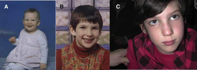
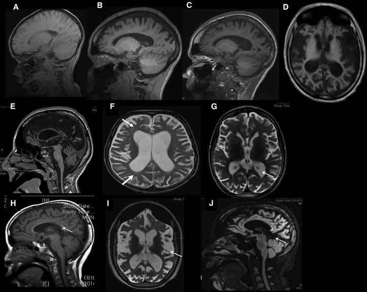
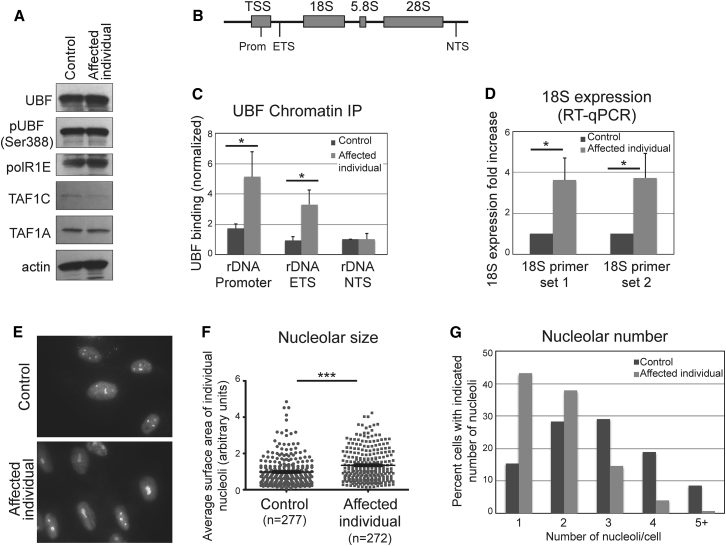
Ribosomal RNA (rRNA) is transcribed from rDNA by RNA polymerase I (Pol I) to produce the 45S precursor of the 28S, 5.8S, and 18S rRNA components of the ribosome. Two transcription factors have been defined for Pol I in mammals, the selectivity factor SL1, and the upstream binding transcription factor (UBF), which interacts with the upstream control element to facilitate the assembly of the transcription initiation complex including SL1 and Pol I. In seven unrelated affected individuals, all suffering from developmental regression starting at 2.5-7 years, we identified a heterozygous variant, c.628G>A in UBTF, encoding p.Glu210Lys in UBF, which occurred de novo in all cases. While the levels of UBF, Ser388 phosphorylated UBF, and other Pol I-related components (POLR1E, TAF1A, and TAF1C) remained unchanged in cells of an affected individual, the variant conferred gain of function to UBF, manifesting by markedly increased UBF binding to the rDNA promoter and to the 5'- external transcribed spacer. This was associated with significantly increased 18S expression, and enlarged nucleoli which were reduced in number per cell. The data link neurodegeneration in childhood with altered rDNA chromatin status and rRNA metabolism.


Figures

Figure 1
Figure 1
Affected Individual 2 at Age 1 Year (A), 6 Years (B), and 10 Years (C)
Figure 2
Figure 2
Progressive Cortical Atrophy Observed in Serial Brain MRI (A–C) Parasagittal T1-weighted brain MRI of affected individual 3 at age 4, 9, and 11 years, respectively. Note progressive cortical atrophy with slight cerebellar atrophy. (D and E) Axial and Midsagittal T1 images of affected individual 4 at age 15 years. Note cortical and cerebellar atrophy. (F) Axial T2 of affected individual 6 at age 8 years. Note cortical atrophy and periventricular white matter hyperintensity (arrows). (G and H) T2 weighted axial and sagittal sequences. Early imaging on affected individual 2 showing modest ventricular dilatation ex-vacuo associated with a thin corpus callous (arrows). (I and J) T2 Weighted axial and sagittal FLAIR sequences in a follow up imaging study repeated after 6 years showing dramatic cortical atrophy and ex-vacuo ventricular dilatation. Note is made of the subdural hygroma below the tentorium in (J).
Figure 3
Figure 3
Mutant UBF Is Hyperactive and Drives Increased Expression of rDNA Genes, Associated with Nucleolar Alterations (A) Western blot analysis of cells of control and affected individual showing that the UBTF mutation does not affect UBF steady state levels, its phosphorylation status, or the levels of the Pol I subunits PolR1E, TAF1A, and TAFC. (B) Schematic representation of the rDNA gene locus, indicating the location of each primer set used: Promoter, external transcribed spacer (ETS), and non-transcribed spacer (NTS). (C) UBF ChIP-qPCR showing that UBF binding to the Promoter and ETS region is significantly increased in cells of the affected individual compared to control cells. Binding was quantified relative to input material, and shown as normalized to the NTS locus in control cells. Bars represent the average of three independent experiments, and error bars represent SD. The asterisk indicates statistical significance (p < 0.05) evaluated using the t test (two-tailed, equal variance). (D) Quantification of 18S expression in cells of affected individual and control, using real-time qPCR, quantified relative to GAPDH expression. Two different 18S primer sets were used. The results are shown as normalized to control cells. Bars represent the average of three independent experiments, and error bars represent SD. The asterisk indicates statistical significance (p < 0.05) evaluated using the t test (two-tailed, equal variance). (E) Representative micrographs of anti-nucleolin immunofluorescence showing altered nucloelar pattern in affected individual cells. (F) Quantification of nucleolar size. Nucleolar area was quantified using ImageJ and statistical analysis was done with Prism (t test, two-tailed, equal variance). Asterisks indicate statistical significance (p < 0.0001). The mean ± SEM is also shown. (G) Quantification of the number of nucleoli in each cell (117 control and 151 affected individual cells were analyzed).

References

    1. Crumrine P.K. Degenerative disorders of the central nervous system. Pediatr. Rev. 2001;22:370–379. - PubMed
    1. Schiffmann R., van der Knaap M.S. Invited article: an MRI-based approach to the diagnosis of white matter disorders. Neurology. 2009;72:750–759. - PMC - PubMed
    1. Tanaka A.J., Cho M.T., Millan F., Juusola J., Retterer K., Joshi C., Niyazov D., Garnica A., Gratz E., Deardorff M. Mutations in SPATA5 Are Associated with Microcephaly, Intellectual Disability, Seizures, and Hearing Loss. Am. J. Hum. Genet. 2015;97:457–464. - PMC - PubMed
    1. Perić S., Glumac J.N., Töpf A., Savić-Pavićević D., Phillips L., Johnson K., Cassop-Thompson M., Xu L., Bertoli M., Lek M. A novel recessive TTN founder variant is a common cause of distal myopathy in the Serbian population. Eur. J. Hum. Genet. 2017;25:572–581. - PMC - PubMed
    1. Ta-Shma A., Zhang K., Salimova E., Zernecke A., Sieiro-Mosti D., Stegner D., Furtado M., Shaag A., Perles Z., Nieswandt B. Congenital valvular defects associated with deleterious mutations in the PLD1 gene. J. Med. Genet. 2016 - PubMed

MeSH terms