. 2017 Aug 3;21(2):256-263.e4.
doi: 10.1016/j.stem.2017.06.016.

Engineered Epidermal Progenitor Cells Can Correct Diet-Induced Obesity and Diabetes

Affiliations

Engineered Epidermal Progenitor Cells Can Correct Diet-Induced Obesity and Diabetes

Jiping Yue et al. Cell Stem Cell. .

Abstract

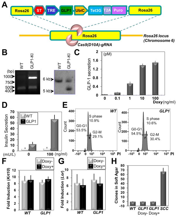
Somatic gene therapy is a promising approach for treating otherwise terminal or debilitating diseases. The human skin is a promising conduit for genetic engineering, as it is the largest and most accessible organ, epidermal autografts and tissue-engineered skin equivalents have been successfully deployed in clinical applications, and skin epidermal stem/progenitor cells for generating such grafts are easy to obtain and expand in vitro. Here, we develop skin grafts from mouse and human epidermal progenitors that were engineered by CRISPR-mediated genome editing to controllably release GLP-1 (glucagon-like peptide 1), a critical incretin that regulates blood glucose homeostasis. GLP-1 induction from engineered mouse cells grafted onto immunocompetent hosts increased insulin secretion and reversed high-fat-diet-induced weight gain and insulin resistance. Taken together, these results highlight the clinical potential of developing long-lasting, safe, and versatile gene therapy approaches based on engineering epidermal progenitor cells.

Keywords: CRISPR; cutaneous gene therapy; diabetes; epidermal progenitor cells; obesity.


Figures

Figure 1
Figure 1. Engineering GLP1-producing skin epidermal progenitor cells with CRISPR
(A) The targeting vector contains two Rosa26 homology arms, flanking the expression cassette for GLP1 (driven by TRE/tet-on promoter). Tet3G (tetracycline transactivator) protein and a selection marker (Puro) are separated by a self-cleavable peptide T2A and driven by a constitutive promoter UbiC (Ubiquitin C promoter). ST: transcriptional stop signal (ST). (B) Integration of the targeting vector into Rosa26 locus is verified by PCR (left panel) and southern blotting (right panel). (C) Secretion of GLP1 in cell culture medium is determined by ELISA (enzyme-linked immunosorbent assay) upon stimulation with doxycycline (Doxy). All error bars represent standard deviation unless otherwise specified. Sample size n=3 independent experiments. (D) Conditioned medium is collected and used to treat starved insulinoma cells. Secretion of insulin in vitro is determined by ELISA. n=4 independent experiments. (E) FACS (fluorescence activated cell sorting) demonstrates similar cell cycle profiles for WT (wild type) and GLP1-expressing epidermal progenitor cells after doxycycline treatment. PI: propidium iodine. (F–G) Western blotting analysis of early (F) and late (G) differentiation marker expression in WT and GLP1-expressing cells upon calcium shift. Band intensity was determined by densitometry and fold of induction is quantified. Krt10: keratin 10; Lor: loricrin. n=4 independent experiments. (H) WT cells or GLP1 cells with or without doxycycline treatment are tested for anchorage independent growth in soft agar. SCC: mouse squamous cell carcinoma cancer initiating cells. n=3 independent experiments.
Figure 2
Figure 2. Stable delivery of GLP1 in vivo through mouse-to-mouse skin transplantation
(A) Images of immunocompetent mice (CD1) grafted with isogenic skin organoids generated from GLP1-expressing cells. Cells were infected with lentivirus encoding Luciferase before grafting. (B) Histological examination of grafted GLP1 skin and adjacent host skin as control (Ctrl). Scale bar=50 μm. (C–E) Sections of grafted skin and adjacent host skin control (ctrl) were immunostained with different antibodies as indicated (Krt14: keratin 14, β4: β4-integrin, CD104). Scale bar=50 μm. Epi: epidermis, Der: dermis, HF: hair follicle. (F) The level of GLP1 in blood is determined by ELISA from mice engrafted with control or GLP1 cells. n=4 different animals. (G) Level of GLP1 in blood is determined by ELISA for 16 weeks after skin engraftment. n=3 different animals.
Figure 3
Figure 3. Expression of GLP1 in epidermal progenitor cells improves body weight and glucose homeostasis in vivo
(A) Images of control and grafted animals fed a regular diet or a HFD (high fat diet). (B) Representative histological staining’s of white fat tissue. Scale bar= 100 μm. (C) Body weight change of different cohorts of mice measured from ~10 weeks of age. n=5 different animals for each group. (D–E) IPGTT (intraperitoneal glucose tolerance test) for control (D) and GLP1 grafted (E) animals. Blood glucose concentrations as a function of time following intraperitoneal injection of glucose showed improved glucose tolerance in GLP1-expressing mice. n=5 different animals. (F–G) ITT (insulin tolerance test). Profile of glucose concentrations (percentage of initial value) as a function of time following intraperitoneal injection of insulin shows reduced insulin resistance in GLP1-expressing mice. n=5 different animals.
Figure 4
Figure 4. Expression of GLP1 in human epidermal progenitor cells with CRISPR
(A) Image of nude mouse grafted with organotypic human skin culture. (B) Sections of grafted skin were immunostained with different antibodies as indicated. Scale bar=50 μm. (C) Integration of the targeting vector into AAVS1 locus is verified by southern blotting. (D) Secretion of GLP1 into the culture medium was determined by the ELISA upon stimulation. n=3 independent experiments. (E) Secretion of insulin upon treatment with conditioned medium was determined by ELISA. n=4 independent experiments. (F) H&E staining of skin organoids developed from control or GLP1-producing human cells. (G) Level of GLP1 was determine by ELISA in blood from control or grafted nude mice. n=4 different animals.

Comment in

  • Engineering Skin with Skinny Genes.
    Böttcher A, Lickert H, Tschöp M. Böttcher A, et al. Cell Stem Cell. 2017 Aug 3;21(2):153-155. doi: 10.1016/j.stem.2017.07.013. Cell Stem Cell. 2017. PMID: 28777938

References

    1. Ahima RS. Digging deeper into obesity. J Clin Invest. 2011;121:2076–2079. - PMC - PubMed
    1. Ashcroft FM, Rorsman P. Diabetes mellitus and the beta cell: the last ten years. Cell. 2012;148:1160–1171. - PMC - PubMed
    1. Blanpain C, Fuchs E. Epidermal stem cells of the skin. Annu Rev Cell Dev Biol. 2006;22:339–373. - PMC - PubMed
    1. Carsin H, Ainaud P, Le Bever H, Rives J, Lakhel A, Stephanazzi J, Lambert F, Perrot J. Cultured epithelial autografts in extensive burn coverage of severely traumatized patients: a five year single-center experience with 30 patients. Burns: journal of the International Society for Burn Injuries. 2000;26:379–387. - PubMed
    1. Centanni JM, Straseski JA, Wicks A, Hank JA, Rasmussen CA, Lokuta MA, Schurr MJ, Foster KN, Faucher LD, Caruso DM, et al. StrataGraft skin substitute is well-tolerated and is not acutely immunogenic in patients with traumatic wounds: results from a prospective, randomized, controlled dose escalation trial. Annals of surgery. 2011;253:672–683. - PMC - PubMed

Publication types

MeSH terms