. 2017 Oct:103:308-317.
doi: 10.1016/j.bone.2017.07.028. Epub 2017 Aug 1.

Stable sulforaphane protects against gait anomalies and modifies bone microarchitecture in the spontaneous STR/Ort model of osteoarthritis

Affiliations

Stable sulforaphane protects against gait anomalies and modifies bone microarchitecture in the spontaneous STR/Ort model of osteoarthritis

Behzad Javaheri et al. Bone. 2017 Oct.

Abstract

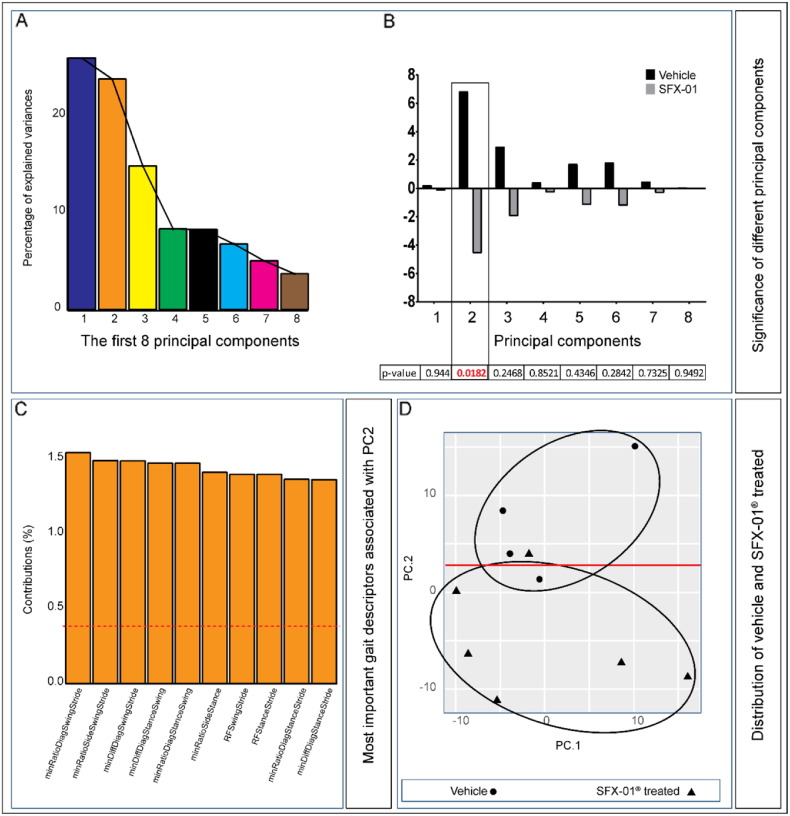
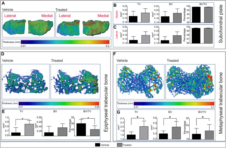
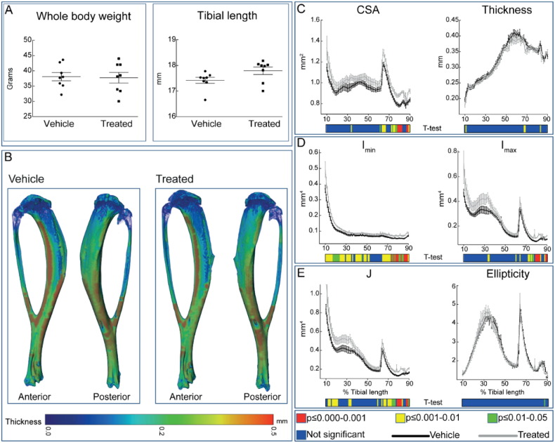
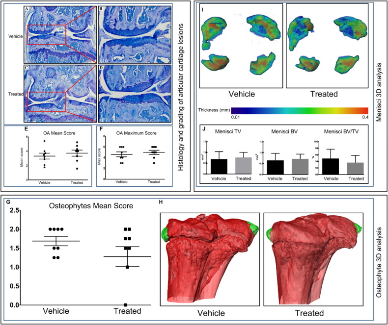
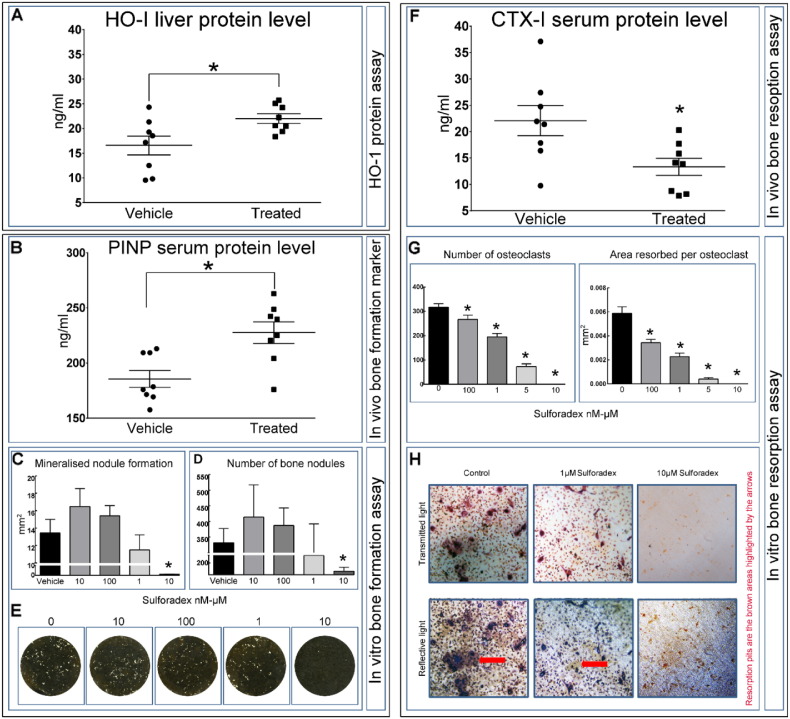
Osteoarthritis (OA), affecting joints and bone, causes physical gait disability with huge socio-economic burden; treatment remains palliative. Roles for antioxidants in protecting against such chronic disorders have been examined previously. Sulforaphane is a naturally occurring antioxidant. Herein, we explore whether SFX-01®, a stable synthetic form of sulforaphane, modifies gait, bone architecture and slows/reverses articular cartilage destruction in a spontaneous OA model in STR/Ort mice. Sixteen mice (n=8/group) were orally treated for 3months with either 100mg/kg SFX-01® or vehicle. Gait was recorded, tibiae were microCT scanned and analysed. OA lesion severity was graded histologically. The effect of SFX-01® on bone turnover markers in vivo was complemented by in vitro bone formation and resorption assays. Analysis revealed development of OA-related gait asymmetry in vehicle-treated STR/Ort mice, which did not emerge in SFX-01®-treated mice. We found significant improvements in trabecular and cortical bone. Despite these marked improvements, we found that histologically-graded OA severity in articular cartilage was unmodified in treated mice. These changes are also reflected in anabolic and anti-catabolic actions of SFX-01® treatment as reflected by alteration in serum markers as well as changes in primary osteoblast and osteoclast-like cells in vitro. We report that SFX-01® improves bone microarchitecture in vivo, produces corresponding changes in bone cell behaviour in vitro and leads to greater symmetry in gait, without marked effects on cartilage lesion severity in STR/Ort osteoarthritic mice. Our findings support both osteotrophic roles and novel beneficial gait effects for SFX-01® in this model of spontaneous OA.

Keywords: Cartilage; Osteoarthritis; SFX-01; STR/Ort; Sulforaphane.


Figures

Fig. 1
Fig. 1
SFX-01® treatment corrects OA-associated gait asymmetry (A) Scree graph displaying percentage of variance explained by each of the first 8 principal components. (B) Average values of each of the first 8 PC's in SFX-01® (grey) and vehicle treated (black) mice and the corresponding p-values (beneath graph) of the differences between these groups. (C) The top 10 gait descriptors that associated with principal component 2 (dotted red line corresponds to the expected value if the contribution where uniform). (D) Scatterplot showing the significant segregation of individual vehicle (closed circles) and SFX-01® (closed triangles) treated mice on the basis of the first 2 PCs.
Fig. 2
Fig. 2
Proximal epiphyseal and metaphyseal tibial bone phenotype of vehicle (black) and SFX-01® treated (grey) mice after 11 weeks of treatment starting at 26 weeks of age. (A) Representative colour-coded thickness images of lateral and medial tibial subchondral plate of vehicle and SFX-01® treated mice. (B and C) Total volume (TV), bone volume (BV) and bone volume fraction (BV/TV) of medial (B) and lateral (C) proximal tibial subchondral plate. (D) Representative colour-coded thickness images of epiphyseal trabecular bone of vehicle and SFX-01® treated mouse tibia. (E) TV, BV and BV/TV of tibial epiphyseal trabecular bone. (F) Representative colour-coded thickness images of metaphyseal trabecular bone of vehicle and SFX-01® treated mice. (g) Ex vivo analyses to determine TV, BV and BV/TV of metaphyseal trabecular bone. Bar graphs represent means ± SEM. Group sizes were n = 8 for vehicle and treated mice. Statistical comparisons: * denotes p ≤ 0.05.
Fig. 3
Fig. 3
Cortical bone phenotype of vehicle (black) and SFX-01® (grey) treated mice. (A) Whole body weight and tibial length of vehicle and SFX-01® treated mice after 11 weeks of treatment. (B) Representative 3D Micro-CT colour-coded images of tibial cortical bone thickness. (C) Bone cross sectional area (CSA) and mean cortical thickness, (D) minimum and maximum second moments of area (Imin and Imax respectively) and (D) ellipticity and J (resistance to torsion) of vehicle and SFX-01® treated STR/Ort mice. Whole bone analyses of cortical bone between 10 and 90% of total tibial length, excluding proximal and distal metaphyseal bone. A two-sample t-test was used to compare means between vehicle KO and SFX-01® treated STR/Ort mice. Line graphs represent means ± SEM. Group sizes were n = 8 for vehicle and treated mice. Graphical heat map summarises statistical differences at specific matched locations along the tibial length, representative of overall effect of SFX-01® treatment. Red p ≤ 0.000–0.001, yellow p ≤ 0.001–0.01, green p ≤ 0.01–0.05 and blue p ≥ 0.05.
Fig. 4
Fig. 4
SFX-01® treatment does not modify different compartments of tibiofemoral complex. (A–C) (lower-power) and (B–D) (higher-power), toluidine blue stained sections of joints from vehicle (A–B) and SFX-01® (C–D) treated STR/Ort mice showing locations of naturally occurring lesions in the articular cartilage of the medial femur compartment of the tibiofemoral joint. Both vehicle and treated groups exhibited localized lesions. (E) and (F), mean and maximum ± SEM lesion severity scores in each compartment of vehicle joints (circle) and treated joints (square). Osteophytes develop in both SFX-01® and vehicle treated STR/Ort mice; toluidine blue stained sections were scored to provide (G) mean number of osteophytes. (H) MicroCT was used to produce representative 3D images of osteophytes (green) in vehicle and SFX-01® treated mice. (I) Representative 3D Micro-CT colour-coded images of meniscal phenotype and (J) ex vivo analyses to determine TV, BV and BV/TV. Group sizes were n = 8 for vehicle and treated mice.
Fig. 5
Fig. 5
SFX-01® treatment activates downstream target (HO-1: heme oxygenase-1) and controls bone remodelling in vivo and in vitro. (A) HO-1 protein level in liver measured by ELISA. (B) In vivo blood serum protein level of procollagen type 1 N-terminal propeptide (P1NP: bone formation marker). (C–D) SFX-01® inhibited bone nodule formation at the highest dose only; no effects were seen at lower concentrations. (E) Representative whole well scans of osteoblast cell layers treated with different concentration of SFX-01®. (F) In vivo blood serum protein level of serum collagen type 1 cross-linked C-telopeptide (CTX-1: bone resorption marker). (G) SFX-01® dose-dependently inhibits osteoclast formation and resorptive activity compared to vehicle. (H) Representative transmitted and reflective light images of osteoclasts treated with 1–10 μM SFX-01®. Graphs represent means ± SEM. Statistical comparisons: * denotes p ≤ 0.05.

Similar articles

Cited by

References

    1. Conaghan P.G. Update on osteoarthritis part 1: current concepts and the relation to exercise. Br. J. Sports Med. 2002;36(5):330–333. - PMC - PubMed
    1. Haseeb A., Haqqi T.M. 146(3) 2013. Immunopathogenesis of Osteoarthritis; pp. 185–196. - PMC - PubMed
    1. Goldring M.B., Goldring S.R. Articular cartilage and subchondral bone in the pathogenesis of osteoarthritis. Ann. N. Y. Acad. Sci. 2010;1192(1):230–237. - PubMed
    1. Hayami T., Pickarski M., Zhuo Y., Wesolowski G.A., Rodan G.A., Duong L.T. Characterization of articular cartilage and subchondral bone changes in the rat anterior cruciate ligament transection and meniscectomized models of osteoarthritis. Bone. 2006;38(2):234–243. - PubMed
    1. Pastoureau P., Chomel A., Bonnet J. Evidence of early subchondral bone changes in the meniscectomized guinea pig. A densitometric study using dual-energy X-ray absorptiometry subregional analysis. Osteoarthr. Cartil. 1999;7(5):466–473. - PubMed