

Review
doi: 10.1016/j.biochi.2017.07.016.
Epub 2017 Aug 1.
TMPRSS2: A potential target for treatment of influenza virus and coronavirus infections
Affiliations
- PMID: 28778717
- PMCID: PMC7116903
- DOI: 10.1016/j.biochi.2017.07.016
Item in Clipboard
Review
TMPRSS2: A potential target for treatment of influenza virus and coronavirus infections
Biochimie.
2017 Nov.
Abstract
Influenza virus and coronavirus epidemics or pandemics have occurred in succession worldwide throughout the early 21st century. These epidemics or pandemics pose a major threat to human health. Here, we outline a critical role of the host cell protease TMPRSS2 in influenza virus and coronavirus infections and highlight an antiviral therapeutic strategy targeting TMPRSS2.
Keywords: Coronavirus; Influenza virus; TMPRSS2; Therapeutics.
Copyright © 2017 Elsevier B.V. and Société Française de Biochimie et Biologie Moléculaire (SFBBM). All rights reserved.
Figures
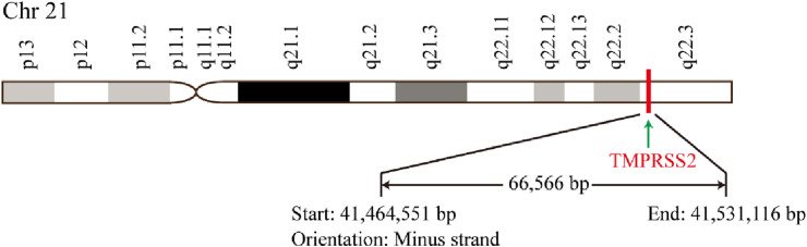
A schematic diagram of TMPRSS2 genomic location.
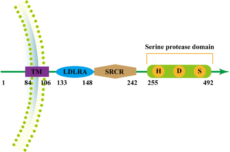
The location and structure of TMPRSS2 protein. TM: transmembrane domain; LDLRA: low-density lipoprotein receptor domain class A; SRCR: Scavenger receptor cysteine-rich domain; Letters H: histidine; Letters D: aspartate; Letters S: serine.
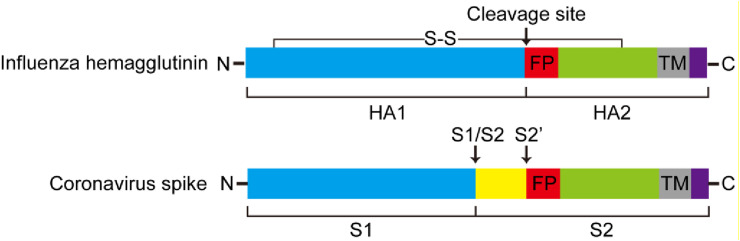
Structure of influenza hemagglutinin and coronavirus spike protein with cleavage sites. Arrows: cleavage site; FP: putative fusion peptide; TM: transmembrane domain; S-S: disulfide bond.
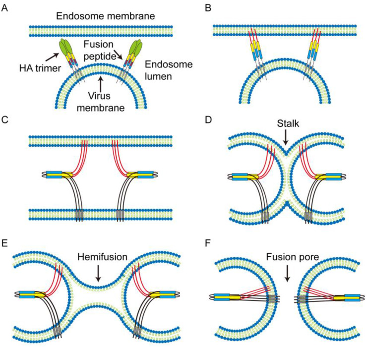
Membrane fusion mediated by hemagglutinin. A) Viral membrane with two representative cleaved neutral pH HA and endosome membrane. B) The acidic environment of the endosome inducing conformational changes results in fusion peptide exposure and insertion into the target membrane. C) Conformational changes drive the viral and cellular membranes close proximity. D) Formation of pre-fusion stalk intermediate. E) Formation of hemifusion intermediate. F) Formation of fusion pore and viral genome is released into the cytoplasm. The Figure is adapted according to Karen J. Cross .
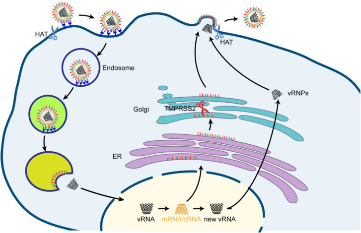
The replication cycle of influenza virus and the proteolytic cleavage of the host proteases. Influenza virus binds to sialic acid-containing cell surface receptors, the bound virus is then endocytosed. During maturation of the endosome, the pH drops initiates the fusion of the viral envelope with the endosomal membrane and the release of the vRNPs into the cytosol. The vRNPs are imported into the nucleus, then transcription and replication proceed. Translation of viral mRNAs is performed by the cellular machinery. Newly formed viral RNAs are exported to the cytosol, assembled with new virus structural proteins, then packaged together at the plasma membrane, and bud off to release new virions. HA is synthesized as precursor that requires cleavage. HA cleavage by membrane-bound proteases (indicated as scissors) can take place in different part and at different time points during the viral life cycle. HA containing a monobasic cleavage site is cleaved by TMPRSS2 in the Golgi apparatus during assembly or cleaved by HAT on the plasma membrane either during attachment and entry into the cell or during budding of virions.
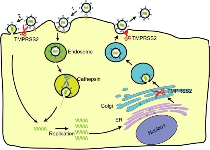
The replication cycle of coronavirus and the proteolytic cleavage of the host proteases. Coronavirus binds to the cellular receptor, resulting in uptake of virions into endosomes (route 1), where the spike protein is activated by cathepsin. The pH drops in endosome initiates the fusion of the viral envelope with the endosomal membrane and the release of the viral genetic material into the cytosol, then RNA transcription, replication and transcription take place. New viral RNA is transported to the endoplasmic reticulum, Golgi intermediate, the site of assembly. Viral RNA and structural proteins assemble and bud into vesicles. Vesicles are transported to the cell surface and release. Alternatively, the spike protein can be activated at the cell surface, resulting in fusion of the viral membrane with the plasma membrane (route 2). Spike is synthesized as precursor that requires cleavage by host proteases. Spike cleavage (indicated as scissors) can take place in different part and at different time points during the viral life cycle. Spike cleavage by cathepsin occurs in the endosome. Spike cleavage by TMPRSS2 takes place in the Golgi or plasma membrane, either during assembly or attachment and release.
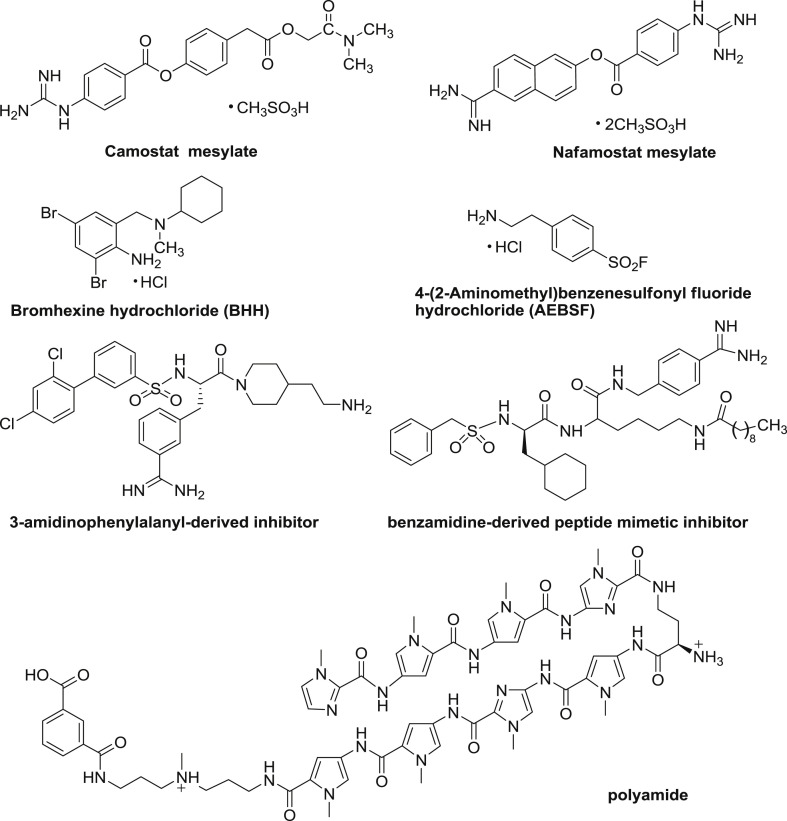
Structures of some small molecular inhibitors for TMPRSS2 and TMPRSS2-related agents.
Similar articles
-
The Proteolytic Activation of (H3N2) Influenza A Virus Hemagglutinin Is Facilitated by Different Type II Transmembrane Serine Proteases.J Virol. 2016 Apr 14;90(9):4298-4307. doi: 10.1128/JVI.02693-15. Print 2016 May. J Virol. 2016. PMID: 26889029 Free PMC article.
-
Repurposing host-based therapeutics to control coronavirus and influenza virus.Drug Discov Today. 2019 Mar;24(3):726-736. doi: 10.1016/j.drudis.2019.01.018. Epub 2019 Jan 31. Drug Discov Today. 2019. PMID: 30711575 Free PMC article. Review.
-
Airway proteases: an emerging drug target for influenza and other respiratory virus infections.Curr Opin Virol. 2017 Jun;24:16-24. doi: 10.1016/j.coviro.2017.03.018. Epub 2017 Apr 14. Curr Opin Virol. 2017. PMID: 28414992 Free PMC article. Review.
-
Tmprss2 knock-out mice are resistant to H10 influenza A virus pathogenesis.J Gen Virol. 2019 Jul;100(7):1073-1078. doi: 10.1099/jgv.0.001274. Epub 2019 May 17. J Gen Virol. 2019. PMID: 31099738
-
Identification of the first synthetic inhibitors of the type II transmembrane serine protease TMPRSS2 suitable for inhibition of influenza virus activation.Biochem J. 2013 Jun 1;452(2):331-43. doi: 10.1042/BJ20130101. Biochem J. 2013. PMID: 23527573
Cited by
-
Interaction between Sars-CoV-2 structural proteins and host cellular receptors: From basic mechanisms to clinical perspectives.Adv Protein Chem Struct Biol. 2022;132:243-277. doi: 10.1016/bs.apcsb.2022.05.010. Epub 2022 Jun 9. Adv Protein Chem Struct Biol. 2022. PMID: 36088078 Free PMC article. Review.
-
ACE2, TMPRSS2 distribution and extrapulmonary organ injury in patients with COVID-19.Biomed Pharmacother. 2020 Nov;131:110678. doi: 10.1016/j.biopha.2020.110678. Epub 2020 Aug 24. Biomed Pharmacother. 2020. PMID: 32861070 Free PMC article. Review.
-
Cell-autonomous immune gene expression is repressed in pulmonary neuroendocrine cells and small cell lung cancer.Commun Biol. 2021 Mar 9;4(1):314. doi: 10.1038/s42003-021-01842-7. Commun Biol. 2021. PMID: 33750914 Free PMC article.
-
The Transmembrane Protease TMPRSS2 as a Therapeutic Target for COVID-19 Treatment.Int J Mol Sci. 2022 Jan 25;23(3):1351. doi: 10.3390/ijms23031351. Int J Mol Sci. 2022. PMID: 35163273 Free PMC article. Review.
-
Improving Soluble Expression of SARS-CoV-2 Spike Priming Protease TMPRSS2 with an Artificial Fusing Protein.Int J Mol Sci. 2023 Jun 22;24(13):10475. doi: 10.3390/ijms241310475. Int J Mol Sci. 2023. PMID: 37445653 Free PMC article.
References
-
- Pamuk S. The black death and the origins of the ‘great divergence’ across Europe, 1300–1600. Eur. Rev. Econ. Hist. 2007;11:289–317.
-
- Cohen J. What's old is new: 1918 virus matches 2009 H1N1 strain. Science. 2010;327:1563–1564. - PubMed
-
- Stock I. Yersinia pestis and plague-an update. Med. Monatsschr. Pharm. 2014;37:441–448. - PubMed
-
- Seto W.H., Tsang D., Yung R.W., Ching T.Y., Ng T.K., Ho M., Ho L.M., Peiris J.S. Advisors of Expert SARS group of Hospital Authority, Effectiveness of precautions against droplets and contact in prevention of nosocomial transmission of severe acute respiratory syndrome (SARS) Lancet. 2003;361:1519–1520. - PMC - PubMed
Publication types
MeSH terms
Substances
LinkOut - more resources
Full Text Sources
Other Literature Sources
Medical
