. 2017 Nov 1;23(21):6529-6540.
doi: 10.1158/1078-0432.CCR-17-0282. Epub 2017 Aug 4.

Targeting RNA-Polymerase I in Both Chemosensitive and Chemoresistant Populations in Epithelial Ovarian Cancer

Affiliations

Targeting RNA-Polymerase I in Both Chemosensitive and Chemoresistant Populations in Epithelial Ovarian Cancer

Robert Cornelison et al. Clin Cancer Res. .

Abstract

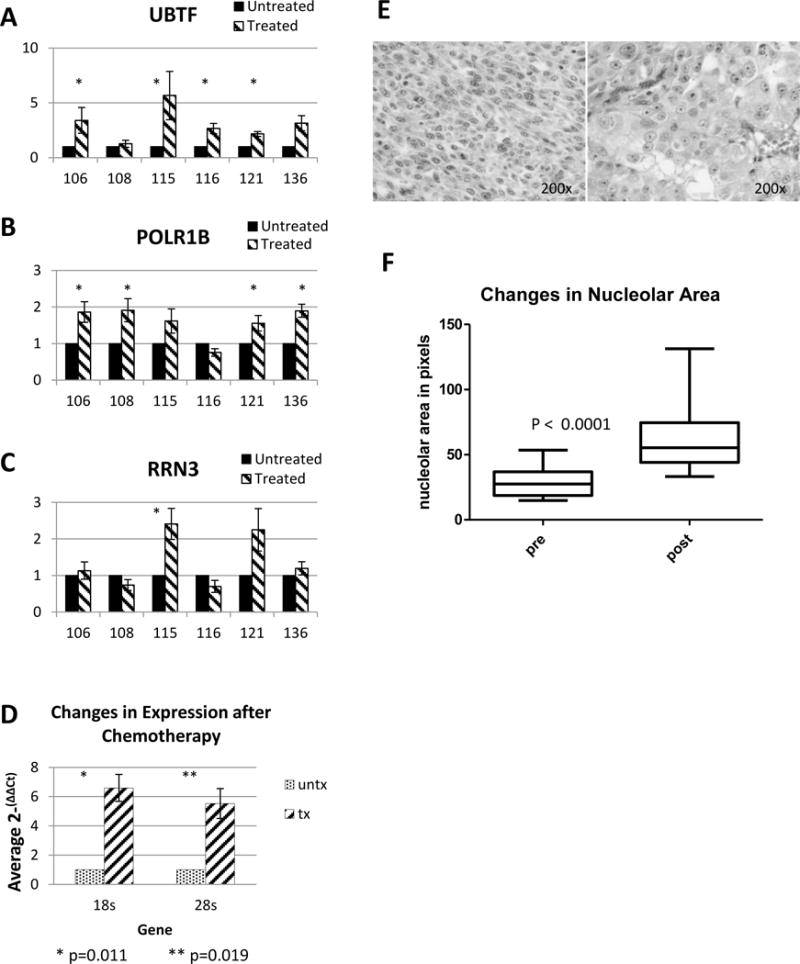
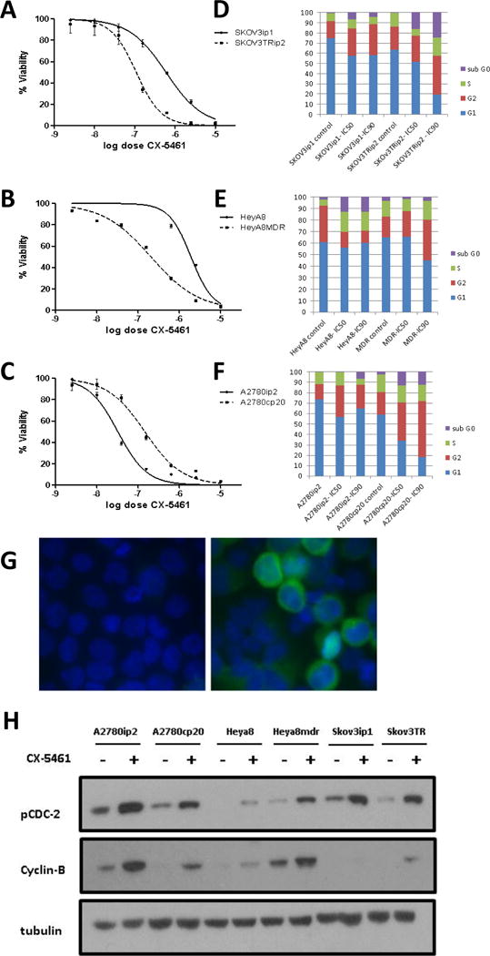
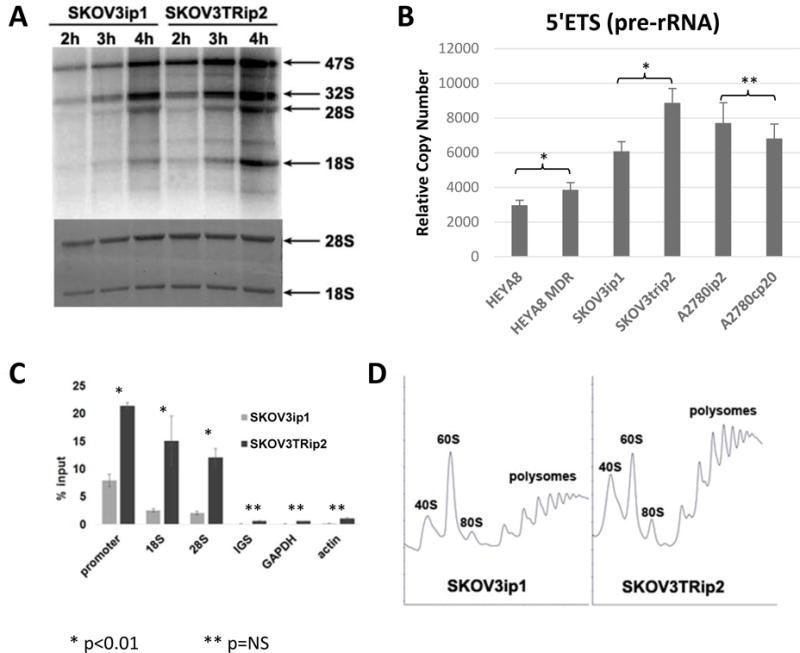
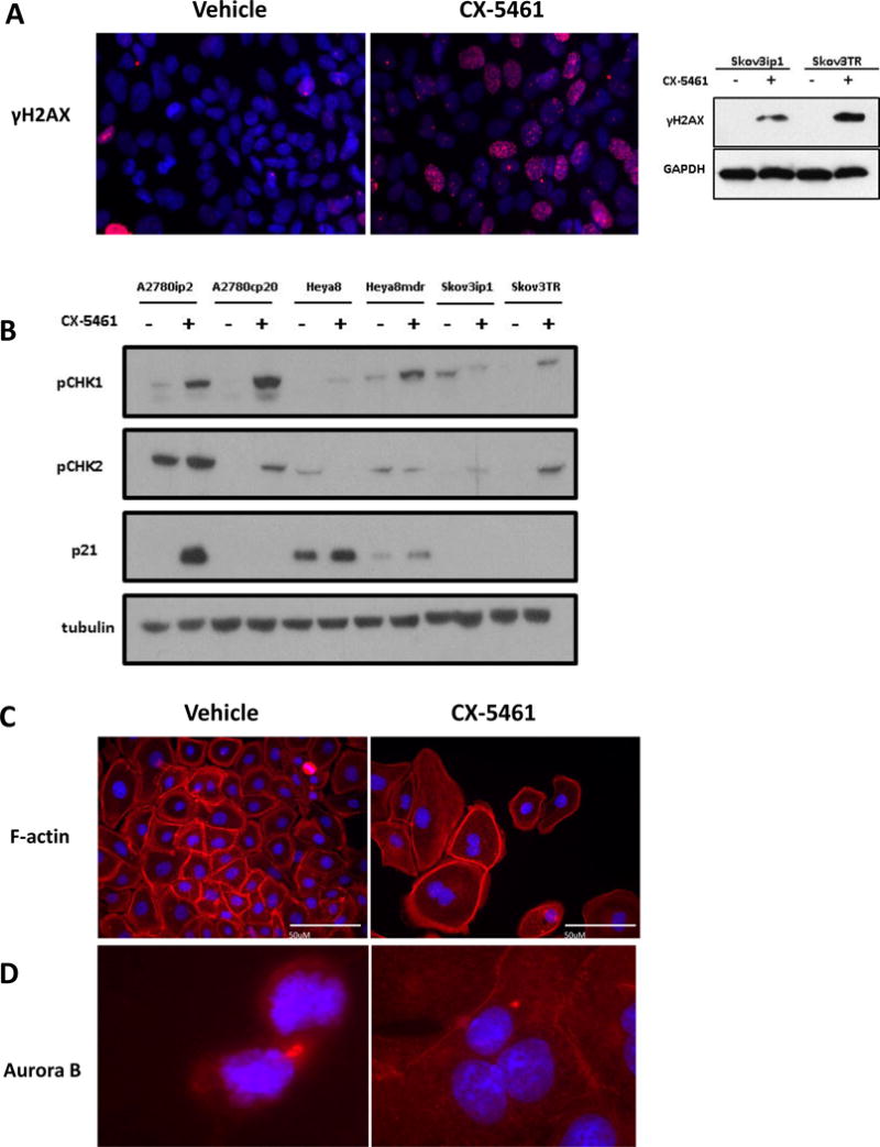
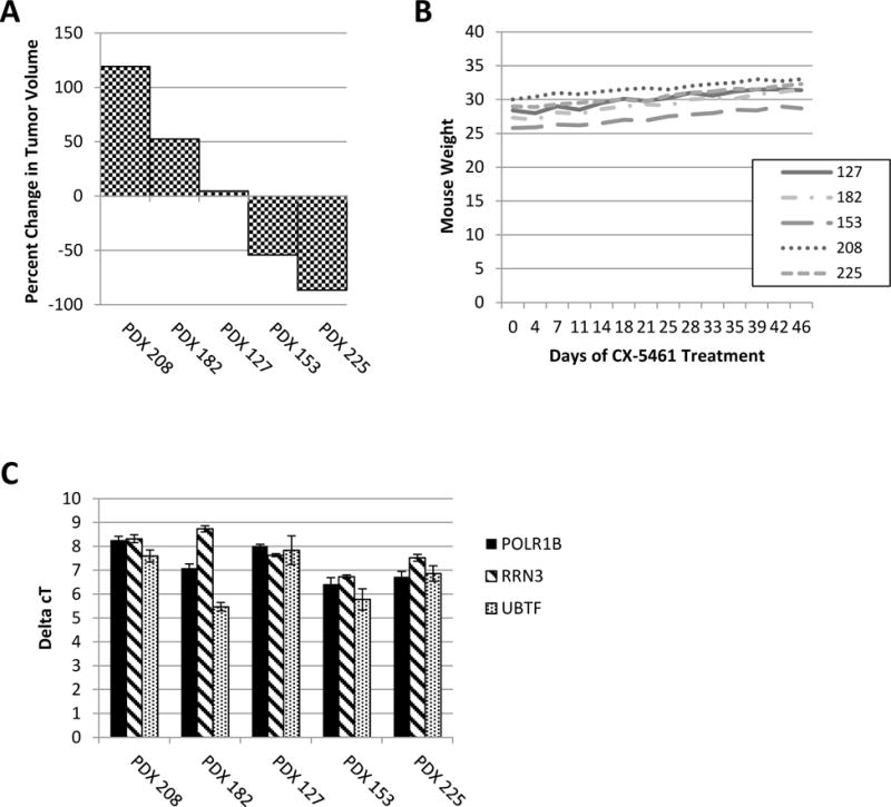
Purpose: A hallmark of neoplasia is increased ribosome biogenesis, and targeting this process with RNA polymerase I (Pol I) inhibitors has shown some efficacy. We examined the contribution and potential targeting of ribosomal machinery in chemotherapy-resistant and -sensitive models of ovarian cancer.Experimental Design: Pol I machinery expression was examined, and subsequently targeted with the Pol I inhibitor CX-5461, in ovarian cancer cell lines, an immortalized surface epithelial line, and patient-derived xenograft (PDX) models with and without chemotherapy. Effects on viability, Pol I occupancy of rDNA, ribosomal content, and chemosensitivity were examined.Results: In PDX models, ribosomal machinery components were increased in chemotherapy-treated tumors compared with controls. Thirteen cell lines were sensitive to CX-5461, with IC50s 25 nmol/L-2 μmol/L. Interestingly, two chemoresistant lines were 10.5- and 5.5-fold more sensitive than parental lines. CX-5461 induced DNA damage checkpoint activation and G2-M arrest with increased γH2AX staining. Chemoresistant cells had 2- to 4-fold increased rDNA Pol I occupancy and increased rRNA synthesis, despite having slower proliferation rates, whereas ribosome abundance and translational efficiency were not impaired. In five PDX models treated with CX-5461, one showed a complete response, one a 55% reduction in tumor volume, and one maintained stable disease for 45 days.Conclusions: Pol I inhibition with CX-5461 shows high activity in ovarian cancer cell lines and PDX models, with an enhanced effect on chemoresistant cells. Effects occur independent of proliferation rates or dormancy. This represents a novel therapeutic approach that may have preferential activity in chemoresistant populations. Clin Cancer Res; 23(21); 6529-40. ©2017 AACR.


Conflict of interest statement

The authors declare no potential conflicts of interest.

Figures

Figure 1
Figure 1. Expression of RNA Polymerase I initiation factors in ovarian cancer PDX models. Comparison of 6 pairs of untreated/treated (Carbo/taxol) PDX tumors showed similar changes in expression profiles. One common pathway was ribosomal synthesis
(A,B,C) qPCR was conducted on 6 pairs of ovarian cancer PDX treated with carboplatin and paclitaxel or control for UBTF, POLR1B, and RRN3 and gene expression was compared to the untreated matched PDX. The tumor cell population surviving initial chemotherapy generally had a greater expression of UBTF, POLR1B, and RRN3. (* = p < 0.05) (D) Total level of ribosomal subunits 18S and 28S was determined. Treated ovarian cancer PDXs had a 37.9-fold increase of 18S (p=0.010) and a 39.0-fold increase of 28S (p=0.019). (E) Comparison of H&E sections with and without treatment, demonstrating marked macronucleoli by (F) Quantification of macronucleoli before and after chemotherapy treatment in PDX models.
Figure 2
Figure 2. Response of ovarian cancer cell lines to CX-5461
Cell viability and cell cycle analysis was conducted on 3 pairs of chemosensitive and chemoresistant ovarian cancer cell lines. (A–C) Dose response curves from MTT assay of each pair. Results show both SKOV3TRip2 and HeyA8MDR resistant lines being more sensitive to CX-5461 treatment than their chemosensitive parental line. (D-F) Cell cycle analysis of SKOV3ip1, SKOV3TRip2, HeyA8, HeyA8MDR, A2780ip2, and A2780cp20 using Propidium Iodide after 48 hr treatment with the control, IC50, or IC90 dose of CX-5461. In general, treatment with CX-5461 resulted in an increase in sub-G0 fraction and a portion of cells in the S/G2 phase. (G) Immunofluorescence assay example verifying G2/M phase cell cycle arrest in A2780cp20 treated cells with 1μM CX-5461 versus vehicle control. Results show an increase in Cyclin-B perinuclear staining in arrested cells compared to untreated control. (H) Western blot analysis verifying G2/M phase arrest in 6 cell lines. Treatment with IC50 dosages of CX-5461 showed an increase in expression of pCDC2(tyr15) and Cyclin-B versus untreated controls at 72 hours.
Figure 3
Figure 3. rRNA synthesis and polymerase occupancy is increased in SKOV3TRip2 cells compared to SKOV3ip1 cells
(A) Cells were pulse labeled with 32P-orthophosphate as a function of time. Total RNA was purified by Trizol, run on a formaldehyde denaturing agarose gel, dried, and visualized by autoradiography. The rate of 32P incorporation into rRNA in SKOV3TRip2 cells was 2.1 fold (+/- 0.3) higher than in SKOV3ip1 cells. (B) rDNA transcription was analyzed by RT-qPCR by targeting short-lived 5′ETS, and the rRNA signal was normalized to the control GAPDH mRNA. (C) CHIP analysis of RPA194 association with rDNA. Pol I molecules were immunoprecipitated by anti-A194 monoclonal antibody. The mean and SD are shown. (D) Polysome profiles were performed by sucrose gradient centrifugation.
Figure 4
Figure 4. CX-5461 induces DNA damage response via ATM/ATR in ovarian cancer cell lines
(A) Immunofluorescent detection of the pH2AX in control SKOV3ip1 cells (red) and cells exposed to 500nM of CX-5461. The punctate nuclear staining illustrated an accumulation of DNA damage foci. Pairs of chemosensitive/chemoresistant ovarian cancer cell lines were treated with their respective IC50 dosages of CX-5461. (B) Western blot analysis shows activation of the pCHK1(ser317) and pCHK2(T68), downstream substrates of ATM and ATR kinases. TP53 wild type cell lines showed a subsequent activation of p21 but drug response was independent of TP53 status. (C) DAPI (blue) labeling showed failed cytokinesis and an accumulation of multinucleated cells after CX-5461 treatment, with F-actin (red) indicating shared cytoplasm. (D) Aurora-B kinase (red) detection showing normal localization to the cleavage furrow during cytokinesis in control SKOV3ip1 cells. After treatment with CX-5461 Aurora-B mislocalization was apparent in an accumulation of multinucleated cells.
Figure 5
Figure 5. Response of ribosomal translation factors after CX-5461 treatment
(A) Waterfall plot displaying percent change of tumor volume in PDXs 208, 182, 127, 153, and 225 after 45 days of treatment with 50mg/kg CX-5461 q3D. (B) Weight of mice through duration of treatment with CX-5461. (C) qPCR of Pol I initiation factors RRN3, POLR1B, and UBTF. ΔCT was calculated. There was not an association between baseline expression of these genes and response to CX-5461.

Similar articles

Cited by

References

    1. Berek JS, Crum C, Friedlander M. Cancer of the ovary, fallopian tube, and peritoneum. Int J Gynaecol Obstet. 2012;119(Suppl):S118–29. - PubMed
    1. Bowtell DD, Böhm S, Ahmed AA, et al. Rethinking ovarian cancer II: reducing mortality from high-grade serous ovarian cancer. Nat Rev Cancer. 2015;15:668–679. - PMC - PubMed
    1. Armstrong DK. Relapsed ovarian cancer: challenges and management strategies for a chronic disease. Oncologist. 2002;7(Suppl 5):20–8. - PubMed
    1. Borst P. Cancer drug pan-resistance: pumps, cancer stem cells, quiescence, epithelial to mesenchymal transition, blocked cell death pathways, persisters or what? Open Biol. 2012;2:120066. - PMC - PubMed
    1. Rubin SC, Randall TC, Armstrong KA, et al. Ten-year follow-up of ovarian cancer patients after second-look laparotomy with negative findings. Obstet Gynecol. 1999;93:21–4. - PubMed

MeSH terms