Review
. 2018 May 1;10(5):a033134.
doi: 10.1101/cshperspect.a033134.

Genetic PrP Prion Diseases

Affiliations
Review

Genetic PrP Prion Diseases

Mee-Ohk Kim et al. Cold Spring Harb Perspect Biol. .

Abstract

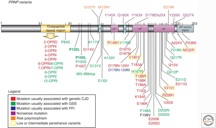
Genetic prion diseases (gPrDs) caused by mutations in the prion protein gene (PRNP) have been classified as genetic Creutzfeldt-Jakob disease, Gerstmann-Sträussler-Scheinker disease, or fatal familial insomnia. Mutations in PRNP can be missense, nonsense, and/or octapeptide repeat insertions or, possibly, deletions. These mutations can produce diverse clinical features. They may also show varying ancillary testing results and neuropathological findings. Although the majority of gPrDs have a rapid progression with a short survival time of a few months, many also present as ataxic or parkinsonian disorders, which have a slower decline over a few to several years. A few very rare mutations manifest as neuropsychiatric disorders, with systemic symptoms that include gastrointestinal disorders and neuropathy; these forms can progress over years to decades. In this review, we classify gPrDs as rapid, slow, or mixed types based on their typical rate of progression and duration, and we review the broad spectrum of phenotypes manifested by these diseases.


Figures

Figure 1.
Figure 1.
Schematic of prion protein gene (PRNP) disease–associated variants. Mutations are color coded based on clinicopathological classification as genetic Creutzfeldt–Jakob disease (gCJD), Gerstmann–Sträussler–Scheinker disease (GSS), familial fatal insomnia (FFI), or nonsense mutations. PRNP mutations present in the UCSF cohort are shown in bold. Most mutations are shown below the gene schematic; nonsense mutations and polymorphisms associated with prion disease risk are above the gene schematic. Low- or intermediate-penetrance variants are based on Minikel et al. (2016) (not all low/intermediate-penetrance variants are shown). For the F198V mutation, the clinical presentation was not classifiable as gCJD, GSS, or FFI (see Table 3), and neuropathology was not reported (Zheng et al. 2008). Variants that are most likely benign (largely based on Minikel et al. 2016) are not included (e.g., G54S, P39L, E196A, R208C) (Beck et al. 2010; Minikel et al. 2016). OPRI, Octapeptide repeat insertion; OPRD, octapeptide repeat deletion. (Reprinted from Takada et al. 2017.)
Figure 2.
Figure 2.
Brain magnetic resonance images (MRIs) in Fast forms of gPrD. (A) Diffusion weighted imaging (DWI) MRI of a genetic Creutzfeldt–Jakob disease (gCJD) 47-yr-old E200K patient 3 mo after onset shows diffuse cortical hyperintensity (cortical ribboning; dashed arrows) of the bilateral frontal and insula (left to right) and temporoparietal cortices. There was also DWI hyperintensity in the bilateral striata (left to right, solid arrows). (B) Attenuation diffusion coefficient (ADC) map of the same gCJD E200K case showed hypointensity in most of the regions that were hyperintense on DWI, confirming reduced diffusion, consistent with prion disease. (C) DWI MRI of an 85-yr-old V180I case 11 mo after onset shows diffuse cortical atrophy and cortical hyperintensity (cortical ribboning; dashed arrows) of the bilateral frontal and cingulate (right to left) and the right temporal and parietal lobes. There was no clear abnormality in the deep nuclei. (D) DWI in a 49-year-old gCJD A224V case 11 mo after the onset shows diffuse cortical ribboning (dashed arrows) greater on the right than the left and hyperintensity in the bilateral deep nuclei (solid arrows) also greater in the right. (E) The ADC map of the same gCJD A224V case showed hypointensity in most of the regions that were bright on DWI, confirming restricted diffusion, which is consistent with CJD/prion disease. Orientation is radiological (left brain is right side of figure). (Reprinted from Takada et al. 2017.)
Figure 3.
Figure 3.
Brain magnetic resonance images (MRIs) in Slow forms of genetic prion disease (gPrD). (A) Fluid-attenuated inversion recovery (FLAIR) brain MRI in a 57-yr-old patient with P102L gPrD possibly 16.5 yr after onset (precise age of onset unclear because of comorbidities) shows mild cerebellar atrophy but no cortical atrophy or typical T2-weighted or diffusion weighted imaging (DWI) hyperintensites (cortical ribboning or deep nuclei hyperintensity), but only several punctate hyperintensities in the white matter consistent with small vessel ischemic vascular disease. DWI/attenuation diffusion coefficient (ADC) map sequences showed no restricted diffusion (not shown). (B) FLAIR brain MRI of with 37-yr-old gPrD 6-octapeptide repeat insertion (OPRI) patient showing diffuse cortical atrophy only 7 mo after reported clinical onset; the amount of atrophy suggests that the disease began years before obvious clinical onset. DWI and ADC map sequences showed no restricted diffusion (not shown). (C) Repeat brain MRI on the same 6-OPRI patient 25 mo later, 32 mo after clinical onset, shows the progression of atrophy. DWI and ADC map sequences still showed no restricted diffusion (not shown). Orientation is radiological. (Reprinted from Takada et al. 2017.)

Similar articles

Cited by

References

    1. 1000 Genomes Project Consortium, Abecasis GR, Auton A, Brooks LD, DePristo MA, Durbin RM, Handsaker RE, Kang HM, Marth GT, McVean GA. 2012. An integrated map of genetic variation from 1,092 human genomes. Nature 491: 56–65. - PMC - PubMed
    1. Alzualde A, Indakoetxea B, Ferrer I, Moreno F, Barandiaran M, Gorostidi A, Estanga A, Ruiz I, Calero M, van Leeuwen FW, et al. 2010. A novel PRNP Y218N mutation in Gerstmann–Sträussler–Scheinker disease with neurofibrillary degeneration. J Neuropathol Exp Neurol 69: 789–800. - PubMed
    1. Beck JA, Mead S, Campbell TA, Dickinson A, Wientjens DP, Croes EA, Van Duijn CM, Collinge J. 2001. Two-octapeptide repeat deletion of prion protein associated with rapidly progressive dementia. Neurology 57: 354–356. - PubMed
    1. Beck G, Kawano T, Naba I, Nishimura T, Sawada J, Hazama T. 2005. A case with a 120 base pair insertional mutation in the prion protein gene: The first case in Japan. J Neurol Neurosurg Psychiatry 76: 756–757. - PMC - PubMed
    1. Beck JA, Poulter M, Campbell TA, Adamson G, Uphill JB, Guerreiro R, Jackson GS, Stevens JC, Manji H, Collinge J, et al. 2010. PRNP allelic series from 19 years of prion protein gene sequencing at the MRC Prion Unit. Hum Mutat 31: E1551–E1563. - PubMed