. 2017 Oct 1;313(4):H839-H853.
doi: 10.1152/ajpheart.00702.2016. Epub 2017 Aug 4.

Modulation of mesenteric collecting lymphatic contractions by σ1-receptor activation and nitric oxide production

Affiliations

Modulation of mesenteric collecting lymphatic contractions by σ1-receptor activation and nitric oxide production

Andrea N Trujillo et al. Am J Physiol Heart Circ Physiol. .

Abstract

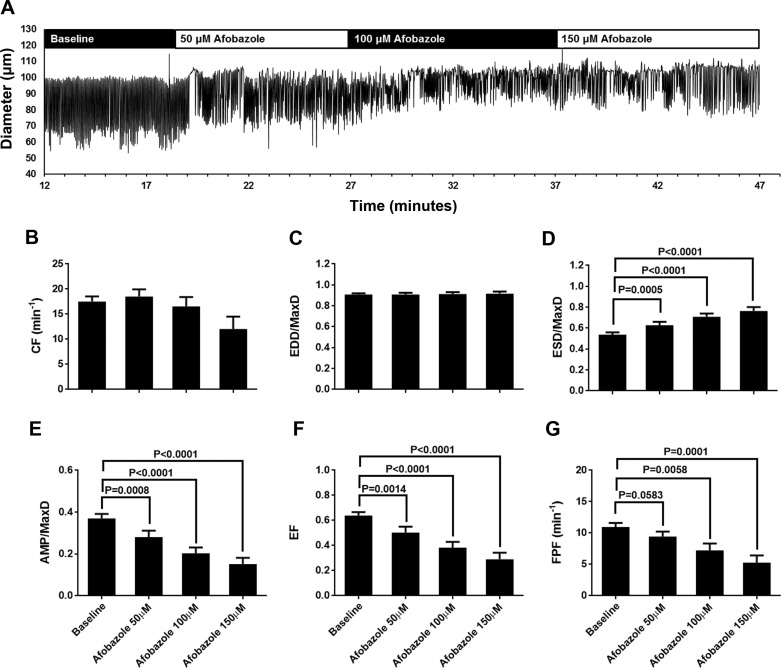
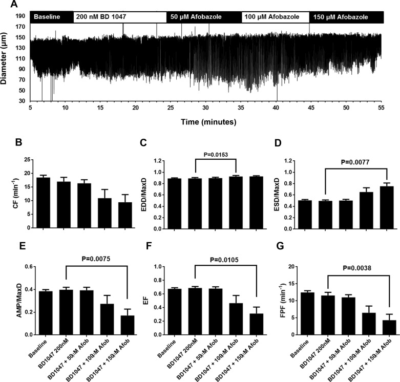
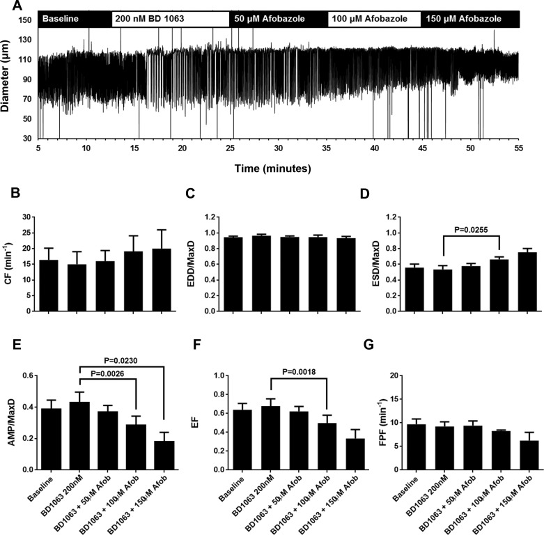
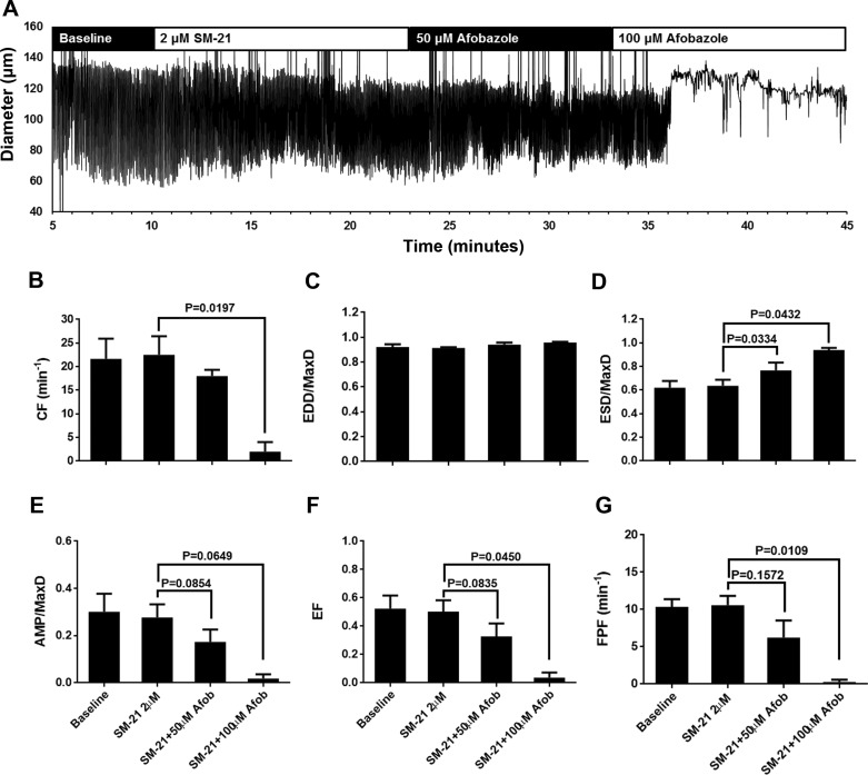
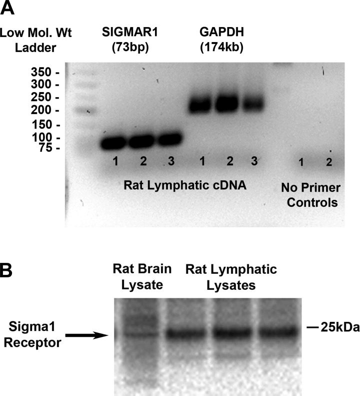
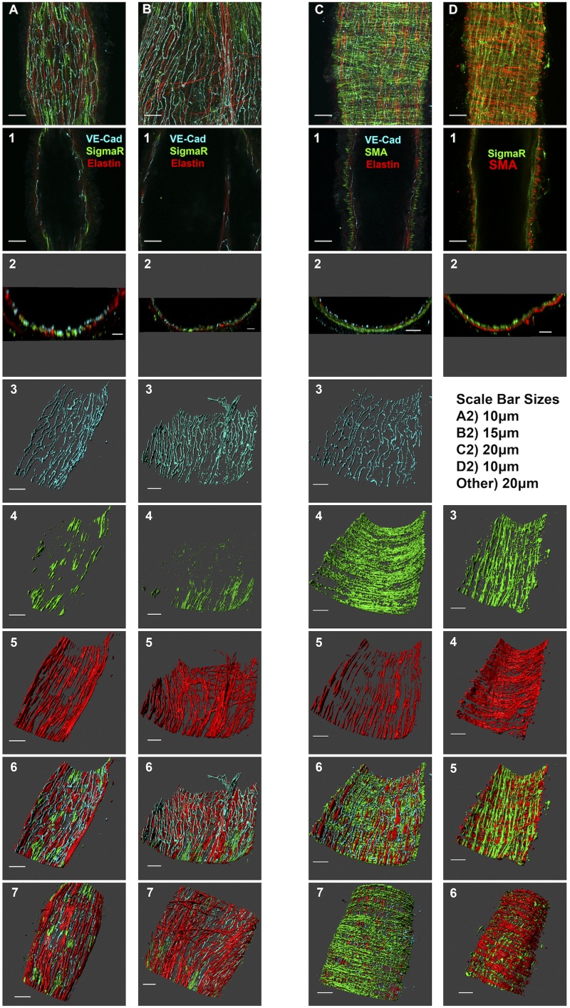
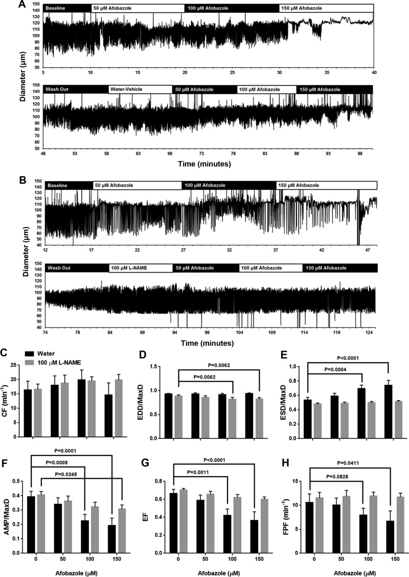
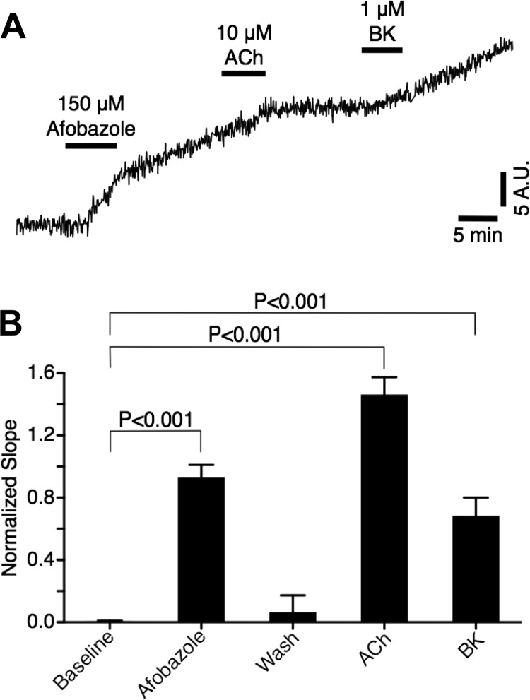
Recently, it has been reported that a σ-receptor antagonist could reduce inflammation-induced edema. Lymphatic vessels play an essential role in removing excess interstitial fluid. We tested the hypothesis that activation of σ-receptors would reduce or weaken collecting lymphatic contractions. We used isolated, cannulated rat mesenteric collecting lymphatic vessels to study contractions in response to the σ-receptor agonist afobazole in the absence and presence of different σ-receptor antagonists. We used RT-PCR and Western blot analysis to investigate whether these vessels express the σ1-receptor and immunofluorescence confocal microscopy to examine localization of the σ1-receptor in the collecting lymphatic wall. Using N-nitro-l-arginine methyl ester (l-NAME) pretreatment before afobazole in isolated lymphatics, we tested the role of nitric oxide (NO) signaling. Finally, we used 4-amino-5-methylamino-2',7'-difluorofluorescein diacetate fluorescence as an indicator to test whether afobazole increases NO release in cultured lymphatic endothelial cells. Our results show that afobazole (50-150 µM) elevated end-systolic diameter and generally reduced pump efficiency and that this response could be partially blocked by the σ1-receptor antagonists BD 1047 and BD 1063 but not by the σ2-receptor antagonist SM-21. σ1-Receptor mRNA and protein were detected in lysates from isolated rat mesenteric collecting lymphatics. Confocal images with anti-σ1-receptor antibody labeling suggested localization in the lymphatic endothelium. Blockade of NO synthases with l-NAME inhibited the effects of afobazole. Finally, afobazole elicited increases in NO production from cultured lymphatic endothelial cells. Our findings suggest that the σ1-receptor limits collecting lymphatic pumping through a NO-dependent mechanism.NEW & NOTEWORTHY Relatively little is known about the mechanisms that govern contractions of lymphatic vessels. σ1-Receptor activation has been shown to reduce the fractional pump flow of isolated rat mesenteric collecting lymphatics. The σ1-receptor was localized mainly in the endothelium, and blockade of nitric oxide synthase inhibited the effects of afobazole.

Keywords: afobazole; collecting lymphatic; lymphatic endothelium; nitric oxide; σ1-receptor.


Figures

Fig. 1.
Fig. 1.
Afobazole modulates rat mesenteric lymphatic contractions. A: representative trace of mesenteric lymphatic pumping in the presence of increasing concentrations of the σ1-receptor agonist afobazole (50–150 µM). B–G: summarized data (means ± SE) from experiments with 19 cannulated lymphatic vessels, each from a different rat. Data represent average values of each parameter obtained during the last 2 min of the baseline period and a 2-min period during each afobazole concentration starting 4 min after administration of afobazole. B and C: 50–150 µM afobazole caused no significant trend in contraction frequency (CF; P = 0.0502) or end-diastolic diameter (EDD) normalized to maximal passive diameter (MaxD; P = 0.5815) as determined by repeated-measures ANOVA. D: 50–150 µM afobazole significantly increased EDD/MaxD. E and F: 50–150 µM afobazole significantly decreased amplitude of contraction (AMP) normalized to MaxD (AMP/MaxD) and ejection fraction (EF). G: 100 or 150 µM afobazole significantly decreased fractional pump flow (FPF) compared with baseline. Data were analyzed by repeated-measures ANOVA with the Geisser-Greenhouse correction; when this test identified a significant trend, multiple comparisons were performed using Dunnett’s test with baseline as the control.
Fig. 2.
Fig. 2.
BD 1047, a specific antagonist of the σ1-receptor, reduced effects of afobazole (Afob) on lymphatic contractions. A: representative trace of vessel diameter as a function of time during baseline, in the presence of BD 1047 (200 nM), and after subsequent addition of 50, 100, and 150 μM afobazole. B–G: summarized data (means ± SE) from 7 isolated lymphatic vessels, each from a different rat. Data represent average values of each parameter obtained during the last 2 min of the baseline and BD 1047 periods and a 2-min period during each afobazole concentration starting 4 min after administration of afobazole. B: pretreatment with BD 1047 followed by afobazole caused an apparent trend in contraction frequency (CF) that was significant (P = 0.0266) when tested by repeated-measures ANOVA. However, multiple comparisons with Dunnett’s test revealed no significant differences between BD 1047 treatment alone and the time points after which afobazole was administered. C: in the presence of BD 1047, treatment with 100 µM (but not 50 or 150 µM) afobazole caused a significant increase in end-diastolic diameter (EDD) normalized to maximal passive diameter (MaxD; 0.891 ± 0.010 in the presence of BD 1047 alone vs. 0.9267 ± 0.018 in the presence of BD 1047 + 100 µM afobazole, mean ± SE, P = 0.0183). D: only 150 µM afobazole caused a significant increase in end-systolic diameter (ESD) normalized to MaxD (ESD/MaxD) in the presence of BD 1047. E–G: only 150 µM afobazole caused a significant decrease in amplitude of contraction (AMP) normalized to MaxD (AMP/MaxD), ejection fraction (EF), and fractional pump flow (FPF) in the presence of BD 1047. Data were analyzed by repeated-measures ANOVA with the Geisser-Greenhouse correction; when this test identified a significant trend, multiple comparisons were performed using Dunnett’s test with the inhibitor alone as the control.
Fig. 3.
Fig. 3.
BD 1063, a specific σ1-receptor antagonist, reduced effects of afobazole (Afob) on lymphatic contraction. A: representative trace of pumping of an isolated, cannulated rat mesenteric lymphatic vessel during baseline, in the presence of BD 1063 (200 nM), and after subsequent addition of 50, 100, and 150 μM afobazole. B–G: summarized data (means ± SE) from 4 isolated lymphatic vessels, each from a different rat. Data represent average values of each parameter obtained during the last 2 min of the baseline and BD 1063 periods and a 2-min period during each afobazole concentration starting 4 min after administration of afobazole. B and C: in the presence of BD 1063, afobazole (50–150 µM) caused no significant trend in contraction frequency (CF; P = 0.2752) or end-diastolic diameter (EDD) normalized to maximal passive diameter (MaxD; P = 0.3963) as determined by repeated-measures ANOVA. D: only 100 µM afobazole caused a significant increase in end-systolic diameter (ESD) normalized to MaxD (ESD/MaxD) in the presence of BD 1063 compared with BD 1063 alone. Treatment with 150 µM afobazole, despite achieving a higher mean than 100 µM afobazole treatment, showed more variability, and compared with BD 1063 alone, produced a P value that was not considered significant. E: only 100 and 150 µM afobazole in the presence of BD 1063 significantly reduced amplitude of contraction (AMP) normalized to MaxD (AMP/MaxD) compared with BD 1063 alone. F: only 100 µM afobazole in the presence of BD 1063 significantly reduced ejection fraction (EF) compared with BD 1063 alone. Despite a lower mean EF, 150 µM afobazole in this case had a greater variance and, compared with BD 1063 alone, produced a P value that was not considered significant. G: no significant changes in fractional pump flow (FPF) were apparent when afobazole was applied at 50–150 µM in the presence of BD 1063 (P = 0.2492, repeated-measures ANOVA). Data were analyzed by repeated-measures ANOVA with the Geisser-Greenhouse correction; when this test identified a significant trend, multiple comparisons were performed using Dunnett’s test with the inhibitor alone as the control.
Fig. 4.
Fig. 4.
SM-21, a specific antagonist of the σ2-receptor, did not block the effects of afobazole (Afob). A: representative trace of an isolated rat mesenteric lymphatic pumping in the presence of SM-21 (2 μM) and afobazole (50 and 100 μM). Lymphatics frequently stopped pumping with 100 µM afobazole in the presence of SM-21, so only 50 and 100 µM afobazole were used for analysis. BG: summarized data (means ± SE) from 4 isolated lymphatic vessels, each from a different rat. Data represent average values of each parameter obtained during the last 2 min of baseline and SM-21 periods and a 2-min period during each afobazole concentration starting 4 min after administration of afobazole. Mean values shown for the 100 µM afobazole groups include lymphatics that stopped pumping. B and C: application of 100 µM afobazole in the presence of SM-21 caused a significant decrease in contraction frequency (CF) and no change in end-diastolic diameter (EDD) normalized to maximal passive diameter (MaxD). D: 50 and 100 μM afobazole significantly elevated end-systolic diameter (ESD) normalized to MaxD (ESD/MaxD) in the presence of SM-21 compared with SM-21 alone. For lymphatics that stopped pumping, diameter/MaxD was used for the ESD/MaxD calculations. E–G: in the presence of SM-21 and compared with SM-21 alone, 100 µM afobazole significantly decreased amplitude of contraction normalized to MaxD (AMP/MaxD), ejection fraction (EF), and fractional pump flow (FPF). For lymphatics that stopped pumping, the value for these parameters was zero. Data were analyzed by repeated-measures ANOVA with the Geisser-Greenhouse correction; when this test identified a significant trend, multiple comparisons were performed using Dunnett’s test with the inhibitor alone as the control.
Fig. 5.
Fig. 5.
σ1-Receptor is expressed in rat mesenteric lymphatics. A: rat mesenteric lymphatics were harvested (10 lymphatic vessels per rat), and total RNA (50 ng) from mesenteric lymphatic vessels (3 samples from 3 different rats) was reverse transcribed into cDNA and RT-PCR was performed with specific primer/probe (FAM) sets for rat SIGMAR1 (NM_030996.1) and rat GAPDH (NM_017008.4). Two cDNA samples were also run with no primers as controls. B: protein lysates (6 samples from 6 rats, only 3 are shown) were prepared for SDS-PAGE and Western blot analysis for the detection of σ1-receptor expression. With use of a rabbit anti-σ1-receptor antibody (Invitrogen) at 0.25 µg/ml (specific for the mouse and rat but not human) and donkey anti-rabbit horseradish peroxidase-conjugated secondary antibody at 1:10,000, a single band was detected at ∼25 kDa in rat lymphatic (30 µg) and rat brain (10 µg) lysates.
Fig. 6.
Fig. 6.
Localization of the σ1-receptor in isolated rat collecting lymphatic vessels. A and B: images of two different collecting lymphatic vessels that were labeled for vascular-endothelial (VE)-cadherin, the σ1-receptor, and elastin. C: labeling for VE-cadherin, elastin, and smooth muscle actin. D: σ1-receptor and smooth muscle actin labeling. Top images show a maximum-intensity z-projection obtained with a ×60 objective. A−D,1: representative confocal slices about halfway through the z-stack, with the target proteins denoted by the colors in the label. A−D,2: orthogonal projections (5 µm thickness) representing a cross-sectional view of the vessel. Images 3–5 in A–C and images 3 and 4 in D show three-dimensional representations of individual channels, with the luminal side of the vessel facing upward. Image 6 in A–C and image 5 in D show three-dimensional composites of all channels, with the luminal side of the vessel facing upward. Image 7 in A–C and image 6 in D show three-dimensional composites of all channels but with the abluminal side of the vessel facing upward. Each labeling scheme is representative of 3 vessels from 3 rats each.
Fig. 7.
Fig. 7.
The nitric oxide synthase inhibitor N-nitro-l-arginine methyl ester (l-NAME) blocks afobazole-induced changes in lymphatic pumping. A: representative trace of a lymphatic vessel first treated for 10 min each with 50, 100, and 150 µM afobazole followed by a washout period for reestablishment of contractions similar to baseline and then treated with vehicle (water) and 50, 100, and 150 µM afobazole. B: representative trace of a lymphatic vessel first treated for 10 min each with 50, 100, and 150 µM afobazole followed by a washout period for reestablishment of contractions similar to baseline and then treated with l-NAME and 50, 100, and 150 µM afobazole. C–H: summarized data (means ± SE) from 5 isolated lymphatic vessels in the water-treated group and 7 isolated lymphatic vessels in the l-NAME-treated group. Each vessel was from a different rat. Data represent average values of each parameter obtained during the last 2 min of baseline and water or l-NAME periods and a 2-min period during each afobazole concentration staring 4 min after the administration of afobazole. C: contraction frequency (CF) did not significantly change in either group (two-way ANOVA, P = 0.1470). D: end-diastolic diameter (EDD) normalized to maximal passive diameter (MaxD) was significantly elevated in l-NAME-treated lymphatics after treatment with 100 and 150 µM afobazole. E: end-systolic diameter (ESD) normalized to MaxD (ESD/MaxD) was significantly elevated in the water- but not l-NAME-treated group after 100 and 150 µM afobazole. F: amplitude of contraction (AMP) normalized to MaxD (AMP/MaxD) was significantly reduced in the water-treated group after 100 and 150 µM afobazole but only after 150 µM afobazole in the l-NAME-treated group. G: ejection fraction (EF) was significantly reduced in the water-treated group after treatment with 100 and 150 µM afobazole but not in the l-NAME-treated group. H: fractional pump flow (FPF) was significantly reduced in the water- but not l-NAME-treated group after 150 µM afobazole. Data were analyzed by repeated-measures two-way ANOVA with the Geisser-Greenhouse correction; when this test identified a significant trend, multiple comparisons were performed using Dunnett’s test.
Fig. 8.
Fig. 8.
Afobazole elevates lymphatic endothelial cell nitric oxide (NO) production. A: trace of change in fluorescence from baseline (F/F0) of a single cultured dermal lymphatic endothelial cell (passage 3) loaded with 4-amino-5-methylamino-2′,7′-difluorofluorescein diacetate (DAF-FM) to detect NO production. DAF-FM is essentially nonfluorescent until it interacts with NO to produce a fluorescent benzotriazole. The time course shows the accumulation of this fluorescent benzotriazole and changes in slope associated with treatments and washouts. Apparent elevations in the slope were visible shortly after afobazole treatment or after treatment with positive controls for endothelium-derived NO production, acetylcholine (ACh) and bradykinin (BK). AU, arbitrary units. B: summarized data from 90 lymphatic endothelial cells that responded to afobazole. Data were analyzed by one-way ANOVA followed by Dunnett’s test, with all groups compared with baseline as control.

Similar articles

Cited by

References

    1. Alon A, Schmidt HR, Wood MD, Sahn JJ, Martin SF, Kruse AC. Identification of the gene that codes for the σ2-receptor. Proc Natl Acad Sci USA 114: 7160–7165, 2017. doi:10.1073/pnas.1705154114. - DOI - PMC - PubMed
    1. Arkill KP, Moger J, Winlove CP. The structure and mechanical properties of collecting lymphatic vessels: an investigation using multimodal nonlinear microscopy. J Anat 216: 547–555, 2010. doi:10.1111/j.1469-7580.2010.01215.x. - DOI - PMC - PubMed
    1. Atchison DJ, Johnston MG. Role of extra- and intracellular Ca2+ in the lymphatic myogenic response. Am J Physiol Regul Integr Comp Physiol 272: R326–R333, 1997. - PubMed
    1. Azuma T, Ohhashi T, Roddie IC. Bradykinin-induced contractions of bovine mesenteric lymphatics. J Physiol 342: 217–227, 1983. doi:10.1113/jphysiol.1983.sp014847. - DOI - PMC - PubMed
    1. Benoit JN, Zawieja DC, Goodman AH, Granger HJ. Characterization of intact mesenteric lymphatic pump and its responsiveness to acute edemagenic stress. Am J Physiol Heart Circ Physiol 257: H2059–H2069, 1989. - PubMed

MeSH terms