. 2017 Oct 17;136(16):1477-1491.
doi: 10.1161/CIRCULATIONAHA.117.028585. Epub 2017 Aug 4.

Experimental Modeling Supports a Role for MyBP-HL as a Novel Myofilament Component in Arrhythmia and Dilated Cardiomyopathy

Affiliations

Experimental Modeling Supports a Role for MyBP-HL as a Novel Myofilament Component in Arrhythmia and Dilated Cardiomyopathy

David Y Barefield et al. Circulation. .

Abstract

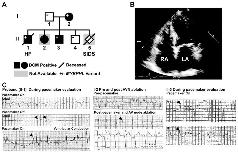
Background: Cardiomyopathy and arrhythmias are under significant genetic influence. Here, we studied a family with dilated cardiomyopathy and associated conduction system disease in whom prior clinical cardiac gene panel testing was unrevealing.

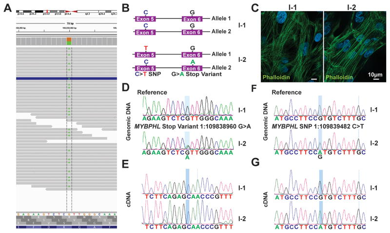
Methods: Whole-genome sequencing and induced pluripotent stem cells were used to examine a family with dilated cardiomyopathy and atrial and ventricular arrhythmias. We also characterized a mouse model with heterozygous and homozygous deletion of Mybphl.

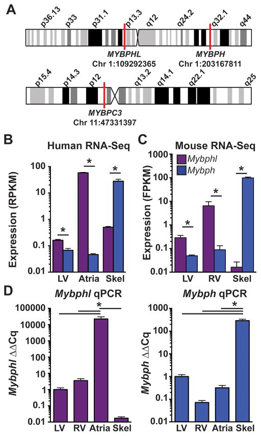
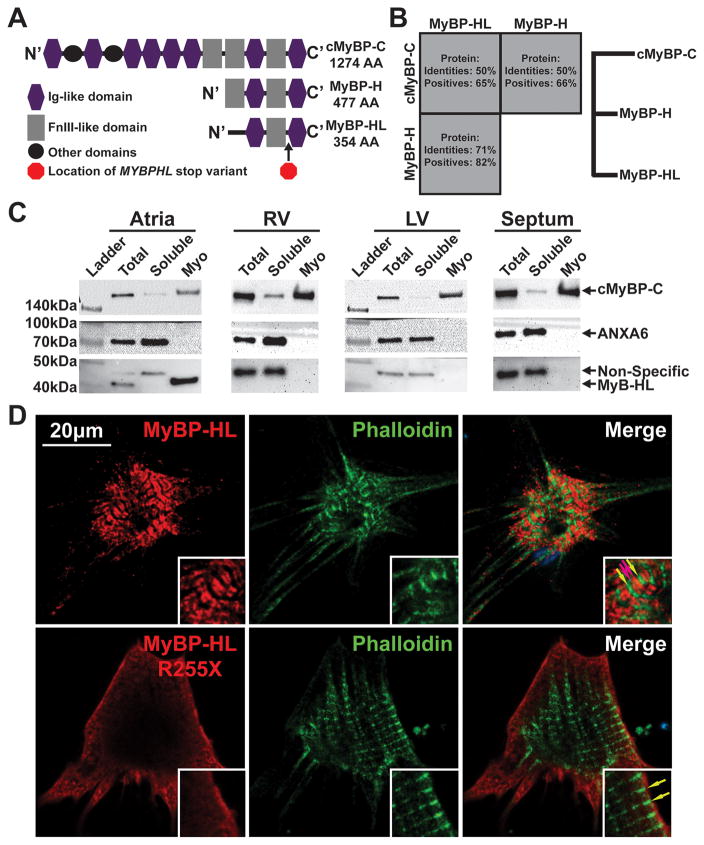
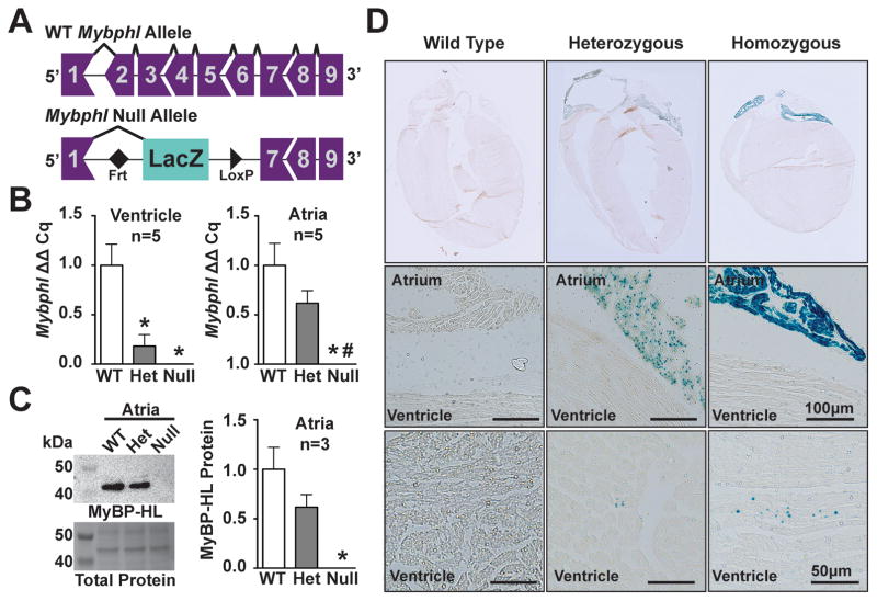
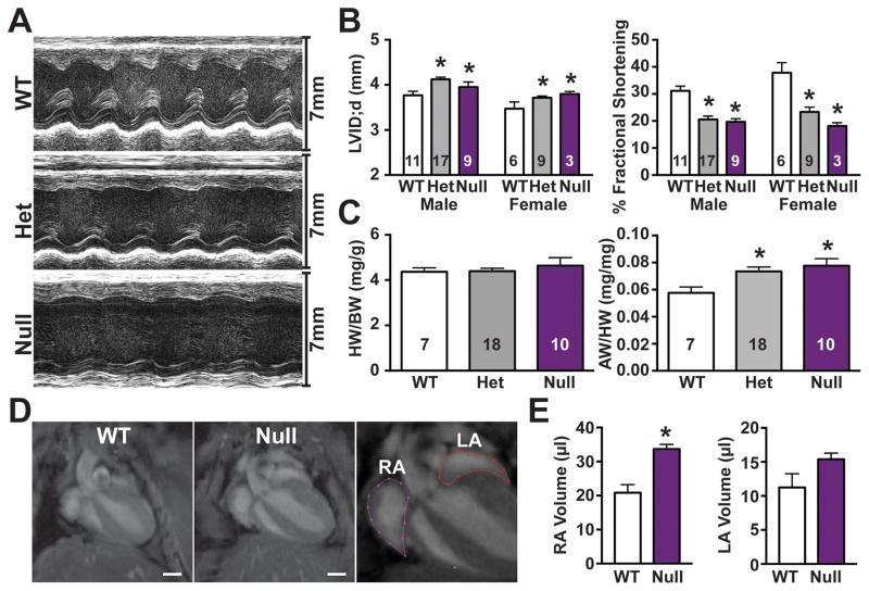
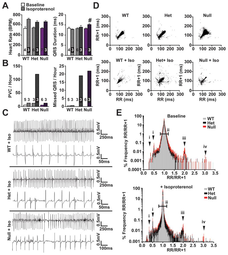
Results: Whole-genome sequencing identified a premature stop codon, R255X, in the MYBPHL gene encoding MyBP-HL (myosin-binding protein-H like), a novel member of the myosin-binding protein family. MYBPHL was found to have high atrial expression with low ventricular expression. We determined that MyBP-HL protein was myofilament associated in the atria, and truncated MyBP-HL protein failed to incorporate into the myofilament. Human cell modeling demonstrated reduced expression from the mutant MYBPHL allele. Echocardiography of Mybphl heterozygous and null mouse hearts exhibited a 36% reduction in fractional shortening and an increased diastolic ventricular chamber size. Atria weight normalized to total heart weight was significantly increased in Mybphl heterozygous and null mice. Using a reporter system, we detected robust expression of Mybphl in the atria, and in discrete puncta throughout the right ventricular wall and septum, as well. Telemetric electrocardiogram recordings in Mybphl mice revealed cardiac conduction system abnormalities with aberrant atrioventricular conduction and an increased rate of arrhythmia in heterozygous and null mice.

Conclusions: The findings of reduced ventricular function and conduction system defects in Mybphl mice support that MYBPHL truncations may increase risk for human arrhythmias and cardiomyopathy.

Keywords: arrhythmias, cardiac; cardiomyopathies; genetics; heart atria; mice; mutation; myofibrils.


Conflict of interest statement

AUTHOR CONFLICTS OF INTEREST/DISCLOSURES: None

Figures

Figure 1
Figure 1. Whole genome sequencing (WGS) identified a premature stop codon in MYBPHL with conduction system abnormalities and DCM
A. WGS on the proband (II-1) and parents (I-1, I-2) identified the MYBPHL R255X variant which was found in multiple family members. B. Four-chamber echocardiogram of I-2 shows enlarged right and left atria (LA, RA). C. Abnormal heart rhythms in multiple family members. ECG recordings from the proband’s pacemaker test months prior to death showing paced rhythm (top, lead 1). In the middle tracing with the pacemaker off, underlying sinus and atrioventricular node dysfunction was evident, as were PVCs (arrow heads). The bottom tracing highlights a paced rhythm and revealed PVCs (arrow head). The center panels show ECG recordings from the affected parent (I-2) show atrial flutter (asterisks) and PVC in the middle tracing. The bottom tracing shows pacing after atrioventricular (AV) node ablation with persistent atrial flutter (asterisks). The right hand panels show ECG tracing from II-3 with a PVC (arrow head) and coarse atrial fibrillation (asterisks).
Figure 2
Figure 2. The mutant MYBPHL allele is suppressed by nonsense-mediated decay
A. Genome viewer shows the alignment of reads at 45x converge over this variant with an even distribution of “A” or “G” reads, consistent with MYBPHL R255X being heterozygous. B. Schematic of both MYBPHL alleles from family members I-1 and I-2. I-2 has the G>A stop variant in exon 6, as well as a synonymous SNP on the opposite allele in exon 5. C. Representative phalloidin stained image of iPSC-derived cardiomyocytes generated from family members I-1 and I-2. D. Sanger sequencing of iPSC-cardiomyocyte genomic DNA confirms the presence of reference sequence in I-1 and the 1:109838960G>A variant in I-2. E. Direct sequencing of RT-PCR-generated cDNA from iPSC-cardiomyocytes from I-1 and I-2 shows expression of only the normal “G” allele (expressed as a “C”) in both I-1 and I-2. This is consistent with no detectable expression from the premature stop allele in I-2. F. Direct sequencing of genomic DNA from iPSC-cardiomyocytes from I-1 and I-2 showed the presence of a benign synonymous C>T SNP in exon 5 of I-2. This variant is found on the opposite allele from R255X. G. Direct sequencing of RT-PCR product generated from iPSC-cardiomyocyte cDNA showed that I-2 only expressed the allele with the C>T SNP, consistent with nonsense mediated decay of the mutant allele.
Figure 3
Figure 3. MYBPHL expression is enriched in human and mouse atria and is distinct from MYBPH
A. Schematic of human chromosomes 1 and 11 show locations of the three genes MYBPHL, MYBPH, and MYBPC3. B. Human RNA-Seq data from the GTEx database of normalized MYBPHL and MYBPH reads in ventricle, atria, and skeletal muscle shows enriched MYBPHL expression in atrial tissue, and enriched MYBPH expression in skeletal muscle (MYBPHL n= 218 LV, 194 atria, 229 skel; MYBPH n= 218 LV, 194 atria, 232 skel). C. RNA-Seq data from mouse tissue shows enrichment of MYBPHL expression in ventricle and enrichment of MYBPH in skeletal muscle (n=6). D. qPCR analysis of wild-type mouse ventricle, atria, and skeletal muscle confirms the cardiac and atrial enrichment of MYBPHL and the enrichment of MYBPH in skeletal muscle (n=4). *= p<0.05 two-way ANOVA with a Bonferroni multiple comparison test (B,C) or one-way ANOVA with a Tukey’s post-hoc test (D).
Figure 4
Figure 4. MYBPHL is structurally similar to other myosin binding proteins and interacts with the myofilament
A. Diagram of the Immunoglobulin (Ig)-like and fibronectin type 3 (FnIII)-like domains that comprise cMyBP-C, MyBP-H, and MyBP-HL; the location of the premature stop in MyBP-HL created by the identified variant is marked between the C’ Ig and FnIII domains. B. Protein sequence conservation and phylogeny between the myosin binding protein family members show that MyBP-H and MyBP-HL are more closely related to each other than to cMyBP-C. C. Immunoblot analysis of total tissue homogenate, soluble protein, and myofilament fractions from atria, right and left ventricle, and ventricular septum detect MyBP-HL in the atrial myofilament, with cMyBP-C and annexin-6 used as myofilament and soluble controls, respectively. D. Immunofluorescence microscopy of neonatal mouse cardiomyocytes transfected with N’-terminally Myc-tagged full-length and R255X MyBP-HL constructs showed myofilament patterned MyBP-HL localization that is not observed with the R255X variant (green and red arrows, insets).
Figure 5
Figure 5. Mybphl null mouse
A. Schematic diagram of the WT Mybphl exons and the Knock-Out Mouse Project (KOMP) gene-trap deletion allele. B. qPCR shows reduction and elimination of Mybphl expression in Het and Null atria and ventricles, respectively (n=4). C. Immnoblot of atrial whole-tissue homogenates confirms reduction and elimination of MyBP-HL protein in Het and Null mice (n=3). D. LacZ staining confirms atrial enrichment of Mybphl expression, with small puncta of LacZ positive cells seen scattered throughout the ventricle. * = p<0.05 vs. WT, # = p<0.05 vs. Het (one-way ANOVA with a Tukey’s multiple comparison test).
Figure 6
Figure 6. Loss of Mybphl causes ventricular dysfunction and atrial enlargement
A. Representative short-axis M-mode echocardiography images. B. M-mode derived fractional shortening and diastolic left ventricular internal diameter shows systolic dysfunction and dilation in heterozygous and null groups, consistent between sexes. C. Heart weight to body weight and atria weight to total heart weight ratios show increased atrial mass in Mybphl heterozygous and null mice of mixed sex. D. MRI of WT and Mybphl null atria at peak systole (left, center) and representative segmentation analysis of atrial volume (right). E. Right atria volume is significantly increased in Mybphl null mice (n=3 female mice). *= p<0.05 vs. WT by two-way ANOVA with a Bonferroni multiple comparison test (B), one-way ANOVA with a Tukey’s multiple comparison test (C), or unpaired t-test (E).
Figure 7
Figure 7. Increased frequency of arrhythmic events in Mybphl mutant mice
A. Conscious ambulatory ECG analysis shows significantly prolonged QRS duration in Mybphl null mice following isoproterenol administration. B. Following isoproterenol, PVCs and nonconducted beats per hour were increased in heterozygous mice. Nonconducted beats were increased in both heterozygous and homozygous mice following isoproterenol. C. Representative ECG traces from each group following isoproterenol administration show an interval with a high rate of PVCs from heterozygous mice and a period with non-conducted beats and sinus arrest in the homozygous traces. D. Poincaré plots showing increased R-R variability in Mybphl heterozygous and null mice that is exacerbated with isoproterenol treatment. E. Histogram of the RR/RR+1 ratio illustrating peaks representing PVCs or early beats (i), single and double skipped beats in sinus rhythm (iii, iv), and overall RR variability illustrated by increased deviation around the 1.0 ratio point (ii) (n=3–5). # = p<0.05 vs. WT + Isoproterenol by two-way ANOVA with a Bonferroni multiple comparison test.

Comment in

Similar articles

Cited by

References

    1. Maron BJ, Towbin JA, Thiene G, Antzelevitch C, Corrado D, Arnett D, Moss AJ, Seidman CE, Young JB American Heart A, Council on Clinical Cardiology HF, Transplantation C, Quality of C, Outcomes R, Functional G, Translational Biology Interdisciplinary Working G, Council on E Prevention. Contemporary Definitions and Classification of the Cardiomyopathies: An American Heart Association Scientific Statement from the Council on Clinical Cardiology, Heart Failure and Transplantation Committee; Quality of Care and Outcomes Research and Functional Genomics and Translational Biology Interdisciplinary Working Groups; and Council on Epidemiology and Prevention. Circulation. 2006;113:1807–1816. doi: 10.1161/CIRCULATIONAHA.106.174287. - DOI - PubMed
    1. McNally EM, Barefield DY, Puckelwartz MJ. The Genetic Landscape of Cardiomyopathy and Its Role in Heart Failure. Cell Metab. 2015;21:174–182. doi: 10.1016/j.cmet.2015.01.013. - DOI - PMC - PubMed
    1. Lakdawala NK, Funke BH, Baxter S, Cirino AL, Roberts AE, Judge DP, Johnson N, Mendelsohn NJ, Morel C, Care M, Chung WK, Jones C, Psychogios A, Duffy E, Rehm HL, White E, Seidman JG, Seidman CE, Ho CY. Genetic Testing for Dilated Cardiomyopathy in Clinical Practice. J Card Fail. 2012;18:296–303. doi: 10.1016/j.cardfail.2012.01.013. - DOI - PMC - PubMed
    1. Herman DS, Lam L, Taylor MR, Wang L, Teekakirikul P, Christodoulou D, Conner L, DePalma SR, McDonough B, Sparks E, Teodorescu DL, Cirino AL, Banner NR, Pennell DJ, Graw S, Merlo M, Di Lenarda A, Sinagra G, Bos JM, Ackerman MJ, Mitchell RN, Murry CE, Lakdawala NK, Ho CY, Barton PJ, Cook SA, Mestroni L, Seidman JG, Seidman CE. Truncations of Titin Causing Dilated Cardiomyopathy. N Engl J Med. 2012;366:619–628. doi: 10.1056/NEJMoa1110186. - DOI - PMC - PubMed
    1. Watkins H, Conner D, Thierfelder L, Jarcho JA, MacRae C, McKenna WJ, Maron BJ, Seidman JG, Seidman CE. Mutations in the Cardiac Myosin Binding Protein-C Gene on Chromosome 11 Cause Familial Hypertrophic Cardiomyopathy. Nat Genet. 1995;11:434–437. doi: 10.1038/ng1295-434. - DOI - PubMed

MeSH terms