. 2017 Sep 26;136(13):1217-1232.
doi: 10.1161/CIRCULATIONAHA.116.026361. Epub 2017 Aug 4.

Disturbed Flow Promotes Arterial Stiffening Through Thrombospondin-1

Affiliations

Disturbed Flow Promotes Arterial Stiffening Through Thrombospondin-1

Chan Woo Kim et al. Circulation. .

Abstract

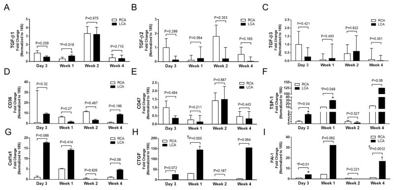
Background: Arterial stiffness and wall shear stress are powerful determinants of cardiovascular health, and arterial stiffness is associated with increased cardiovascular mortality. Low and oscillatory wall shear stress, termed disturbed flow (d-flow), promotes atherosclerotic arterial remodeling, but the relationship between d-flow and arterial stiffness is not well understood. The objective of this study was to define the role of d-flow on arterial stiffening and discover the relevant signaling pathways by which d-flow stiffens arteries.

Methods: D-flow was induced in the carotid arteries of young and old mice of both sexes. Arterial stiffness was quantified ex vivo with cylindrical biaxial mechanical testing and in vivo from duplex ultrasound and compared with unmanipulated carotid arteries from 80-week-old mice. Gene expression and pathway analysis was performed on endothelial cell-enriched RNA and validated by immunohistochemistry. In vitro testing of signaling pathways was performed under oscillatory and laminar wall shear stress conditions. Human arteries from regions of d-flow and stable flow were tested ex vivo to validate critical results from the animal model.

Results: D-flow induced arterial stiffening through collagen deposition after partial carotid ligation, and the degree of stiffening was similar to that of unmanipulated carotid arteries from 80-week-old mice. Intimal gene pathway analyses identified transforming growth factor-β pathways as having a prominent role in this stiffened arterial response, but this was attributable to thrombospondin-1 (TSP-1) stimulation of profibrotic genes and not changes to transforming growth factor-β. In vitro and in vivo testing under d-flow conditions identified a possible role for TSP-1 activation of transforming growth factor-β in the upregulation of these genes. TSP-1 knockout animals had significantly less arterial stiffening in response to d-flow than wild-type carotid arteries. Human arteries exposed to d-flow had similar increases TSP-1 and collagen gene expression as seen in our model.

Conclusions: TSP-1 has a critical role in shear-mediated arterial stiffening that is mediated in part through TSP-1's activation of the profibrotic signaling pathways of transforming growth factor-β. Molecular targets in this pathway may lead to novel therapies to limit arterial stiffening and the progression of disease in arteries exposed to d-flow.

Keywords: disturbed flow; thrombospondins; vascular stiffness.


Figures

Figure 1
Figure 1. Partial carotid ligation invokes disturbed flow to the intima and down-regulates shear sensitive genes
A. Carotid artery duplex ultrasound demonstrates flow reversal and depressed velocities in the left carotid artery (LCA) that underwent partial ligation. Right carotid artery (RCA) was not manipulated and demonstrates unidirectional flow. (Note differences in the scale bars on the y axis [LCA: −50, 0, +50mm/s; RCA: −150, 0, +150 mm/s at 1 d post-ligation and −200, 0, +200 mm/s at 42 d post-ligation, respectively]). B. Using velocity data points from the ultrasound images, the LCA had significantly diminished flow rates compared to the RCA through 6 weeks post partial carotid ligation. C. (left panel) Intimal RNA demonstrates EC-enrichment by the selective and segregated PECAM-1 expression from that of α-SMA or CD11b at 24 hours. (middle panel) RNA from the media/adventitia of these arteries demonstrate increased α-SMA. (right panel) D-flow conditions were then internally validated 24 hours after partial ligation by the significant down-regulation of the shear sensitive genes KLK10 and KLF2 in the LCA. n=3; mean ± standard error of the mean (sem); unpaired Student’s t-test with significance at P<.05. D. WSS in the RCA is consistent with stable flow (s-flow) throughout the experiment. However, the LCA manifests disturbed flow (d-flow) with low and oscillatory WSS acutely and low WSS throughout the latest time point of experiments in this work (6 weeks). *P<0.05; †<.01; ‡<.001
Figure 2
Figure 2. D-flow and aging stiffen arteries similarly
A/B. Pressure diameter and compliance curves demonstrate that the carotid arteries of 12-week-old animals who had their LCA exposed to d-flow (solid black circles) were significantly stiffer than the contralateral RCA (open circle), which were not manipulated and exposed to s-flow. C/D. We then compared carotid arteries from 12-week-old mice exposed to d-flow to un-manipulated (s-flow) carotid arteries from aged (80-week-old animals) animals. Due to the enlarged diameter of the older arteries, the pressure diameter curve of the aged carotid arteries demonstrated greater dilation than the 12-week-old mice exposed to d-flow beginning at 60mmHg. However, the compliance curves were very similar without any statistically significant differences in compliance between the 12-week-old d-flow carotid arteries and that of the unmanipulated aged murine carotid arteries. N=6; mean ± sem; unpaired Student’s t-test with significance (§) at P<.0029 and P<.0031 respectively. E. Structurally, the arterial stiffening from d-flow was due to increased collagen deposition, which was found in the LCA (d-flow) compared to RCA (s-flow), and not due to changes in elastin, which were similar between groups. n=12 RCA; n=14 LCA; mean ± sem; unpaired students t-test with significance set at P<.05. *P<0.05; †<.01; ‡<.001
Figure 3
Figure 3. TGF-β and TGFβR expression in the carotid arterial wall in response to d-flow in vivo
A/D. TGF-β1 is downregulated in the endothelium (but not media/adventitia) in response to d-flow. B–F. TGF-β2 and TGF-β3 are not changed in the endothelium or media/adventitia under d-flow. G–L. TGFBR1-3 are not changed by d-flow in the endothelium or media/adventitia in vivo. N=3; mean ± sem; paired Students t-test with significance set at P<.05. *P<0.05; †<.01; ‡<.001
Figure 4
Figure 4. Acute and chronic exposure to d-flow induces TSP-1 expression in vivo
A/B. TSP-1 expression was increased in the d-flow LCA versus s-flow RCA in the endothelium and media/adventitia. n=3; mean ± sem; paired students t-test; *p<0.05. C. TSP-1 staining (Red: TSP-1; Blue: DAPI) in the mouse carotid artery (L: lumen of artery) is increased under d-flow. C1 and C2: Cross section IHC staining for TSP-1 in the RCA and LCA demonstrate increased TSP-1 in the LCA (d-flow) at day 3 post partial carotid ligation. C3 and C4: Enface IHC staining in the endothelium of the RCA and LCA demonstrates increased TSP-1 in the LCA at day 3 post partial carotid ligation. D/E. Increased TSP-1 expression in the endothelium and media/adventitia under chronic d-flow conditions in the lesser curvature (LC) compared to the greater curvature (GC) of the aortic arch, which is s-flow naturally. F/G. CD36 and CD47 expression were not significantly changed in the endothelium under d-flow conditions. N=4; mean ± sem; paired Student’s t-test with significance set at P<.05. Scale: 20 μm. *P<0.05; †<.01; ‡<.001
Figure 5
Figure 5. Temporal gene changes in the endothelial RNA under d-flow
A–E. TGF-β1 is increased at 1 week in the LCA, but there were no other significant differences in TGF-β1-3, CD36 or CD47 identified over these time points (day 3, week 1, 2, and 4). F. TSP1 was significantly increased in the LCA at day 3 and week 1 and demonstrated a trend toward significance at week 4. G. Col1a1 was not significantly increased at these time points. H/I. CTGF was significantly increased at one week, and PAI1 was significantly increased at day 3 and week 4. n=3–4; mean ± sem; unpaired Student’s t-test with significance set at P<.05. *P<0.05; †<.01; ‡<.001
Figure 6
Figure 6. D-flow increases pro-fibrotic gene expression in human aortic ECs (HAEC) in vitro and LSKL down-regulates these genes in vitro and in vivo
[Differences between OS and either static (ST) and LS are demonstrated above ST and LS, while differences between OS +LSKL and OS + SLLK are listed above their respective columns.] A. Putative mechanism by which TSP-1 can activate TGF-β and how the small peptide LSKL competitively inhibits TSP-1 activation of TGF-β. B. ECs under oscillatory shear (OS) have increased TSP-1 expression compared to ECs under static (ST) and laminar shear (LS) conditions. TSP-1 expression is not affected by LSKL treatment or the control peptide (SLLK). C. TGF-β1 was also upregulated in ECs by OS and not affected by LSKL or SLLK. D. Col1a1 was upregulated by OS over ST and LS, and LSKL treatment significantly decreased Col1a1 expression under OS conditions compared to SLLK treatment. E. CTGF is significantly upregulated by OS (compared to ST and LS), and LSKL treatment significantly decreased CTGF expression under OS conditions compared to SLLK. F. PAI-1 is significantly upregulated by OS (compared to ST and LS) and LSKL treatment significantly decreased PAI-1 expression under OS conditions compared to SLLK. [N=5-6; mean±sem.; ANOVA with Tukey’s post-hoc test with significance set at P<.05. *P<0.05; †<.01; ‡<.001 G–I. In vivo, LSKL globally decreased expression of pro-fibrotic genes compared to SLLK control treatment in both the RCA (s-flow) and LCA (d-flow). Here only CTGF was still significantly upregulated in the LSKL treatment group by d-flow. J. TSP-1 expression the LCA of LSKL treated animals was still significantly increased compared to the RCA of LSKL treated animals. K–O. Again, there were no significant differences in TGF-β or TGFβR expression under d-flow or s-flow conditions (LCA versus RCA) in either the LSKL or SLLK groups. However, there were contrasting trends with greater TGF-β1 expression in SLLK treated animals and greater TGF-βR1-3 expression in the LSKL treated animals. P/Q. There were not any flow mediated differences in the expression of CD36 or CD47 identified within groups, but there was a trend toward increased CD47 in the LSKL treated animals compared to SLLK in both the LCA and RCA. n=3–4; mean±sem; paired Student’s t-test with significance set at P<.05. *P<0.05; †<.01; ‡<.001
Figure 7
Figure 7. D-flow induced stiffening is significantly attenuated in TSP-1 KO animals
A/B. Pressure-diameter and compliance curves from ex vivo cylindrical biaxial mechanical testing of LCA and RCA from C57BL/6 (C57) animals demonstrates that the LCA is significantly stiffer than the RCA in this strain as well (N=5). C/D. Similar but less prominent differences were seen in the TSP-1 KO animals (N=7). E/F. When comparing the pressure-diameter and compliance curves of the LCA (exposed to d-flow) from TSP-1 KO and C57 animals, the TSP-1 KO was significantly less stiff by pressure diameter curves beginning at 70mmHg and by compliance curves at 40, 60, and 70 mmHg (N=5–7). mean ± sem; unpaired Student’s t-test with significance (§) at P<.0029 and P<.0031 respectively. G. For in vivo validation, we calculated Green’s strain Eθθ from ultrasound imaging to identify the time course of stiffening in these animals. Here we identified increased arterial stiffness (compared to TSP-1 KO) in the C57BL/6 arteries exposed to d-flow to occur between weeks 2 and 3 (N=9). H. Similar to young WT arteries in vivo, the LCA of 80-week old animals exposed to d-flow were significantly stiffer than RCA after 4 weeks (N=8). I. H&E staining of C57BL/6 and TSP-1 KO arteries under 4x and 20x magnification demonstrates increased myointimal hyperplasia in the LCA of C57 mice; this was not found in the TSP-1 KO mice. Elastin was visualized by autofluorescence, and we did not demonstrate differences in the elastin orientation or composition in this model. J. Graphical representation of the intima/media ratio demonstrates significant increase in myointimal hyperplasia in the C57 (but not TSP-1) arteries under d-flow (LCA) compared to s-flow conditions (RCA). N=7–8; mean±sem; unpaired Student’s t-test with significance set at P<.05. [scale bar for 20X is 100 microns; scale bar for 40X is 20 microns] *P<0.05; †<.01; ‡<.001
Figure 8
Figure 8. TSP-1 and collagen genes are increased in human arteries exposed to d-flow
A–D. Human peripheral arteries with inline (non-obstructive) flow have s-flow, while those arteries distal to obstruction have d-flow. A. Inline flow is demonstrated in a representative patient’s superficial femoral artery on arterial duplex. B/C. Distal to the occlusion there is flow reversal, which is manifest as retrograde mixed with antegrade filling of the posterior tibial artery and consistent with oscillatory WSS. D. Modeling of WSS in these images during antegrade flow demonstrates low WSS in the posterior tibial artery (distal to occlusion) compared to normal WSS in the superficial femoral artery with inline flow, representing s-flow. E. TSP-1 is significantly increased in human arteries under d-flow conditions compared to s-flow (n=4 for s-flow human arteries, n=6 for d-flow human arteries; mean±sem; unpaired Student’s t-test with significance set at P<.05. F. IHC demonstrates increased TSP-1 expression in d-flow regions of human arteries compared to s-flow regions (L: lumen of artery; scale bar is 500 μm). G–I. Col1a1 is increased under d-flow in our murine model in the endothelium and the media/adventitia of the LCA compared to RCA (s-flow). Col4a1 and Col16a were not significantly affected by d-flow in our murine model. N=3; mean±sem; paired t-test with significance set at P<.05. J–L. In human arteries, both Col1a1 and Col4a1 are upregulated under d-flow compared to s-flow conditions. Col16a1 is not altered by d-flow human arteries. N=4 for s-flow human arteries, N=6 for d-flow human arteries; mean±sem; unpaired Student’s t-test with significance set at P<.05. *P<0.05; †<.01; ‡<.001

Similar articles

Cited by

References

    1. Huveneers S, Daemen MJ, Hordijk PL. Between Rho(k) and a hard place: the relation between vessel wall stiffness, endothelial contractility, and cardiovascular disease. Circ Res. 2015;116:895–908. - PubMed
    1. Vlachopoulos C, Aznaouridis K, Stefanadis C. Prediction of cardiovascular events and all-cause mortality with arterial stiffness: a systematic review and meta-analysis. J Am Coll Cardiol. 2010;55:1318–1327. - PubMed
    1. Mitchell GF, Hwang SJ, Vasan RS, Larson MG, Pencina MJ, Hamburg NM, Vita JA, Levy D, Benjamin EJ. Arterial stiffness and cardiovascular events: the Framingham Heart Study. Circulation. 2010;121:505–511. - PMC - PubMed
    1. London GM, Marchais SJ, Guerin AP, Pannier B. Arterial stiffness: pathophysiology and clinical impact. Clin Exp Hypertens. 2004;26:689–699. - PubMed
    1. Rammos C, Hendgen-Cotta UB, Deenen R, Pohl J, Stock P, Hinzmann C, Kelm M, Rassaf T. Age-related vascular gene expression profiling in mice. Mech Ageing Dev. 2014;135:15–23. - PubMed

MeSH terms

Substances