Review
. 2018 Jun 1;8(6):a031666.
doi: 10.1101/cshperspect.a031666.

Regulation of Energy Metabolism by Bone-Derived Hormones

Affiliations
Review

Regulation of Energy Metabolism by Bone-Derived Hormones

Paula Mera et al. Cold Spring Harb Perspect Med. .

Abstract

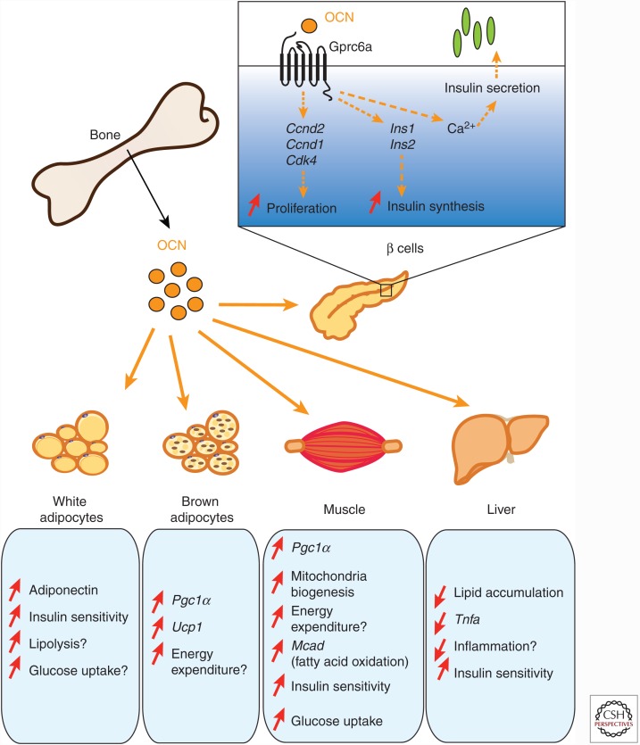
Like many other organs, bone can act as an endocrine organ through the secretion of bone-specific hormones or "osteokines." At least two osteokines are implicated in the control of glucose and energy metabolism: osteocalcin (OCN) and lipocalin-2 (LCN2). OCN stimulates the production and secretion of insulin by the pancreatic β-cells, but also favors adaptation to exercise by stimulating glucose and fatty acid (FA) utilization by the muscle. Both of these OCN functions are mediated by the G-protein-coupled receptor GPRC6A. In contrast, LCN2 influences energy metabolism by activating appetite-suppressing signaling in the brain. This action of LCN2 occurs through its binding to the melanocortin 4 receptor (MC4R) in the paraventricular nucleus of the hypothalamus (PVN) and ventromedial neurons of the hypothalamus.


Figures

Figure 1.
Figure 1.
Endocrine functions of osteocalcin (OCN). Once released in the bloodstream, undercarboxylated bioactive OCN affects glucose metabolism mainly in two ways. First, OCN directly affects β-cell function by binding to the receptor GPRC6A and increasing their capacity to proliferate as well as to synthetize and secrete insulin. Second, OCN improves insulin sensitivity and energy expenditure through multiple mechanisms. OCN stimulates energy expenditure by increasing mitochondrial biogenesis in the muscle and by regulating the expression of genes implicated in energy consumption in brown adipose tissue and skeletal muscle. OCN also affects insulin sensitivity possibly by increasing adiponectin expression in white fat and decreasing lipid accumulation and inflammation in steatotic liver. A direct impact of OCN as an insulin-sensitizing hormone is speculative and remains to be established.
Figure 2.
Figure 2.
Regulation of osteocalcin (OCN) activity. (A) Once γ-carboxylated and secreted by osteoblasts, OCN is stored in the bone extracellular matrix in an inactive form. To fulfill its beneficial effects on glucose metabolism, OCN has to be activated, that is, decarboxylated. This is accomplished through osteoclastic bone resorption that generates the acid pH necessary for OCN decarboxylation. (B) Insulin signaling in osteoblasts affects OCN activity by increasing bone resorption through osteoprotegerin (Opg) down-regulation. Because OCN stimulates insulin secretion, a feedforward loop exists between OCN and insulin activity. Esp, a tyrosine phosphatase, negatively regulates insulin receptor signaling and decreases OCN activity.
Figure 3.
Figure 3.
Mechanism of action of osteocalcin (OCN) in skeletal muscle during exercise. During exercise, bioactive OCN is released by osteoblasts and binds to the receptor GPRC6A in myofibers where it promotes the uptake and utilization of nutrients. First, OCN favors the expression of fatty acid (FA) transporters and stimulates β-oxidation. Second, OCN favors the translocation of the glucose transporter GLUT4 to the plasma membrane. This, in turn, allows the increase in glucose uptake and catabolism.
Figure 4.
Figure 4.
A cross talk between bone, via osteocalcin (OCN), and skeletal muscle, via interleukin 6 (IL-6), promotes adaptation to exercise. Circulating levels of bioactive OCN increases during exercise. OCN signals in myofibers through GPRC6A, in which it induces the expression of Il6 and the increase in circulating levels of this myokine. IL-6 signals back to skeletal muscle where it favors glucose and fatty acid (FA) utilization. IL-6 also stimulates FA production in the white adipose tissue and glucose production in the liver. Furthermore, IL-6 signals to bone to increase the production of bioactive OCN.
Figure 5.
Figure 5.
Regulation of food intake by lipocalin-2 (LCN2). Following feeding, LCN2 is released by osteoblasts and accumulates in the hypothalamus where it binds the melanocortin 4 receptor (MC4R) in the paraventricular nucleus of the hypothalamus (PVN) and ventromedial neurons of the hypothalamus (VMH). Activation of MC4R by LCN2 induces cAMP and the expression of Crh, Trh, Sim1, and Bdnf so as to suppress food intake.

Similar articles

Cited by

References

    1. Alkharfy KM, Al-Daghri NM, Vanhoutte PM, Krishnaswamy S, Xu A. 2012. Serum retinol-binding protein 4 as a marker for cardiovascular disease in women. PLoS ONE 7: e48612. - PMC - PubMed
    1. Bonneau J, Ferland G, Karelis AD, Doucet E, Faraj M, Rabasa-Lhoret R, Ferron M. 2017. Association between osteocalcin γ-carboxylation and insulin resistance in overweight and obese postmenopausal women. J Diabetes Complications 31: 1027–1034. - PubMed
    1. Borges BC, Rorato R, Avraham Y, da Silva LE, Castro M, Vorobiav L, Berry E, Antunes-Rodrigues J, Elias LL. 2011. Leptin resistance and desensitization of hypophagia during prolonged inflammatory challenge. Am J Physiol Endocrinol Metab 300: E858–E869. - PubMed
    1. Cornish J, Callon KE, Bava U, Lin C, Naot D, Hill BL, Grey AB, Broom N, Myers DE, Nicholson GC, et al. 2002. Leptin directly regulates bone cell function in vitro and reduces bone fragility in vivo. J Endocrinol 175: 405–415. - PubMed
    1. Das SK, Sharma NK, Elbein SC. 2010. Analysis of osteocalcin as a candidate gene for type 2 diabetes (T2D) and intermediate traits in Caucasians and African Americans. Dis Markers 28: 281–286. - PMC - PubMed

Publication types

Grants and funding