Genetic background-dependent role of Egr1 for eyelid development
- PMID: 28778995
- PMCID: PMC5576810
- DOI: 10.1073/pnas.1705848114
Genetic background-dependent role of Egr1 for eyelid development
Abstract
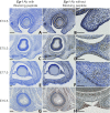
EGR1 is an early growth response zinc finger transcription factor with broad actions, including in differentiation, mitogenesis, tumor suppression, and neuronal plasticity. Here we demonstrate that Egr1-/- mice on the C57BL/6 background have normal eyelid development, but back-crossing to BALB/c background for four or five generations resulted in defective eyelid development by day E15.5, at which time EGR1 was expressed in eyelids of WT mice. Defective eyelid formation correlated with profound ocular anomalies evident by postnatal days 1-4, including severe cryptophthalmos, microphthalmia or anophthalmia, retinal dysplasia, keratitis, corneal neovascularization, cataracts, and calcification. The BALB/c albino phenotype-associated Tyrc tyrosinase mutation appeared to contribute to the phenotype, because crossing the independent Tyrc-2J allele to Egr1-/- C57BL/6 mice also produced ocular abnormalities, albeit less severe than those in Egr1-/- BALB/c mice. Thus EGR1, in a genetic background-dependent manner, plays a critical role in mammalian eyelid development and closure, with subsequent impact on ocular integrity.
Keywords: Egr1; eyelid development; genetic background-specific effects; ocular abnormalities; tyrosinase.
Conflict of interest statement
The authors declare no conflict of interest.
Figures


Similar articles
-
Eyelid closure in embryogenesis is required for ocular adnexa development.Invest Ophthalmol Vis Sci. 2014 Nov 6;55(11):7652-61. doi: 10.1167/iovs.14-15155. Invest Ophthalmol Vis Sci. 2014. PMID: 25377219 Free PMC article.
-
Defective eyelid leading edge cell migration in C57BL/6-corneal opacity mice with an "eye open at birth" phenotype.Genet Mol Res. 2016 Aug 26;15(3). doi: 10.4238/gmr.15036741. Genet Mol Res. 2016. PMID: 27706598
-
Egr1 gene knockdown affects embryonic ocular development in zebrafish.Mol Vis. 2006 Oct 26;12:1250-8. Mol Vis. 2006. PMID: 17110908
-
Wakayama Symposium: Epithelial-mesenchymal interactions in eyelid development.Ocul Surf. 2012 Oct;10(4):212-6. doi: 10.1016/j.jtos.2012.07.005. Epub 2012 Jul 25. Ocul Surf. 2012. PMID: 23084141 Review.
-
Microphthalmia and associated abnormalities in inbred black mice.Lab Anim Sci. 1994 Dec;44(6):551-60. Lab Anim Sci. 1994. PMID: 7898027 Review.
Cited by
-
Reconstruction strategy in isolated complete Cryptophthalmos: a case series.BMC Ophthalmol. 2019 Jul 31;19(1):165. doi: 10.1186/s12886-019-1170-6. BMC Ophthalmol. 2019. PMID: 31366340 Free PMC article.
-
Egr-1 is a key regulator of the blood-brain barrier damage induced by meningitic Escherichia coli.Cell Commun Signal. 2024 Jan 17;22(1):44. doi: 10.1186/s12964-024-01488-y. Cell Commun Signal. 2024. PMID: 38233877 Free PMC article.
-
Gene profiling involved in fate determination of salivary gland type in mouse embryogenesis.Genes Genomics. 2018 Jun 22. doi: 10.1007/s13258-018-0715-z. Online ahead of print. Genes Genomics. 2018. PMID: 29934934
-
New Insights on the Regulatory Gene Network Disturbed in Central Areolar Choroidal Dystrophy-Beyond Classical Gene Candidates.Front Genet. 2022 May 17;13:886461. doi: 10.3389/fgene.2022.886461. eCollection 2022. Front Genet. 2022. PMID: 35656327 Free PMC article.
References
-
- Gómez-Martín D, Díaz-Zamudio M, Galindo-Campos M, Alcocer-Varela J. Early growth response transcription factors and the modulation of immune response: Implications towards autoimmunity. Autoimmun Rev. 2010;9:454–458. - PubMed
-
- Forsdyke DR. cDNA cloning of mRNAS which increase rapidly in human lymphocytes cultured with concanavalin-A and cycloheximide. Biochem Biophys Res Commun. 1985;129:619–625. - PubMed
-
- Sukhatme VP, et al. A zinc finger-encoding gene coregulated with c-fos during growth and differentiation, and after cellular depolarization. Cell. 1988;53:37–43. - PubMed
-
- DeLigio JT, Zorio DA. Early growth response 1 (EGR1): A gene with as many names as biological functions. Cancer Biol Ther. 2009;8:1889–1892. - PubMed
-
- Nguyen HQ, Hoffman-Liebermann B, Liebermann DA. The zinc finger transcription factor Egr-1 is essential for and restricts differentiation along the macrophage lineage. Cell. 1993;72:197–209. - PubMed
Publication types
MeSH terms
Substances
LinkOut - more resources
Full Text Sources
Other Literature Sources
Molecular Biology Databases

