

Bidirectional Modulation of Intrinsic Excitability in Rat Prelimbic Cortex Neuronal Ensembles and Non-Ensembles after Operant Learning
- PMID: 28779019
- PMCID: PMC5588469
- DOI: 10.1523/JNEUROSCI.3761-16.2017
Bidirectional Modulation of Intrinsic Excitability in Rat Prelimbic Cortex Neuronal Ensembles and Non-Ensembles after Operant Learning
Abstract
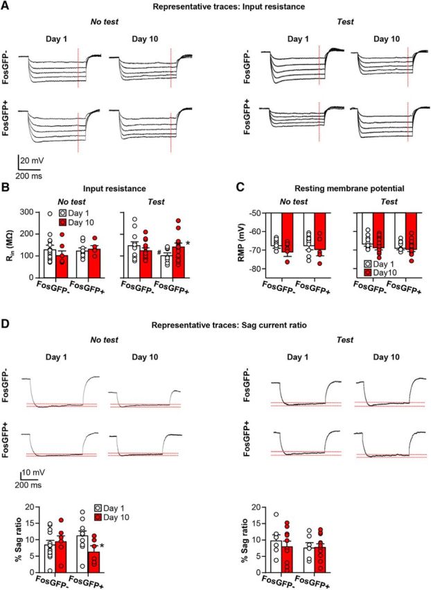
Learned associations between environmental stimuli and rewards drive goal-directed learning and motivated behavior. These memories are thought to be encoded by alterations within specific patterns of sparsely distributed neurons called neuronal ensembles that are activated selectively by reward-predictive stimuli. Here, we use the Fos promoter to identify strongly activated neuronal ensembles in rat prelimbic cortex (PLC) and assess altered intrinsic excitability after 10 d of operant food self-administration training (1 h/d). First, we used the Daun02 inactivation procedure in male FosLacZ-transgenic rats to ablate selectively Fos-expressing PLC neurons that were active during operant food self-administration. Selective ablation of these neurons decreased food seeking. We then used male FosGFP-transgenic rats to assess selective alterations of intrinsic excitability in Fos-expressing neuronal ensembles (FosGFP+) that were activated during food self-administration and compared these with alterations in less activated non-ensemble neurons (FosGFP-). Using whole-cell recordings of layer V pyramidal neurons in an ex vivo brain slice preparation, we found that operant self-administration increased excitability of FosGFP+ neurons and decreased excitability of FosGFP- neurons. Increased excitability of FosGFP+ neurons was driven by increased steady-state input resistance. Decreased excitability of FosGFP- neurons was driven by increased contribution of small-conductance calcium-activated potassium (SK) channels. Injections of the specific SK channel antagonist apamin into PLC increased Fos expression but had no effect on food seeking. Overall, operant learning increased intrinsic excitability of PLC Fos-expressing neuronal ensembles that play a role in food seeking but decreased intrinsic excitability of Fos- non-ensembles.SIGNIFICANCE STATEMENT Prefrontal cortex activity plays a critical role in operant learning, but the underlying cellular mechanisms are unknown. Using the chemogenetic Daun02 inactivation procedure, we found that a small number of strongly activated Fos-expressing neuronal ensembles in rat PLC play an important role in learned operant food seeking. Using GFP expression to identify Fos-expressing layer V pyramidal neurons in prelimbic cortex (PLC) of FosGFP-transgenic rats, we found that operant food self-administration led to increased intrinsic excitability in the behaviorally relevant Fos-expressing neuronal ensembles, but decreased intrinsic excitability in Fos- neurons using distinct cellular mechanisms.
Keywords: Fos; electrophysiology; intrinsic plasticity; motivated behavior; reward learning; transgenic rat.
Copyright © 2017 the authors 0270-6474/17/378845-12$15.00/0.
Figures


Similar articles
-
Distinct Fos-Expressing Neuronal Ensembles in the Ventromedial Prefrontal Cortex Mediate Food Reward and Extinction Memories.J Neurosci. 2016 Jun 22;36(25):6691-703. doi: 10.1523/JNEUROSCI.0140-16.2016. J Neurosci. 2016. PMID: 27335401 Free PMC article.
-
Changes in Appetitive Associative Strength Modulates Nucleus Accumbens, But Not Orbitofrontal Cortex Neuronal Ensemble Excitability.J Neurosci. 2017 Mar 22;37(12):3160-3170. doi: 10.1523/JNEUROSCI.3766-16.2017. Epub 2017 Feb 17. J Neurosci. 2017. PMID: 28213443 Free PMC article.
-
Separate vmPFC Ensembles Control Cocaine Self-Administration Versus Extinction in Rats.J Neurosci. 2019 Sep 11;39(37):7394-7407. doi: 10.1523/JNEUROSCI.0918-19.2019. Epub 2019 Jul 22. J Neurosci. 2019. PMID: 31331999 Free PMC article.
-
Learning-induced intrinsic and synaptic plasticity in the rodent medial prefrontal cortex.Neurobiol Learn Mem. 2020 Mar;169:107117. doi: 10.1016/j.nlm.2019.107117. Epub 2019 Nov 23. Neurobiol Learn Mem. 2020. PMID: 31765801 Free PMC article. Review.
-
Using c-fos to study neuronal ensembles in corticostriatal circuitry of addiction.Brain Res. 2015 Dec 2;1628(Pt A):157-73. doi: 10.1016/j.brainres.2014.11.005. Epub 2014 Nov 11. Brain Res. 2015. PMID: 25446457 Free PMC article. Review.
Cited by
-
BEHAVIORAL AND NEUROBIOLOGICAL MECHANISMS OF PAVLOVIAN AND INSTRUMENTAL EXTINCTION LEARNING.Physiol Rev. 2021 Apr 1;101(2):611-681. doi: 10.1152/physrev.00016.2020. Epub 2020 Sep 24. Physiol Rev. 2021. PMID: 32970967 Free PMC article. Review.
-
Drug-taking in a socio-sexual context enhances vulnerability for addiction in male rats.Neuropsychopharmacology. 2019 Feb;44(3):503-513. doi: 10.1038/s41386-018-0235-1. Epub 2018 Oct 6. Neuropsychopharmacology. 2019. PMID: 30337639 Free PMC article.
-
Cell-Type-Specific Changes in Intrinsic Excitability in the Subiculum following Learning and Exposure to Novel Environmental Contexts.eNeuro. 2019 Jan 4;5(6):ENEURO.0484-18.2018. doi: 10.1523/ENEURO.0484-18.2018. eCollection 2018 Nov-Dec. eNeuro. 2019. PMID: 30627661 Free PMC article.
-
Necessity and recruitment of cue-specific neuronal ensembles within the basolateral amygdala during appetitive reversal learning.Neurobiol Learn Mem. 2022 Oct;194:107663. doi: 10.1016/j.nlm.2022.107663. Epub 2022 Jul 21. Neurobiol Learn Mem. 2022. PMID: 35870716 Free PMC article.
-
What does the Fos say? Using Fos-based approaches to understand the contribution of stress to substance use disorders.Neurobiol Stress. 2018 Jun 2;9:271-285. doi: 10.1016/j.ynstr.2018.05.004. eCollection 2018 Nov. Neurobiol Stress. 2018. PMID: 30450391 Free PMC article. Review.
References
-
- Brons JF, Woody CD (1980) Long-term changes in excitability of cortical neurons after Pavlovian conditioning and extinction. J Neurophysiol 44:605–615. - PubMed
Publication types
MeSH terms
LinkOut - more resources
Full Text Sources
Other Literature Sources