. 2017 Aug 4;7(1):7345.
doi: 10.1038/s41598-017-07433-z.

Pathogenicity and peramivir efficacy in immunocompromised murine models of influenza B virus infection

Affiliations

Pathogenicity and peramivir efficacy in immunocompromised murine models of influenza B virus infection

Philippe Noriel Q Pascua et al. Sci Rep. .

Abstract

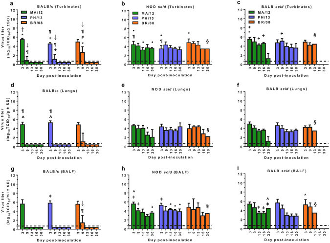
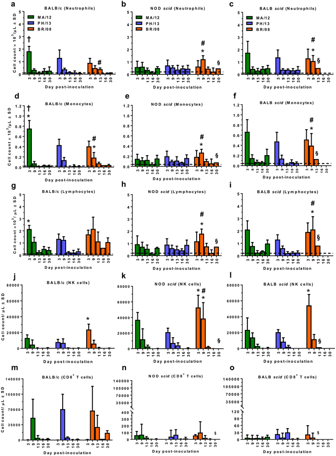
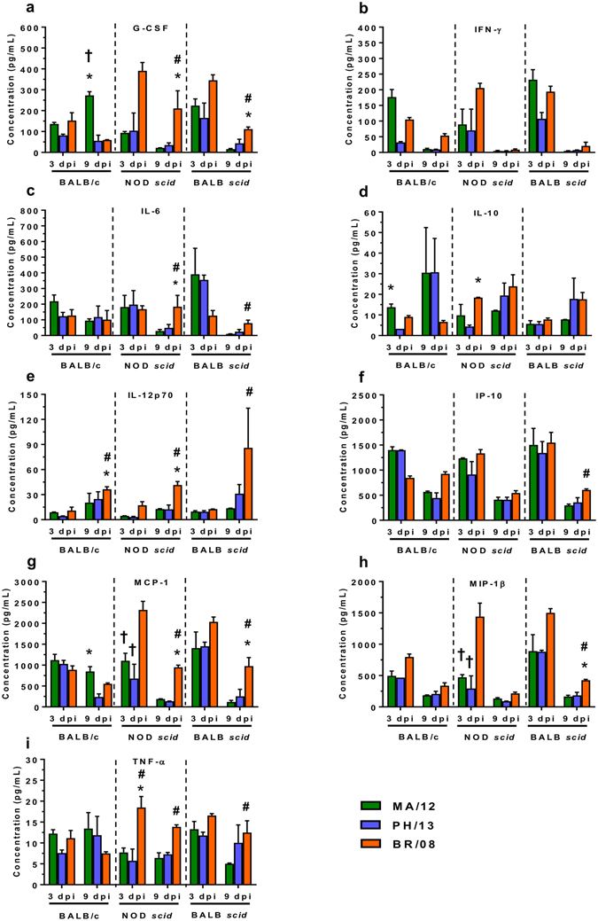
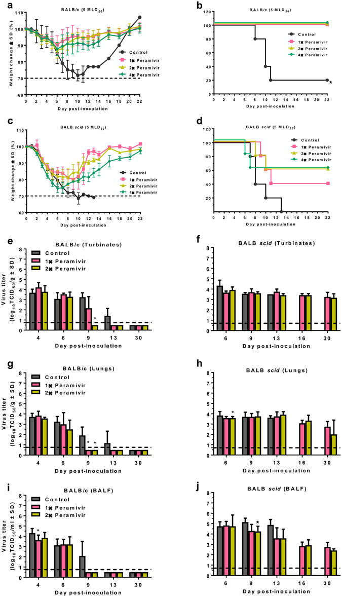
Influenza B viruses are important human pathogens that remain inadequately studied, largely because available animal models are poorly defined. Here, we developed an immunocompromised murine models for influenza B virus infection, which we subsequently used to study pathogenicity and to examine antiviral efficacy of the neuraminidase inhibitor peramivir. We studied three influenza B viruses that represent both the Yamagata (B/Massachusetts/2/2012 and B/Phuket/3073/2013) and Victoria (B/Brisbane/60/2008, BR/08) lineages. BR/08 was the most pathogenic in genetically modified immunocompromised mice [BALB scid and non-obese diabetic (NOD) scid strains] causing lethal infection without prior adaptation. The immunocompromised mice demonstrated prolonged virus shedding with modest induction of immune responses compared to BALB/c. Rather than severe virus burden, BR/08 virus-associated disease severity correlated with extensive virus spread and severe pulmonary pathology, stronger and persistent natural killer cell responses, and the extended induction of pro-inflammatory cytokines and chemokines. In contrast to a single-dose treatment (75 mg/kg/day), repeated doses of peramivir rescued BALB scid mice from lethal challenge with BR/08, but did not result in complete virus clearance. In summary, we have established immunocompromised murine models for influenza B virus infection that will facilitate evaluations of the efficacy of currently available and investigational anti-influenza drugs.


Conflict of interest statement

The authors declare that they have no competing interests.

Figures

Figure 1
Figure 1
Morbidity and mortality of immunocompetent and immunocompromised mice inoculated with influenza B viruses. Female 6-week-old BALB/c, NOD scid, and BALB scid mice (n = 5/group) were lightly anesthetized with isoflurane and inoculated intranasally with 104 (af) or 105 (gl) TCID50/mouse of influenza MA/12 (green line), PH/13 (blue line), or BR/08 (orange line) virus. The graphs show the weight loss (ac,g–i) and survival (df,jl) of the inoculated mice, which were monitored and observed daily. Dashed lines indicate endpoint for mortality (30% of initial weight). *P < 0.05, relative to PH/13; # P < 0.05, relative to MA/12, as determined by one-way ANOVA with Bonferroni’s multiple comparison post-test. The probabilities for survival were determined by Kaplan-Meier and log-rank tests, and the differences in weight were analyzed by one-way ANOVA.
Figure 2
Figure 2
Influenza B virus load and duration of replication in immunocompetent and immunocompromised mice. BALB/c, NOD scid, and BALB scid mice were lightly anesthetized with isoflurane and inoculated intranasally with 104 TCID50/mouse of influenza PH/13 (blue bars), MA/12 (green bars), or BR/08 (orange bars) virus. Virus titers were determined in the nasal turbinates (ac), lungs (df), and BALF (gi) of mice (n = 3/group/time-point) at 3, 9, 13, 16, and 30 dpi by TCID50 assays in MDCK cells. The bars represent the mean virus titers ± SD (log10TCID50/mL) for each time-point. Dashed lines indicate the minimum level of virus detection (0.75 log10TCID50/mL). *P < 0.05, relative to PH/13; # P < 0.05, relative to MA/12; P < 0.05, relative to BR/08; + P < 0.05 for virus titers in nasal turbinates versus lungs; ǂ P < 0.05 for virus titers in nasal turbinates versus BALF; ^P < 0.05 for virus titers in lungs versus BALF; P < 0.05 for virus titers in BALB/c versus NOD scid mice; P < 0.05 for virus titers in BALB/c versus BALB scid mice; and P < 0.05 for virus titers in NOD scid mice versus BALB scid mice, as determined by one-way ANOVA with Bonferroni’s multiple comparison post-test. §Detection in one mouse only.
Figure 3
Figure 3
Induction of virus-induced inflammatory correlates in BALF of immunocompetent and immunocompromised mice inoculated with influenza B viruses. BALB/c, NOD scid, and BALB scid mice were inoculated with viruses as described in the legend for Fig. 2. Inflammatory cell counts [neutrophils (ac), monocytes (df), and lymphocytes (gi)] were determined in the BALF of virus-inoculated mice at 3, 9, 13, 16, and 30 dpi by automatic cell counter and lymphocyte subpopulations [NK (jl) and CD8+ T (mo) cells] were differentiated by flow cytometry. Bars represent mean values ± SD (n = 3/group/time-point). * P < 0.05, relative to PH/13; # P < 0.05, relative to MA/12; and P < 0.05, relative to BR/08, as determined by one-way ANOVA with Bonferroni’s multiple comparison post-test. §Detection in one mouse only. Dashed line indicates the cell counts in control mice inoculated with sterile 1× PBS and sacrificed at 6 dpi.
Figure 4
Figure 4
Histopathologic changes in the lungs of immunocompetent and immunocompromised mice inoculated with influenza B viruses. BALB/c, NOD scid, and BALB scid mice were inoculated with viruses as described in the legend for Fig. 2. Pulmonary lesions were evaluated at 6 dpi (n = 4/virus/mouse strain). The panels show the extent of virus infection and histopathology of influenza MA/12, PH/13, and BR/08 viruses in the lungs of immunocompetent BALB/c (a,d,g) and immunocompromised NOD scid (b,e,h) and BALB scid (c,f,i) mice. A representative section is shown for each virus and mouse strain. The green lines designate total lung areas measured; red-shaded areas designate bronchioles/alveoli with active virus infection (i.e., containing antigen-positive epithelial cells). The average percentage of the total lung field containing antigen-positive epithelial cells are shown in red texts and the average severity score ± SE for inflammation are shown in black texts. *P < 0.05, relative to BR/08 scores, as determined by one-way ANOVA with Bonferroni’s multiple comparison post-test.
Figure 5
Figure 5
Induction of pulmonary cytokine/chemokine responses in immunocompetent and immunocompromised mice inoculated with influenza B viruses. BALB/c, NOD scid, and BALB scid mice were inoculated with viruses as described in the legend for Fig. 2. The expression levels of 25 cytokines and chemokines were assayed in lung homogenates at 3 and 9 dpi. Bars represent mean values ± SE (n = 5–6/group/time-point). * P < 0.05, relative to PH/13; # P < 0.05, relative to MA/12; and P < 0.05, relative to BR/08, as determined by one-way ANOVA with Bonferroni’s multiple comparison post-test.
Figure 6
Figure 6
Efficacy of peramivir treatment on survival of immunocompetent and immunocompromised mice inoculated with influenza BR/08 virus. BALB/c and BALB scid mice (n = 5/group) were lightly anesthetized with isoflurane and inoculated with 5 MLD50 (2.5 × 105 TCID50/mouse) of influenza BR/08 virus. The NAI peramivir was administered by IM injection at a dose of 75 mg/kg/day (0.1 mL/mouse) once a day in a single-dose (1×: +24 hpi), double-dose (2×: +24 and +72 hpi), or four-dose (4×: +24, +72, +120, and +168 hpi) regimen. Control animals received IM injection of sterile RNAse-free water in a four-dose regimen (4×: +24, +72, +120, and +168 hpi). The graphs show the weight loss (a,c) and survival (b,d) of inoculated mice, which were monitored and observed daily, up to 30 dpi. The bar charts show the effect of peramivir treatment (1×: +24 hpi or 2×: +24 and +72 hpi) on virus titers in nasal turbinates (e,f), lungs (g,h), and BALFs (i,j) of BALB/c and BALB scid mice (n = 3/group/time-point) inoculated with BR/08 virus at 4, 6, 9, 13, 16 and 30 dpi. Virus titers were determined by TCID50 assays in MDCK cells. *P < 0.05, compared between control and peramivir-treated mice inoculated with BR/08 virus. Probabilities for survival were determined by the Kaplan-Meier and log-rank tests, and the differences in weight were analyzed by one-way ANOVA.

Similar articles

Cited by

References

    1. Englund J, Feuchtinger T, Ljungman P. Viral infections in immunocompromised patients. Biol Blood Marrow Transplant. 2011;17:S2–5. doi: 10.1016/j.bbmt.2010.11.008. - DOI - PMC - PubMed
    1. Ison MG. Influenza prevention and treatment in transplant recipients and immunocompromised hosts. Influenza Other Respir Viruses. 2013;7(Suppl 3):60–66. doi: 10.1111/irv.12170. - DOI - PMC - PubMed
    1. Kim YJ, Boeckh M, Englund JA. Community respiratory virus infections in immunocompromised patients: hematopoietic stem cell and solid organ transplant recipients, and individuals with human immunodeficiency virus infection. Semin Respir Crit Care Med. 2007;28:222–242. doi: 10.1055/s-2007-976494. - DOI - PubMed
    1. Kunisaki KM, Janoff EN. Influenza in immunosuppressed populations: a review of infection frequency, morbidity, mortality, and vaccine responses. Lancet Infect Dis. 2009;9:493–504. doi: 10.1016/S1473-3099(09)70175-6. - DOI - PMC - PubMed
    1. Memoli MJ, et al. The natural history of influenza infection in the severely immunocompromised vs nonimmunocompromised hosts. Clin Infect Dis. 2014;58:214–224. doi: 10.1093/cid/cit725. - DOI - PMC - PubMed

Publication types

MeSH terms

LinkOut - more resources