. 2017 Aug 4;7(1):7305.
doi: 10.1038/s41598-017-07848-8.

Targeting primary acute myeloid leukemia with a new CXCR4 antagonist IgG1 antibody (PF-06747143)

Affiliations

Targeting primary acute myeloid leukemia with a new CXCR4 antagonist IgG1 antibody (PF-06747143)

Yanyan Zhang et al. Sci Rep. .

Abstract

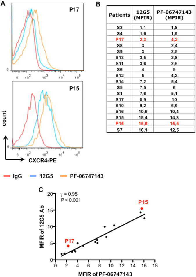
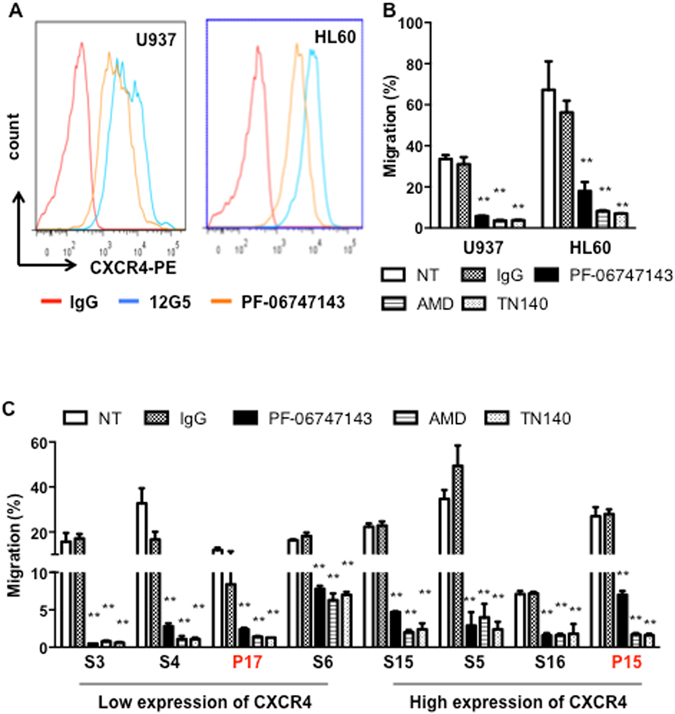
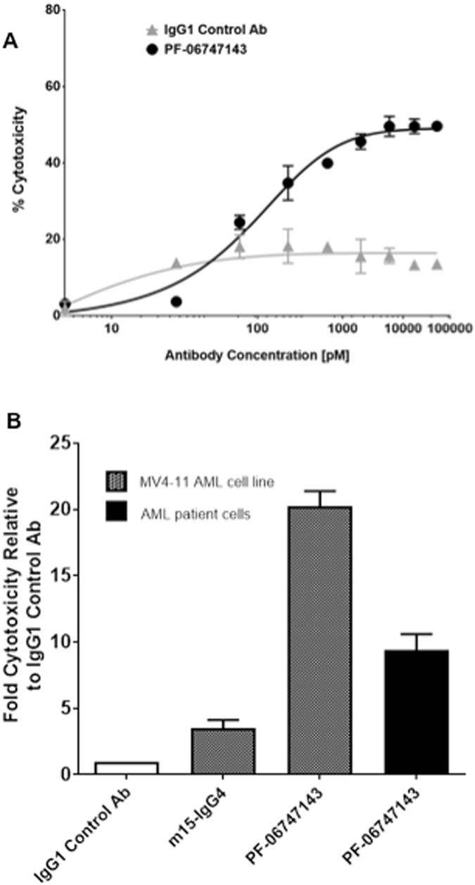
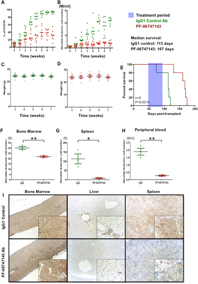
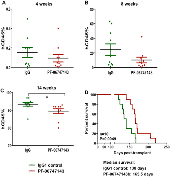
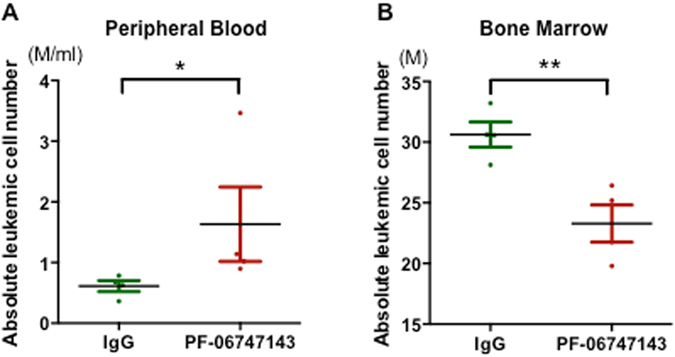
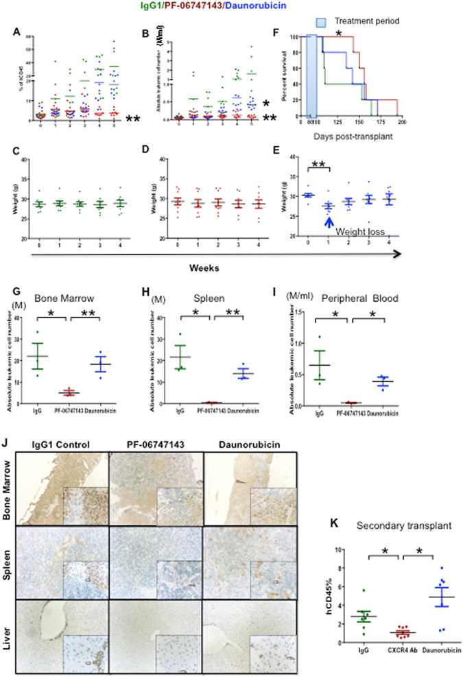
The chemokine receptor CXCR4 mediates cell anchorage in the bone marrow (BM) microenvironment and is overexpressed in 25-30% of patients with acute myeloid leukemia (AML). Here we have shown that a new CXCR4 receptor antagonist IgG1 antibody (PF-06747143) binds strongly to AML cell lines and to AML primary cells inhibiting their chemotaxis in response to CXCL12. PF-06747143 also induced cytotoxicity in AML cells via Fc-effector function. To characterize the effects of PF-06747143 on leukemia progression, we used two different patient-derived xenograft (PDX) models: Patient 17CXCR4-low and P15CXCR4-high models, characterized by relatively low and high CXCR4 expression, respectively. Weekly administration of PF-06747143 to leukemic mice significantly reduced leukemia development in both models. Secondary transplantation of BM cells from PF-06747143-treated or IgG1 control-treated animals showed that leukemic progenitors were also targeted by PF-06747143. Administration of a single dose of PF-06747143 to PDX models induced rapid malignant cell mobilization into the peripheral blood (PB). These findings support evaluation of this antibody in AML therapy, with particular appeal to patients resistant to chemotherapy and to unfit patients, unable to tolerate intensive chemotherapy.


Conflict of interest statement

Y.G., P.L., D.T., S.-H.L., T.S., V.R.F. and F.P. were employees of Pfizer, Inc. when the studies were conducted. The other authors declared no potential conflicts of interests.

Figures

Figure 1
Figure 1
PF-06747143 binds to CXCR4 in AML primary cells similarly to 12G5. (A) Representative histograms of flow cytometry comparing 12G5 (a commercially available CXCR4 Ab clone) and PF-06747143 CXCR4 antibody binding to patient 17 (P17CXCR4-low) and patient 15 (P15CXCR4-high) primary cells. These two patients were chosen for subsequent studies using PF-06747143. (B) Quantitative analyses of fluorescence intensity were performed on CD45+CD33+ cells of primary AML samples. The two patients chosen for subsequent studies using PF-06747143 were marked in red. Mean fluorescence intensity ratio (MFIR) for 12G5 and PF-06747143 were calculated by dividing the mean fluorescence intensity (MFI) of CXCR4 by the MFI of the respective nonspecific isotype control. (C) Correlation between 12G5 and PF-06747143 CXCR4 antibody binding to patient primary cells.
Figure 2
Figure 2
PF-06747143 inhibits CXCL12-driven migration of leukemic cell lines and primary AML cells from patients. (A) Cell surface CXCR4 of U937 and HL-60 was measured by flow cytometry with PF-06747143 and 12G5 antibodies. (B,C) Effect of PF-06747143 (10 μg/ml), IgG1 (10 μg/ml), AMD3100 (20 μM) or TN140 (5 μM) on chemotactic responses to 100 ng/ml CXCL12. Leukemic cell lines (B), primary AML cells (C). **P < 0.01(student’s t-test).
Figure 3
Figure 3
PF-06747143 induces AML cell death by ADCC or ADCP. (A) ADCC activity was evaluated by incubating PF-06747143, or IgG1 control Ab, at increasing concentrations with AML primary BM cells, in the presence of NK92 158 V effector cells. (B) ADCC or ADCP activity was also evaluated incubating 100 nM of PF-06747143, IgG control Ab, or a human IgG4 backbone (m15-IgG4) in the AML MV4-11 cell line or AML primary BM cells, in the presence of PBMC purified from a healthy donor, as effector cells.
Figure 4
Figure 4
PF-06747143 treatment inhibits leukemia growth in an AML PDX model expressing low CXCR4. Mice were engrafted with P17CXCR4-low as described in Supplemental Figure 1A. After leukemia establishment, at 7 weeks, the mean percentage of AML cells in the peripheral blood was 7%. Mice were then divided into two groups, 8 mice/group, which received weekly subcutaneous treatment with IgG1 control or PF-06747143 Ab for the treatment period indicated. Twenty-four hours after each weekly treatment, percentages of hCD45+ cells (A) and absolute leukemic cell numbers (B) calculated by the equation: total cell number x % of human CD45+ cells in the blood were evaluated. Tumor burden in the blood at the end of treatment is significantly different between PF-06747143 and IgG1 control group. **P < 0.01 assessed by test of Mann-Whitney. (C) and (D) Body weights are shown. (E) Survival analysis was performed (n = 5 animals/group). Treatment was performed between Days 49 and 89, as indicated in the blue box. Statistical difference: p = 0.0018, between IgG1 and PF-06747143 treated mice (log-rank test). After seven weeks treatment, 3 mice per group were sacrificed and absolute leukemic cells in BM (F), spleen (G) and blood (H) were evaluated. *P < 0.05; **P < 0.01 assessed by the Mann-Whitney test. Data for each animal is shown and the horizontal bar represents the mean. (I) At the end of treatment, the femur with BM, spleen, and liver were collected and stained immunohistochemically with anti-human mitochondria membrane antibody. The sections were counterstained with hematoxylin to enhance cellular morphology. Punctate nuclear staining of human AML cells is indicated by an arrow in each image. Photos were acquired at 10X magnification for large panels and 40X magnification for the inserts.
Figure 5
Figure 5
PF-06747143 treatment reduced the leukemic secondary transplantation potential. After 7 weekly treatments (Day 47 from the beginning of treatment), BM of IgG1 control and PF-06747143 antibody-treated mice (n = 3 mice/group) were flushed in 1 mL of PBS, pooled together, and 100 μl injected per mice for secondary transplantations (n = 10 mice/group). The percentages (A,B,C) of hCD45+ cells were evaluated in the blood of secondary mice at different time points (4, 8 and 14 weeks). Data for each animal is shown and the horizontal bar represents the mean. *P < 0.05 assessed by the Mann-Whitney test. Survival of secondary transplanted mice was analyzed (D). Statistical difference: p = 0.0049, between IgG control and PF-06747143 treated mice (log rank test).
Figure 6
Figure 6
PF-06747143 induces mobilization of leukemic cells in blood. Mice implanted with CXCR4-low (P17CXCR4-low) AML patient cells were treated subcutaneously with a single dose of PF-06747143 or IgG1 Control Ab at 10 mg/kg. Absolute leukemic cell numbers in PB (A) and BM (B) were evaluated at 4 hrs post-dose (n = 3 or 4 animals/group). Data for each animal is shown and the horizontal bar represents the mean. *P < 0.05; **P < 0.01 determined by Mann-Whitney test.
Figure 7
Figure 7
PF-06747143 treatment leads to leukemia regression in a PDX model expressing high CXCR4. Leukemic mice reconstituted with P15CXCR4-high cells were treated subcutaneously with 10 mg/kg weekly doses of IgG1 Control Ab or PF-06747143, for five weeks. Daunorubicin was administered intravenously at 2.5 mg/kg every other day, for 1st week of study (8 mice/group). 24 hours after each injection, percentages (A) and absolute leukemic hCD45+ cell numbers (B) were evaluated in the blood of IgG1 control Ab, PF-06747143 and daunorubicin-treated mice. **P < 0.01, PF-06747143 vs IgG1 control. *P < 0.05, Daunorubicin vs IgG1 Control assessed by Mann-Whitney test. Body weights (C,D,E). Significant weight loss was observed after 1 week of treatment with Daunorubicin. **P < 0.01 assessed by Mann-Whitney test. Survival analysis were performed (F) (n = 5/group), with treatment period (between Day 72 and Day 101) as indicated in blue. Statistical difference: *p = 0.042 between IgG1 control and PF-06747143 treated mice (log-rank test). After 5 weeks treatment, the hCD45+ absolute leukemic cell numbers were evaluated in the bone marrow (G), spleen (H) and the peripheral blood (I) of IgG1 control Ab, PF-06747143 and Daunorubicin treated mice. Data for each animal is represented and the horizontal bar is the average. *P < 0.05; **P < 0.01 assessed by Mann-Whitney test. (J) At the end of treatment, the femur, spleen and liver were collected and stained immunohistochemically with anti-human mitochondria membrane antibody. The sections were counterstained with hematoxylin. Punctate nuclear staining of human AML cells is indicated by an arrow in each image. Photos were acquired at 10X magnification for large panels and 40X magnification for the inserts. (K) After 5 weeks treatment BM cell suspension from IgG1 control Ab-, PF-06747143- or daunorubicin-treated mice were implanted to secondary mice. Eight weeks later, the percentage of human CD45+ cells in the blood of reconstituted mice was analyzed by cytometry. Data are mean ± SD (n = 7 or 8 animals/group). *P < 0.05 assessed by t-test.

References

    1. Aiuti A, Webb IJ, Bleul C, Springer T, Gutierrez-Ramos JC. The chemokine SDF-1 is a chemoattractant for human CD34+ hematopoietic progenitor cells and provides a new mechanism to explain the mobilization of CD34+ progenitors to peripheral blood. J Exp Med. 1997;185:111–120. doi: 10.1084/jem.185.1.111. - DOI - PMC - PubMed
    1. Peled A, et al. The chemokine SDF-1 stimulates integrin-mediated arrest of CD34(+) cells on vascular endothelium under shear flow. J Clin Invest. 1999;104:1199–1211. doi: 10.1172/JCI7615. - DOI - PMC - PubMed
    1. Peled A, et al. The chemokine SDF-1 activates the integrins LFA-1, VLA-4, and VLA-5 on immature human CD34(+) cells: role in transendothelial/stromal migration and engraftment of NOD/SCID mice. Blood. 2000;95:3289–3296. - PubMed
    1. Imai K, et al. Selective secretion of chemoattractants for haemopoietic progenitor cells by bone marrow endothelial cells: a possible role in homing of haemopoietic progenitor cells to bone marrow. Br. J. Haematol. 1999;106:905–11. doi: 10.1046/j.1365-2141.1999.01644.x. - DOI - PubMed
    1. Trampont PC, et al. CXCR4 acts as a costimulator during thymic beta-selection. Nat. Immunol. 2010;11:162–70. doi: 10.1038/ni.1830. - DOI - PMC - PubMed

Publication types

MeSH terms

Substances