. 2017 Aug 4;7(1):7340.
doi: 10.1038/s41598-017-07494-0.

Bee-derived antibacterial peptide, defensin-1, promotes wound re-epithelialisation in vitro and in vivo

Affiliations

Bee-derived antibacterial peptide, defensin-1, promotes wound re-epithelialisation in vitro and in vivo

Marcela Bucekova et al. Sci Rep. .

Abstract

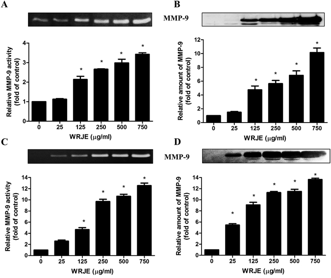
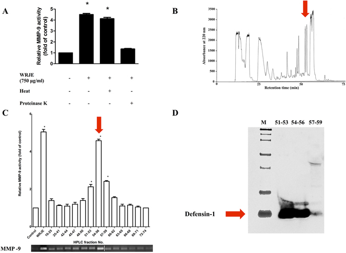
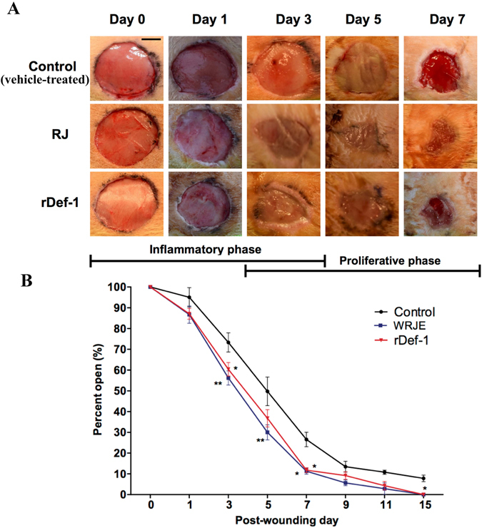
Royal jelly (RJ) has successfully been used as a remedy in wound healing. RJ has multiple effects, including antibacterial, anti-inflammatory and immunomodulatory activities, in various cell types. However, no component(s) (other than antibacterial) have been identified in RJ-accelerated wound healing. In this study, we demonstrate that keratinocytes are responsible for the elevated production of matrix metalloproteinase-9 (MMP-9) after incubation with a water extract of RJ. Furthermore, the keratinocyte migration and wound closure rates were significantly increased in the presence of RJ extract. MMP-9 production was reduced significantly following proteinase K treatment but remained stable after heat treatment, indicating that active component(s) have a proteinous character. To identify the component responsible for inducing MMP-9 production, RJ extract was fractionated using C18 RP-HPLC. In fractions exhibiting stimulatory activity, we immunochemically detected the bee-derived antibacterial peptide, defensin-1. Defensin-1 was cloned, and recombinant peptide was produced in a baculoviral expression system. Defensin-1 stimulated MMP-9 secretion from keratinocytes and increased keratinocyte migration and wound closure in vitro. In addition, defensin-1 promoted re-epithelisation and wound closure in uninfected excision wounds. These data indisputably demonstrate that defensin-1, a regular but concentration variable factor found in honey and RJ, contributes to cutaneous wound closure by enhancing keratinocyte migration and MMP-9 secretion.


Conflict of interest statement

The authors declare that they have no competing interests.

Figures

Figure 1
Figure 1
The effect of a water royal jelly extract (WRJE) on matrix metalloproteinase 9 (MMP-9) secretion and proteolytic activity in HaCaT cells (A,B) and human epidermal keratinocytes (HEK) (C,D). HaCaT and HEK cells were treated with different doses of WRJE for 72 h. (A,C) Conditioned equal volumes of the culture media were collected and subjected to gelatine zymography. Densitometric quantification of MMP-9 activity in culture media is presented. (B,D) Conditioned equal volumes of the concentrated culture media were subjected to 10% SDS-PAGE gels. MMP-9 (92 kDa) was detected by Western blotting and quantified densitometrically. The gels were run under the same experimental conditions. Shown are cropped gels/blots. (The gels/blots with indicated cropping lines are shown in Supplementary Fig. S2). Data are expressed as means and SEMs of three independent measurements. Asterisks indicate a significant difference from the untreated group, *P < 0.001.
Figure 2
Figure 2
In vitro wound healing properties of a water royal jelly extract (WRJE) in HaCaT cells. Scratch wound analyses were performed in confluent monolayers of HaCaT cells. (A) Wounded cells were treated with different doses of WRJE for 24 h. (B) Scratch wounding with HaCaT cells was also conducted by pre-treating HaCaTcells with 10 μg/ml mitomycin C for 2 h and subsequent treatment with WRJE for 24 h. (C) Wound closure rates were determined as the difference between wound width at 0 and 24 h. (D) HaCaT cell migration was evaluated by the transwell assay plate (8 μm pore size, ThinCert™ 24 Well Cell Culture Inserts), where the cells were treated with different doses of WRJE for 24 h. Asterisks indicate a significant difference from the untreated group, *P < 0.001. Scale bar = 300 μm. NS: non-significant.
Figure 3
Figure 3
Characterisation and purification of the royal jelly (RJ) component responsible for elevated MMP-9 production. (A) Heat and proteinase K treatment was performed by incubation of water royal jelly extract (WRJE) at 100 °C for 5 min and incubation with 150 μg/ml proteinase K for 1 h at 40 °C followed by heating to 98 °C for 10 min to inactivate the enzyme. Treated WRJE was incubated with HaCaT cells and conditioned equal volumes of the culture media were collected and subjected to gelatine zymography. Densitometric quantification of MMP-9 activity in culture media is presented. (B) Heat-treated WRJE was fractionated by a reverse phase-high performance liquid chromatography (RP-HPLC) on a C18 column (250 × 4.6 mm, 5 μm) at a flow rate 0.3 ml/min, with elution using a 10–90% gradient of acetonitrile (containing 0.1% (v/v) trifluoroacetic acid) for 85 min. (C) The HPLC fractions were assayed for MMP-9 induction. (D) HPLC fractions with maximal MMP-9 activity (51 to 59 min) were used for identification of MMP-9 inducer and were subjected to 16.5% Tricine-SDS-PAGE gels. Defensin-1 (5.5 kDa) was detected by Western blotting using a rabbit polyclonal anti-honeybee defensin-1 antibody diluted 1:2000 in blocking buffer. Horseradish peroxidise-conjugated secondary antibodies were applied. (The gels/blots with indicated cropping lines are shown in Supplementary Fig. S3). White line in gel indicates the place where two gels were spliced together. Data are expressed as means and SEMs of three independent measurements. Asterisks indicate a significant difference from the untreated group, *P < 0.001.
Figure 4
Figure 4
Induction of matrix metalloproteinase 9 (MMP-9) in HaCaT cells using recombinant defensin-1 (rDef-1). Relative amount of MMP-9 and its activity is presented following incubation of HaCaT cells for 72 h with rDef-1 at concentrations ranging from 0.05 to 0.5 µg/ml. (A) Conditioned equal volumes of the culture media were collected and subjected to gelatine zymography. Densitometric quantification of MMP-9 activity in culture media is presented. (B) Conditioned equal volumes of the concentrated culture media were subjected to 10% SDS-PAGE gels. MMP-9 (92 kDa) was detected by Western blotting and quantified densitometrically. The gels were run under the same experimental conditions. Shown are cropped gels/blots. (The gels/blots with indicated cropping lines are shown in Supplementary Fig. S4). Data are expressed as means and SEMs of three independent measurements. Asterisks indicate a significant difference from the untreated group, *P < 0.001.
Figure 5
Figure 5
In vitro wound healing properties of a recombinant defensin-1 (rDef-1) in HaCaT cells. Scratch wound analysis was performed with confluent monolayers of HaCaT cells. (A,B) Wounded cells were treated either with rDef-1 at two concentrations (0.05 and 0.5 µg/ml) or with rDef-1 at 0.5 µg/ml after 2 h pre-treatment with mitomycin C, for 24 h. Wound closure rate was determined as the difference between wound width at 0 and 24 h. Symbol “#” indicate a significant difference from the rDef-1 group without mitomycin C, # P < 0.001. (D) HaCaT cell migration was evaluated by the transwell assay plate (8 μm pore size, ThinCert™ 24 Well Cell Culture Inserts), where the cells were treated with rDef-1 at two concentrations (0.05 and 0.5 µg/ml) for 24 h. Data are expressed as means and SEMs of three independent measurements. Asterisks indicate a significant difference from the untreated group, *P < 0.001. Scale bar = 300 μm.
Figure 6
Figure 6
Typical macroscopic appearance of royal jelly (RJ) and recombinant defensin-1 (rDef-1) treated wounds compared with vehicle (carboxymethyl cellulose)-treated control wounds. RJ and rDef-1 treated wounds exhibited more rapid wound closure, especially at day 7 post-wounding (A). Wound healing curve (B) demonstrating significant differences between treated and control wounds. Data are expressed as means and SEMs of 4 to 20 independent measurements. Asterisks indicate a significant difference from the control (vehicle-treated) group, *P < 0.01, **P < 0.05. Scale bar = 5 mm.
Figure 7
Figure 7
Representative pictures of haematoxylin and eosin-stained sections of wounds (n = 4 per time points) at days 3, 7 and 15 post-wounding exhibiting differences in epithelisation between vehicle (carboxymethyl cellulose)-treated and rDef-1- and RJ-treated wounds. Blue arrows indicate inflammatory cells. Black arrows indicate fibroblasts. Green asterisks indicate new blood vessels. Scale bar = 100 and 10 μm.

Similar articles

Cited by

References

    1. Fray JM, Dickinson RP, Huggins JP, Occleston NL. A potent, selective inhibitor of matrix metalloproteinase-3 for the topical treatment of chronic dermal ulcers. J. Med. Chem. 2003;46:3514–3525. doi: 10.1021/jm0308038. - DOI - PubMed
    1. Rayment EA, Upton Z, Shooter GK. Increased matrix metalloproteinase-9 (MMP-9) activity observed in chronic wound fluid is related to the clinical severity of the ulcer. Br J Dermatol. 2008;158:951–961. doi: 10.1111/j.1365-2133.2008.08462.x. - DOI - PubMed
    1. Haydak MH. Honey bee nutrition. Annu. Rev. Entomol. 1970;15:143–156. doi: 10.1146/annurev.en.15.010170.001043. - DOI
    1. Fujiwara S, et al. A potent antibacterial protein in royal jelly. Purification and determination of the primary structure of royalisin. J. Biol. Chem. 1990;265:11333–11337. - PubMed
    1. Fontana R, et al. Jelleines: a family of antimicrobial peptides from the Royal Jelly of honeybees (Apis mellifera) Peptides. 2004;25:919–928. doi: 10.1016/j.peptides.2004.03.016. - DOI - PubMed

Publication types