. 2017 Aug 4;7(1):7272.
doi: 10.1038/s41598-017-07683-x.

Epilation induces hair and skin pigmentation through an EDN3/EDNRB-dependent regenerative response of melanocyte stem cells

Affiliations

Epilation induces hair and skin pigmentation through an EDN3/EDNRB-dependent regenerative response of melanocyte stem cells

Huirong Li et al. Sci Rep. .

Abstract

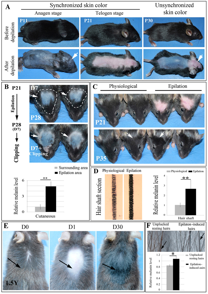
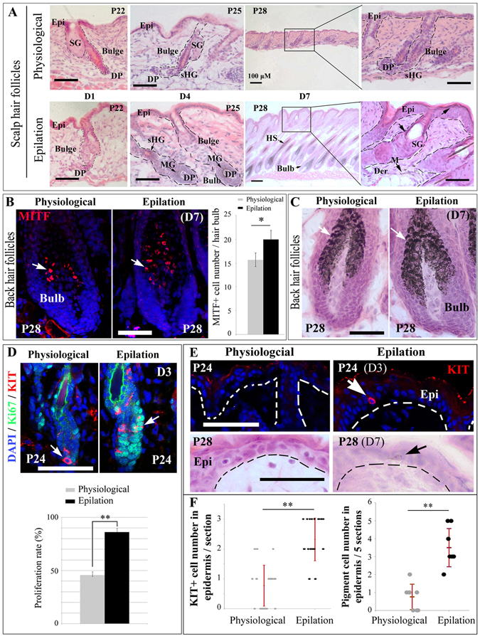
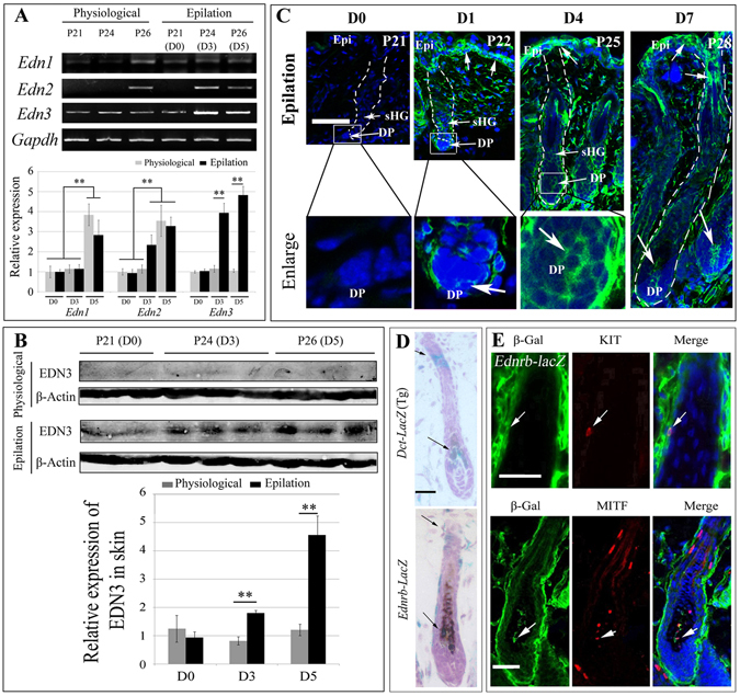
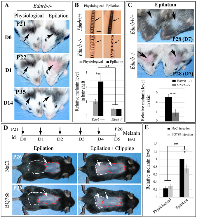
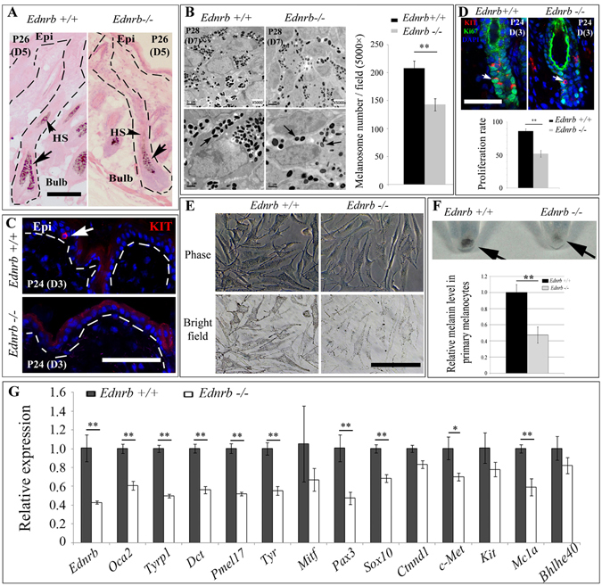
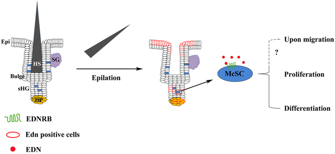
In response to various types of injury, melanocyte stem cells (McSCs) located in the bulge of hair follicles can regenerate mature melanocytes for hair and skin pigmentation. How McSCs respond to injury, however, remains largely unknown. Here we show that after epilation of mice, McSCs regenerate follicular and epidermal melanocytes, resulting in skin and hair hyperpigmentation. We further show that epilation leads to endogenous EDN3 upregulation in the dermal papilla, the secondary hair germ cells, and the epidermis. Genetic and pharmacological disruption of the EDN3 receptor EDNRB in vivo significantly blocks the effect of epilation on follicular and epidermal melanocyte regeneration as well as skin and hair hyperpigmentation. Taken together, these results indicate that epilation induces McSCs activation through EDN3/EDNRB signaling and in turn leads to skin and hair hyperpigmentation. The findings suggest that EDN/EDNRB signaling may serve as a potential therapeutic target to promote repigmentation in hypopigmentation disorders.


Conflict of interest statement

The authors declare that they have no competing interests.

Figures

Figure 1
Figure 1
Epilation-induced skin and hair hyperpigmentation in C57BL/6 mice. (A) Images of control mice at the indicated ages before (upper panels) and immediately after (lower panels) hair clipping to reveal skin pigmentation. Note that during the first postnatal hair cycle, the colors of scalp and back skin differ. Arrows point to different color of scalp skin at P11 and P30. (B) Scalp of mice epilated at P21 and observed at P28 before (upper panels) or after clipping along the white dotted line (lower panels). Arrows point to the skin color of the clipped area. (C) Scalp hair pigmentation after epilation at P21. Note hyperpigmentation 14 days after epilation. Arrows indicate the different color between physiological hairs and epilation-induced regenerated hairs. (D) P35 scalp hair shafts and melanin levels from control mice and mice epilated at P21. (E) Back hairs of a 1.5 year-old mouse before and after epilation. Arrows indicate the change of hair color before and after epilation. (F) Back hair shafts (upper panel) and melanin levels (lower panel) from the epilated area and the surrounding area of a 1.5 year-old mouse 30 days after epilation. Arrows indicate the different color between physiological hairs and epilation-induced regenerated hairs. *Indicates p < 0.05, **indicates p < 0.01.
Figure 2
Figure 2
Epilation stimulates McSC proliferation, leads to regeneration of epidermal melanocytes and induces expression of melanogenesis-related genes. (A) H&E staining of distinct stages of scalp hair cycling 1, 4, and 7 days after epilation. C57BL/6 mice were epilated at P21 and the histology of hair follicles was analyzed at the indicated days. (B,C) Anti-MITF immunostaining (B) and H&E staining (C) of back hair follicles from control mice at P28 and age-matched mice epilated at P21. Arrows indicate the MITF+ cells (B) and melanin granules in the hair bulb (C). The 3D images of anti-MITF immunostaining are shown in Fig. S10. (D) Immunostaining of anti-KIT and Ki67 in back hair follicles of C57BL/6J mice 3 days after epilation. Note that epilation induces proliferation of McSCs. Arrows indicate the KIT+ cells in the hair bulge. (E) Anti-KIT immunostaining of back skin from control mice at P24 and age-matched mice epilated at P21 (upper panels) and H&E staining of back skin (lower panels) on day 7 after epilation. Note KIT-positive cells in the epidermis 3 days after epilation and pigmented melanocytes in the epidermis 7 days after epilation. Arrows indicate the KIT+ cell (upper panels) and pigmented cell (lower panels). (F) graphs show the number of Kit+ (left panel) and pigmented cells (right panel) in the epidermis 3 days and 7 days after epilation, respectively. DP, dermal papilla; Epi, epidermis; Der, dermis; M, melanocyte; MG, melanin granules; SG, sebaceous gland; sHG, secondary hair germ. Bar, 50 μm. *Indicates p < 0.05, **indicates p < 0.01.
Figure 3
Figure 3
Epilation induces expression of endothelin-3 (Edn3) in C57BL/6 J mice. (A) RT-PCR and qRT-PCR analysis for Edns expression changes in skin after epilation. Mice were epilated at P21 and gene expression was analyzed at the indicated days. Full-length gels are shown in Fig. S11. (B) Western blotting analysis for protein expression of EDN3 in the skin. Full-length gels are shown in Fig. S12. (C) Immunofluorescence of EDN3 on scalp skin at 0, 1, 4, and 7 days after epilation. Arrows indicate EDN3+ cells in the hair follicles. (D) X-gal staining of hair follicles from Dct-lacZ transgenic mice (upper panels) and Ednrb lacZ/+ mice (lower panels). Arrows indicate the lacZ+ cells in the hair bulges and bulbs. (E) Immunohistochemistry of β-Gal, KIT, and MITF in pigmented hair follicles from postnatal day 7 Ednrb lacZ/+ mice. Arrows indicate KIT and β-Gal co-labeled cells in the bulge (upper panels) and MITF and β-Gal co-labeled cells in the hair bulb (lower panels). Epi, epidermis; DP, dermal papilla; sHG, secondary hair germ. Bar: 50 μm. **Indicates p < 0.01.
Figure 4
Figure 4
Genetic and pharmacological disruption of Ednrb blocks epilation-induced hair and skin hyperpigmentation. (A) Hair pigmentation of Ednrb−/− mice before and after epilation at P21. Arrows point to the epilated (right panels) and unepilated (left panels) pigmented hairs of Ednrb−/− scalp. (B) Histological section of hairs (upper panels) and melanin content (lower panel) of the hair shafts of wildtype and Ednrb−/− mice before and after epilation. Arrows indicate the decrease of melanin in the hair shaft of Ednrb−/− mice. (C) Pigmentation of P28 scalp of wildtype and Ednrb−/− mice after epilation at P21. Arrows point to the epilated area of wildtype (upper panels) and Ednrb−/− mice (lower panels). (D, E) Back skin of P26 mice epilated at P21 and intracutaneously injected with either physiological saline (upper panels) or BQ788 (lower panels) on the days indicated by the vertical arrows (D). The white arrows point to the skin color of epilated and clipped areas. (E) The graph shows relative melanin levels of the back skin. Note that on day 5 after epilation along the red dotted line, clipping along the white dotted line shows the surrounding hairs as controls for physiological regeneration of hair follicular pigmentation. id, intradermal injection. *indicates p < 0.05; **indicates p < 0.01.
Figure 5
Figure 5
Loss of EDNRB blocks McSC migration into epidermis, reduces melanocyte proliferation in hair bulb and decreases expression of some melanogenesis-related genes. (A) Histology of scalp hair follicles from wildtype and Ednrb−/− mice 5 days post-epilation. Arrows indicate melanin granules in the hair bulb and pigmented hair bulb. (B) Melanosomes of wildtype and Ednrb−/− mice in the hair bulbs 7 days post-epilation revealed by transmission electron microscopy (TEM). Arrows indicate the melanosome. (C) Anti-KIT immunostaining of scalp from wildtype and Ednrb−/− mice 3 days after epilation. Note lack of KIT-positive cells in Ednrb−/− epidermis 3 days after epilation. (D) Anti-KIT and Ki67 immunostaining of hair follicles from wildtype and Ednrb−/− mice 3 days post-epilation (upper panels) and the quantitative graph of melanocyte proliferation in the hair bulb (lower panel). (E) Images of primary cultures of melanocytes from wildtype and Ednrb−/− mice and (F) corresponding cell pellets (upper panel) and melanin content (lower panel). Arrows point to the cell pellet. (G) qRT-PCR analysis for expression of melanogenesis-related genes in scalp skin of wildtype and Ednrb−/− mice 7 days after epilation. Epi, epidermis; HS, hair shaft. Bar, 50 μm. *Indicates p < 0.05; **indicates p < 0.01.
Figure 6
Figure 6
Schematic illustration of the role of EDN/EDNRB signaling in epilation-induced skin and hair pigmentation. After epilation, expression of EDNs is increased in epidermis, secondary hair germ and dermal papilla. McSCs respond to the increased EDNs secreted from surrounding cells, resulting in activation of the EDNRB signaling pathway. As a result, McSCs proliferate, likely migrate along the hair follicles into epidermis and differentiate into mature melanocytes for melanogenesis. Epi, epidermis; DP, dermal papilla; HS, hair shaft; sHG, secondary hair germ.

Similar articles

Cited by

References

    1. Saldana-Caboverde A, Kos L. Roles of endothelin signaling in melanocyte development and melanoma. Pigment cell & melanoma research. 2010;23:160–170. doi: 10.1111/j.1755-148X.2010.00678.x. - DOI - PMC - PubMed
    1. Hou L, Pavan WJ. Transcriptional and signaling regulation in neural crest stem cell-derived melanocyte development: do all roads lead to Mitf? Cell research. 2008;18:1163–1176. doi: 10.1038/cr.2008.303. - DOI - PubMed
    1. Yamaguchi, Y. & Hearing, V. J. Melanocytes and their diseases. Cold Spring Harbor perspectives in medicine 4, doi:10.1101/cshperspect.a017046 (2014). - PMC - PubMed
    1. Nishimura EK, et al. Dominant role of the niche in melanocyte stem-cell fate determination. Nature. 2002;416:854–860. doi: 10.1038/416854a. - DOI - PubMed
    1. Nishimura EK. Melanocyte stem cells: a melanocyte reservoir in hair follicles for hair and skin pigmentation. Pigment cell & melanoma research. 2011;24:401–410. doi: 10.1111/j.1755-148X.2011.00855.x. - DOI - PubMed

Publication types