. 2017 Aug 4;7(1):7282.
doi: 10.1038/s41598-017-07304-7.

cAMP receptor protein regulates mouse colonization, motility, fimbria-mediated adhesion, and stress tolerance in uropathogenic Proteus mirabilis

Affiliations

cAMP receptor protein regulates mouse colonization, motility, fimbria-mediated adhesion, and stress tolerance in uropathogenic Proteus mirabilis

Yi-Lin Tsai et al. Sci Rep. .

Abstract

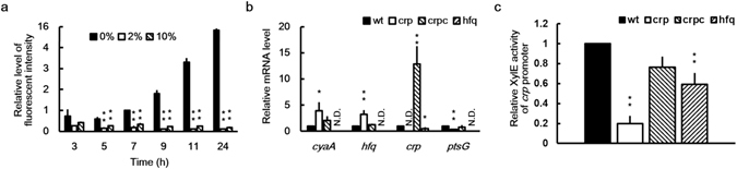
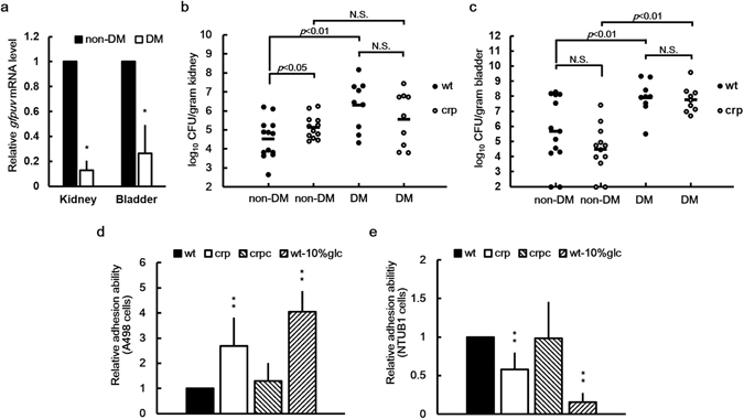
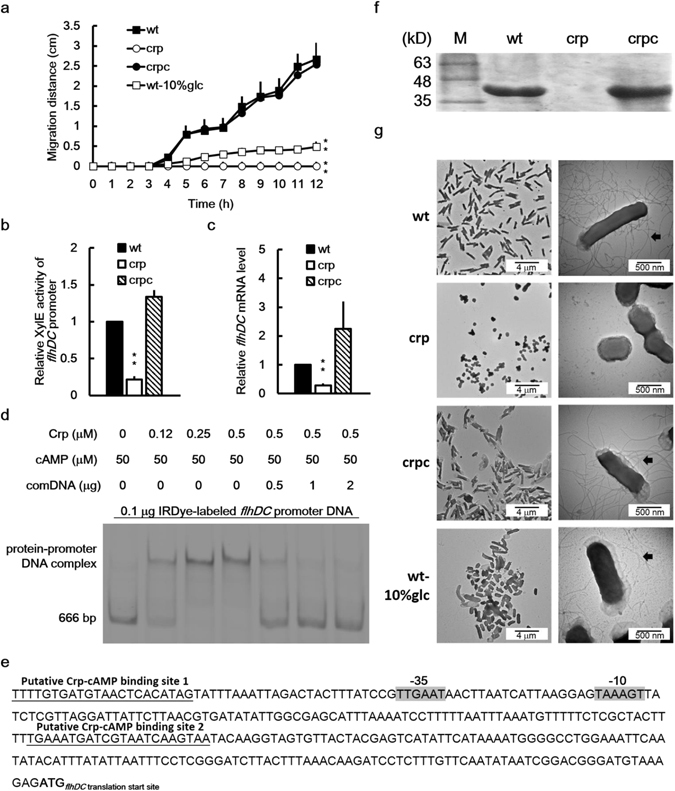
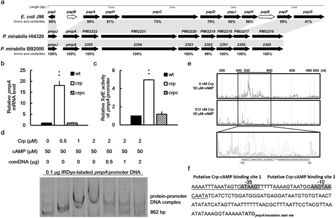
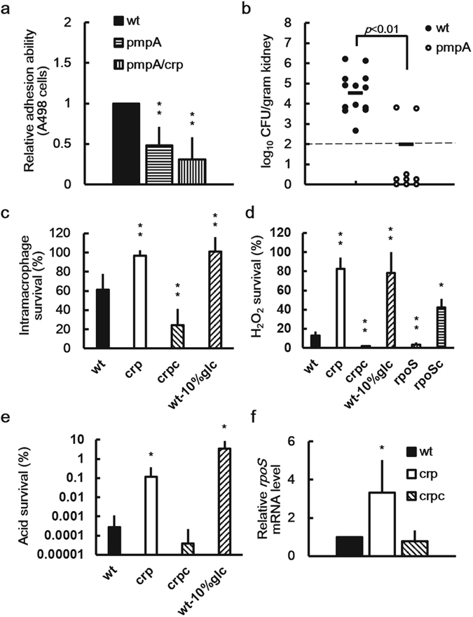
Cyclic AMP receptor protein (Crp) is a major transcriptional regulator in bacteria. This study demonstrated that Crp affects numerous virulence-related phenotypes, including colonization of mice, motility, fimbria-mediated adhesion, and glucose stress tolerance in uropathogenic Proteus mirabilis. Diabetic mice were more susceptible to kidney colonization by wild-type strain than nondiabetic mice, in which the crp mutant exhibited increased kidney colonization. Loss of crp or addition of 10% glucose increased the P. mirabilis adhesion to kidney cells. Direct negative regulation of pmpA (which encodes the major subunit of P-like fimbriae) expression by Crp was demonstrated using a reporter assay and DNase I footprinting. Moreover, the pmpA/crp double mutant exhibited reduced kidney adhesion comparable to that of the pmpA mutant, and mouse kidney colonization by the pmpA mutant was significantly attenuated. Hence, the upregulation of P-like fimbriae in the crp mutant substantially enhanced kidney colonization. Moreover, increased survival in macrophages, increased stress tolerance, RpoS upregulation, and flagellum deficiency leading to immune evasion may promote kidney colonization by the crp mutant. This is the first study to elucidate the role of Crp in the virulence of uropathogenic P. mirabilis, underlying mechanisms, and related therapeutic potential.


Conflict of interest statement

The authors declare that they have no competing interests.

Figures

Figure 1
Figure 1
Crp activity was inhibited by high glucose levels; cyaA, hfq, ptsG, and crp itself were regulated by Crp, and crp was regulated by Hfq in P. mirabilis. (a) Crp activity increased in the absence of glucose and decreased by 2% and 10% (2000 and 10,000 mg/dL) glucose. The plac-gfpuv reporter plasmid-transformed wild-type P. mirabilis was grown in LB broth with or without glucose, and the fluorescent intensity of GFPuv was monitored after incubation for 3, 5, 7, 9, 11, and 24 h. The fluorescent intensity in the absence of glucose at 7 h was set at 1, and all other data are expressed relative to this value after being normalized against the OD600 value. The data are the averages and standard deviations of three independent experiments. The significant difference between the presence and absence of glucose at each time point was determined using the Student’s t test (**P < 0.01). (b) Crp regulated the expression of cyaA, hfq, and ptsG mRNA, and crp expression was regulated by Hfq in P. mirabilis. Overnight bacterial cultures were diluted in LB broth and incubated for 7 h before total RNA was prepared for measuring mRNA levels of cyaA, hfq, crp, and ptsG in the wild-type strain, crp mutant, crp-complemented strain, or hfq mutant by using real-time RT-PCR. The mRNA level for the wild-type strain was set at 1, and all other data are expressed relative to this value. N.D., not determined. (c) crp promoter activities of wild-type P. mirabilis, crp mutant, crp-complemented strain, and hfq mutant. The activities of XylE in the crp-xylE reporter plasmid-transformed P. mirabilis strains were determined using the reporter assay at 7 h after incubation. The value obtained for the wild-type strain was set at 1. In (b) and (c), the data are the averages and standard deviations of three independent experiments. Significant difference from the wild-type strain is indicated with an asterisk (*P < 0.05; **P < 0.01 by using Student’s t test). wt, wild-type; crp, crp mutant; crpc, crp-complemented strain; hfq, hfq mutant.
Figure 2
Figure 2
Crp activity, urinary tract colonization, and urothelial cell adhesion of P. mirabilis were affected by the diabetic environment, loss of crp or the high glucose level. (a) Crp activity of P. mirabilis was decreased in the kidneys and bladders of diabetic mice. Six-week-old female mice were injected transurethrally with overnight cultures of the plac-gfpuv reporter plasmid-transformed wild-type P. mirabilis strains (107 CFU per mouse). On day 3 after injection, the mice were sacrificed, and bladder and kidney samples were collected to quantify the mRNA content of gfpuv using real-time RT-PCR. The mRNA level for each organ of nondiabetic mice was set at 1. The data are the averages and standard deviations of three independent experiments. The significant difference between nondiabetic mice (non-DM) and diabetic mice (DM) was observed using Student’s t test (*P < 0.05). Kidney (b) and bladder (c) colonization in DM and non-DM by wild-type P. mirabilis (wt) or crp mutant (crp). Diabetic or nondiabetic ICR mice (at least nine mice per group) were inoculated transurethrally with overnight cultures of bacteria at a dose of 107 CFU per mouse. Bacterial loads (CFU) in the kidneys and bladders were determined on day 3 after inoculation. Horizontal bars indicate the average for each group, and the limit of detection was 100 CFU/g organ. Significant differences were determined using the Mann–Whitney test, except for the difference between colonization by the wild-type strain and crp mutant in the kidneys of nondiabetic mice as determined using the Student’s t test. N.S., no significant difference. Adhesion of wild-type in the absence (wt) or presence of 10% glucose (wt-10%glc), the crp mutant (crp), and the crp-complemented strain (crpc) to kidney (d) and bladder (e) epithelial cells. Adhesion abilities to A498 cells and NTUB1 cells were determined as described in the Materials and Methods section. The adhesion ability of the wild-type strain was set at 1 and other data are relative to this value. The data represent the averages and standard deviations of three independent experiments. The significant difference from the wild-type strain is indicated with an asterisk (**P < 0.01, using Student’s t test).
Figure 3
Figure 3
Loss of crp or high glucose reduced swarming, production of flagellin/flagella and flhDC expression in P. mirabilis. (a) Swarming migration of the wild-type in the absence or presence of 10% glucose, crp mutant and the crp-complemented strain. An aliquot (5 μl) of overnight cultures was inoculated centrally onto the swarming plate. The migration distance was measured hourly after inoculation. The significant difference from wild-type is indicated with an asterisk at 12 h. (b) Promoter activities of flhDC in wild-type, crp mutant, and crp-complemented strain. The activities of XylE in the flhDC-xylE reporter plasmid-transformed bacterial strains were determined using the reporter assay at 7 h after incubation. The value obtained for the wild-type was set at 1. (c) The flhDC mRNA levels in the wild-type, crp mutant, and crp-complemented strain. The flhDC mRNA amounts were quantified using real-time RT-PCR at 7 h after incubation. The value obtained for the wild-type was set at 1. In (a),(b) or (c), the data represent the averages and standard deviations of three independent experiments. Significant difference from the wild-type is indicated with an asterisk (**P < 0.01 by Student’s t test). (d) The binding of P. mirabilis Crp-cAMP to the flhDC promoter region revealed using an EMSA. IRDye-labeled DNA fragments (0.1 μg) of the flhDC promoter region (666 bp) obtained by PCR were incubated with the purified His-tagged Crp (0–0.5 μM) in the presence of cAMP. The protein-DNA complex was resolved on a 5% non-denaturing polyacrylamide gel and the gel image was obtained by the quantitative infrared fluorescent imaging system. The unlabeled flhDC promoter DNA acted as a competitor to verify the binding specificity. comDNA, competitive DNA fragments. (e) A diagram showing the Crp-cAMP binding sites upstream of flhDC gene. The putative Crp-cAMP binding sequences are underlined and the putative −10 and −35 promoter sequences of sigma 70 are shadowed. (f) The flagellin levels of wild-type, crp mutant and crp-complemented strain. Flagellin levels were examined after seeding on the swarming plates for 4 h by the SDS-PAGE. The band of flagellin is 40 kD. M, molecular weight marker. (g) TEM pictures of wild-type in the absence or presence of 10% glucose, crp mutant and the crp-complemented strain. Bacterial cultures were applied onto a carbon-coated grid, cells were stained with 1% PTA and TEM pictures were taken. Flagella are indicated by arrows. In (a–c,f and g), wt, wild-type; wt-10%glc, wild-type with 10% glucose; crp, crp mutant; crpc, crp-complemented strain. Full-length gels for (d) and (f) are shown in Supplementary Fig. S8.
Figure 4
Figure 4
Regulation of pmpA expression by P. mirabilis Crp-cAMP. (a) The P-like fimbrial gene locus (pmp) similar to the well characterized pap locus in UPEC J96 in P. mirabilis genome. An amino acid sequence analysis of the pmp locus in P. mirabilis HI4320 (accession no. AM942759) and its counterparts in E. coli J96 (accession no. ALIN02000070) and P. mirabilis BB2000 (accession no. NC_022000) was performed using position-specific iterative BLAST. The nine proteins of the pmp locus in P. mirabilis HI4320 or BB2000 are similar to the PapI, PapA, PapH, PapC, PapD, PapJ, PapK, PapF and PapG in E. coli J96 with corresponding genes in shadows. The percent amino acid similarities between P. mirabilis HI4320 and E. coli J96 or P. mirabilis BB2000 were shown below each gene. The white arrows represent genes that are not found in either P. mirabilis HI4320 or P. mirabilis BB2000. (b) Loss of crp increased the pmpA mRNA level. The pmpA mRNA levels of the wild-type, crp mutant, and crp-complemented strain were quantified using real-time RT-PCR at 24 h after incubation. The value obtained for the wild-type was set at 1. (c) Loss of crp increased the promoter activity of pmpA. The activities of XylE in the pmpA-xylE reporter plasmid-transformed wild-type, crp mutant, and crp-complemented strain were determined using the reporter assay at 7 h after incubation. The value obtained for the wild-type was set at 1. In (b and c), the data represent the averages and standard deviations of three independent experiments. The significant difference from the wild-type was determined by Student’s t test (**P < 0.01). wt, wild-type; crp, crp mutant; crpc, crp-complemented strain. (d) The binding of P. mirabilis Crp-cAMP to pmpA promoter region revealed using an EMSA. The EMSA was performed as described in Fig. 3d, except that IRDye-labeled pmpA promoter region DNA fragments (862 bp) were incubated with 0–2 μM of the purified His-tagged Crp. (e) Identification of the Crp-cAMP binding site in the pmpA promoter region by a DNase I footprinting assay. FAM was used to label the pmpA promoter DNA fragment and the DNA fragment was incubated with cAMP (50 μM) with or without the recombinant Crp (512 nM) followed by DNase I treatment. The mixture was subject to electrophoresis. The fluorescence intensity of the FAM-labeled DNA fragment (ordinate) was plotted against the sequence length of the fragment. Two Crp-cAMP binding sites (underlined) located between −95 and −142 upstream of pmpA start codon were shown in an expanded view. (f) A diagram showing the Crp-cAMP binding sites upstream of pmpA gene. The putative Crp-cAMP binding sequences are underlined and the putative −10 and −35 promoter sequences of sigma 70 are shadowed. The full-length gel for (d) is shown in Supplementary Fig. S8.
Figure 5
Figure 5
Loss of pmpA abolished the increased kidney adhesion of crp mutant and decreased kidney colonization, and crp mutant displayed enhanced stress tolerance and upregulation of RpoS. (a) Adhesion of wild-type P. mirabilis, pmpA mutant or pmpA/crp double mutant to A498 cells. Adhesion abilities were determined as in Fig. 2. (b) Mouse kidney colonization by the wild-type or pmpA mutant. Colonization was determined as in Fig. 2b. (c) Intramacrophage survival of the wild-type in the absence or presence of 10% glucose, crp mutant and crp-complemented strain. THP-1 cells were infected with bacteria for 30 min at an MOI of 10. Intramacrophage survival was determined as the percentage of viable bacteria that survived in the macrophages for 4 h versus those for 1 h after outside bacteria were killed with streptomycin. (d) Survival in H2O2 of wild-type in the absence or presence of 10% glucose, crp mutant, crp-complemented strain, rpoS mutant and rpoS-complemented strain. Overnight cultures were regrown and adjusted to a density of 108 cells/ml before cells were exposed to 30 mM H2O2 for 20 min at 37 °C. The percent cell survival was determined by colony counting relative to the untreated control. (e) Survival in acid of wild-type in the absence or presence of 10% glucose, crp mutant and crp-complemented strain. Overnight bacterial cultures were regrown before cells were challenged with acid (pH 3.0) for 2 h. The percent cell survival was obtained as in (d). (f) Loss of crp increased the rpoS mRNA level. The mRNA amounts in the wild-type, crp mutant and crp-complemented strain were quantified sing real-time RT-PCR at 3 h after incubation. The value obtained for the wild-type was set at 1. In (a,cf), the data represent the averages and standard deviations of three independent experiments. The significant difference from the wild-type was determined by Student’s t test (*P < 0.05; **P < 0.01). wt, wild-type; pmpA, pmpA mutant; crp, crp mutant; pmpA/crp, pmpA/crp double mutant; crpc, crp-complemented strain; wt-10%glc, wild-type with 10% glucose; rpoS, rpoS mutant; rpoSc, rpoS-complemented strain.

Similar articles

Cited by

References

    1. Shimada T, Fujita N, Yamamoto K, Ishihama A. Novel roles of cAMP receptor protein (CRP) in regulation of transport and metabolism of carbon sources. PLoS One. 2011;6:e20081. doi: 10.1371/journal.pone.0020081. - DOI - PMC - PubMed
    1. Gosset G, Zhang Z, Nayyar S, Cuevas WA, Saier MH. Transcriptome analysis of Crp-dependent catabolite control of gene expression in Escherichia coli. J. Bacteriol. 2004;186:3516–3524. doi: 10.1128/JB.186.11.3516-3524.2004. - DOI - PMC - PubMed
    1. Zheng D, Constantinidou C, Hobman JL, Minchin SD. Identification of the CRP regulon using in vitro and in vivo transcriptional profiling. Nucleic Acids Res. 2004;32:5874–5893. doi: 10.1093/nar/gkh908. - DOI - PMC - PubMed
    1. Green J, et al. Cyclic-AMP and bacterial cyclic-AMP receptor proteins revisited: adaptation for different ecological niches. Curr. Opin. Microbiol. 2014;18:1–7. doi: 10.1016/j.mib.2014.01.003. - DOI - PMC - PubMed
    1. Heroven AK, Dersch P. Coregulation of host-adapted metabolism and virulence by pathogenic yersiniae. Front. Cell Infect. Microbiol. 2014;4:146. doi: 10.3389/fcimb.2014.00146. - DOI - PMC - PubMed

Publication types

MeSH terms