. 2017 Aug 4;7(1):7326.
doi: 10.1038/s41598-017-06469-5.

CDK5 Regulatory Subunit-Associated Protein 1-like 1 Negatively Regulates Adipocyte Differentiation through Activation of Wnt Signaling Pathway

Affiliations

CDK5 Regulatory Subunit-Associated Protein 1-like 1 Negatively Regulates Adipocyte Differentiation through Activation of Wnt Signaling Pathway

Kazumi Take et al. Sci Rep. .

Abstract

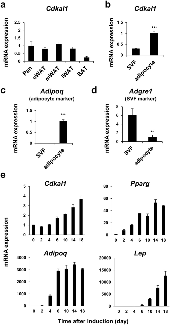
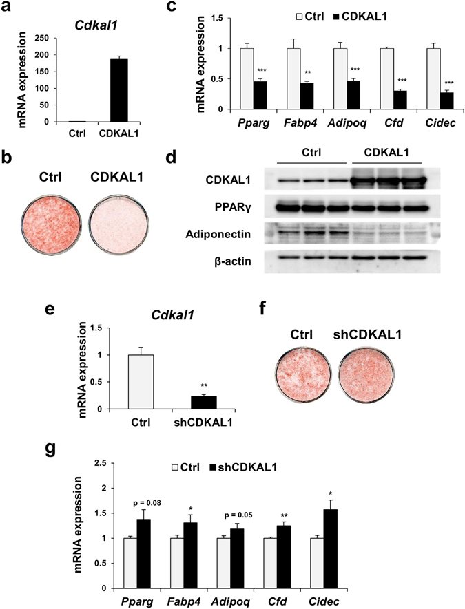
CDK5 Regulatory Subunit-Associated Protein 1-like 1 (CDKAL1) was identified as a susceptibility gene for type 2 diabetes and body mass index in genome-wide association studies. Although it was reported that CDKAL1 is a methylthiotransferase essential for tRNALys(UUU) and faithful translation of proinsulin generated in pancreatic β cells, the role of CDKAL1 in adipocytes has not been understood well. In this study, we found that CDKAL1 is expressed in adipose tissue and its expression is increased during differentiation. Stable overexpression of CDKAL1, however, inhibited adipocyte differentiation of 3T3-L1 cells, whereas knockdown of CDKAL1 promoted differentiation. CDKAL1 increased protein levels of β-catenin and its active unphosphorylated form in the nucleus, thereby promoting Wnt target gene expression, suggesting that CDKAL1 activated the Wnt/β-catenin pathway-a well-characterized inhibitory regulator of adipocyte differentiation. Mutant experiments show that conserved cysteine residues of Fe-S clusters of CDKAL1 are essential for its anti-adipogenic action. Our results identify CDKAL1 as novel negative regulator of adipocyte differentiation and provide insights into the link between CDKAL1 and metabolic diseases such as type 2 diabetes and obesity.


Conflict of interest statement

The authors declare that they have no competing interests.

Figures

Figure 1
Figure 1
Expression levels of CDKAL1 in adipocytes. (a) Expression levels of Cdkal1 in tissues of C57BL/6 J mice. eWAT, epididymal white adipose tissue (WAT); mWAT, mesenchymal WAT; iWAT, inguinal WAT; BAT, brown adipose tissue; Panc, pancreas (n = 4 [BAT], 7 [Pancreas] or 8 [(others]). Expression of Cdkal1 (b), Adipoq (c) and Adgre1 (d) in the stromal vascular fraction (SVF) and adipocytes fraction isolated from eWAT of C57BL/6 J mice (n = 4, **p < 0.01, ***p < 0.001 vs SVF group by Student’s t-test). (e) Gene expression levels during adipocyte differentiation of 3T3-L1 cells: upper left, Cdkal1; upper right, Pparg; lower left, Adipoq; lower right, Lep (n = 3).
Figure 2
Figure 2
CDKAL1 suppresses adipocyte differentiation. (ad) Overexpression of CDKAL1 in 3T3-L1 adipocytes. (a) Expression levels of Cdkal1 mRNA levels. (b) Plate view of Oil Red O staining (day 7). (c) Expression levels of adipocyte specific genes (n = 3, *p < 0.05, **p < 0.01, ***p < 0.001 vs control group by Student’s t-test). (d) Western blotting of CDKAL1, PPARγ, adiponectin and β-actin. (eg) Knockdown of CDKAL1 by using short hairpin RNA (shRNA) targeting CDKAL1 (shCDKAL1) in 3T3-L1 adipocytes. (e) Expression levels of Cdkal1 mRNA levels. (f) Plate view of Oil Red O staining. (g) Expression levels of adipocyte specific genes (n = 3, *p < 0.05, **p < 0.01 vs control group by Student’s t-test).
Figure 3
Figure 3
Suppression of PPARγ expression by CDKAL1 contributes to the anti-adipogenic action of CDKAL1. (a,b) Time course of gene expression levels of early adipogenic regulators, C/EBPβ and C/EBPδ (a) and late regulators, PPARγ and C/EBPα (b) during adipocyte differentiation of 3T3-L1 cells expressing CDKAL1 (n = 3). (c,d) Effect of PPARγ agonist pioglitazone on lipid accumulation (c) and gene expression (d) in differentiated 3T3-L1 cells expressing CDKAL1 (n = 3). (e,f) Effect of coexpression of PPARγ on lipid accumulation (e) and gene expression (f) in differentiated 3T3-L1 cells expressing CDKAL1 (n = 3, **p < 0.005, ***p < 0.0005 vs control group by Dunnett’s test).
Figure 4
Figure 4
Activation of Wnt/β -catenin signaling pathway by CDKAL1. (a) Western blotting of β-catenin in 3T3-L1 expressing CDKAL1 on day 0 and day 6 of differentiation. β-actin was used as an internal control for protein loading. The experiments were repeated multiple times and representative blot was shown. (b) Western blotting of active (unphosphorylated) β-catenin in the nuclear fraction in 3T3-L1 expressing CDKAL1. (c,d) The effect of stable knockdown of CDKAL1 by shRNA in 3T3-L1 cells on protein levels of active β-catenin in total lysate (c) and in the nuclear fraction (d). The inset graph in (d) shows quantification of the active β-catenin (*p < 0.05 vs control group by Student’s t-test). (e) Western blotting of β-catenin and inactive form of GSK-3β (phosphorylated at Ser 9) in 3T3-L1 expressing CDKAL1. (f) Measurement of Wnt signaling activities by TOPFLASH reporter assay in 3T3-L1 cells. FOPFLASH reporter was used as the negative control (n = 6, *p < 0.05 vs control group by Student’s t-test). (g) Gene expression changes of Wnt target genes in 3T3-L1 cells expressing CDKAL1 (12 hours after the initiation of differentiation, n = 3, ***p < 0.001 vs control group by Student’s t-test).
Figure 5
Figure 5
Knockdown of β-catenin compromised the suppression of expression of adipogenic genes by CDKAL1. Gene expression levels of (a) CDKAL1 and β-catenin (Ctnnb) and (b) adipogenic marker genes in 3T3-L1 adipocytes coexpressing CDKAL1 and shRNA targeting β-catenin (n = 3, *p < 0.05, **p < 0.01, ***p < 0.001 vs control group by Student’s t-test; #p < 0.05, ##p < 0.01 vs CDKAL1 group by Student’s t-test).
Figure 6
Figure 6
Conserved cysteine residues in the amino terminal region of CDKAL1 are essential for its anti-adipogenic action. (a) Diagram of constructs used in the experiment. The numbers indicate the amino acid residues from the N-terminus of CDKAL1. (b) Plate view of Oil Red O staining of differentiated 3T3-L1 cells expressing CDKAL1 with point mutations (Ctrl, control). (c) Gene expression levels in differentiated 3T3-L1 cells expressing CDKAL1 with the point mutations (n = 3, *p < 0.025, **p < 0.005, ***p < 0.0005 vs control group by Dunnett’s test). (d) Western-blotting analysis of β-catenin protein in differentiated 3T3-L1 adipocytes expressing CDKAL1 with the point mutations. (e) Measurement of tRNALys(UUU) modification index in 3T3-L1 cells expressing shCDKAL1 (*p < 0.05, **p < 0.01 vs control group by Student’s t-test) and in 3T3-L1 cells expressing wild type CDKAL1 (n = 3).

Similar articles

Cited by

References

    1. Steinthorsdottir V, et al. A variant in CDKAL1 influences insulin response and risk of type 2 diabetes. Nat. Genet. 2007;39:770–5. doi: 10.1038/ng2043. - DOI - PubMed
    1. Zeggini E, et al. Replication of genome-wide association signals in UK samples reveals risk loci for type 2 diabetes. Science. 2007;316:1336–41. doi: 10.1126/science.1142364. - DOI - PMC - PubMed
    1. Scott LJ, et al. A genome-wide association study of type 2 diabetes in Finns detects multiple susceptibility variants. Science. 2007;316:1341–5. doi: 10.1126/science.1142382. - DOI - PMC - PubMed
    1. Saxena R, et al. Genome-wide association analysis identifies loci for type 2 diabetes and triglyceride levels. Science. 2007;316:1331–6. doi: 10.1126/science.1142358. - DOI - PubMed
    1. Okada Y, et al. Common variants at CDKAL1 and KLF9 are associated with body mass index in east Asian populations. Nat. Genet. 2012;44:302–6. doi: 10.1038/ng.1086. - DOI - PMC - PubMed

Publication types