. 2017 Aug 4;7(1):7343.
doi: 10.1038/s41598-017-07566-1.

Epigenetic regulation of starvation-induced autophagy in Drosophila by histone methyltransferase G9a

Affiliations

Epigenetic regulation of starvation-induced autophagy in Drosophila by histone methyltransferase G9a

Phan Nguyen Thuy An et al. Sci Rep. .

Abstract

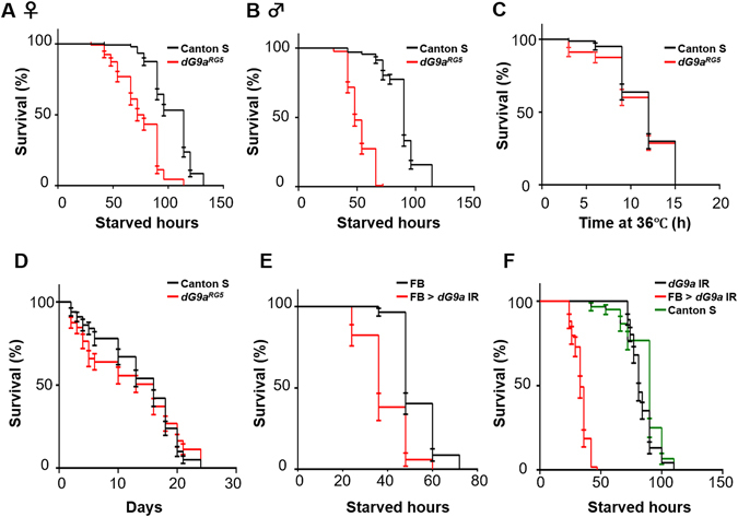
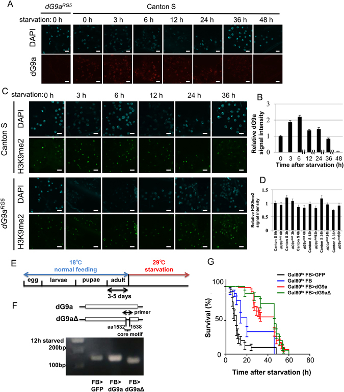
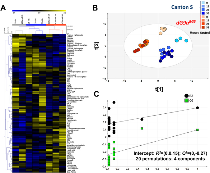
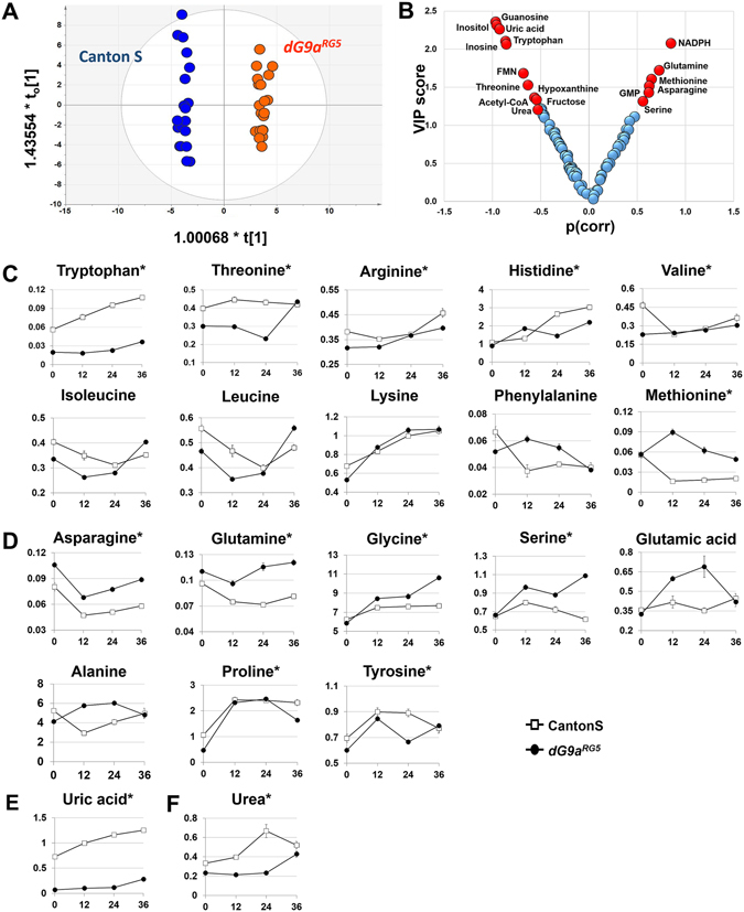
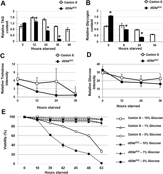
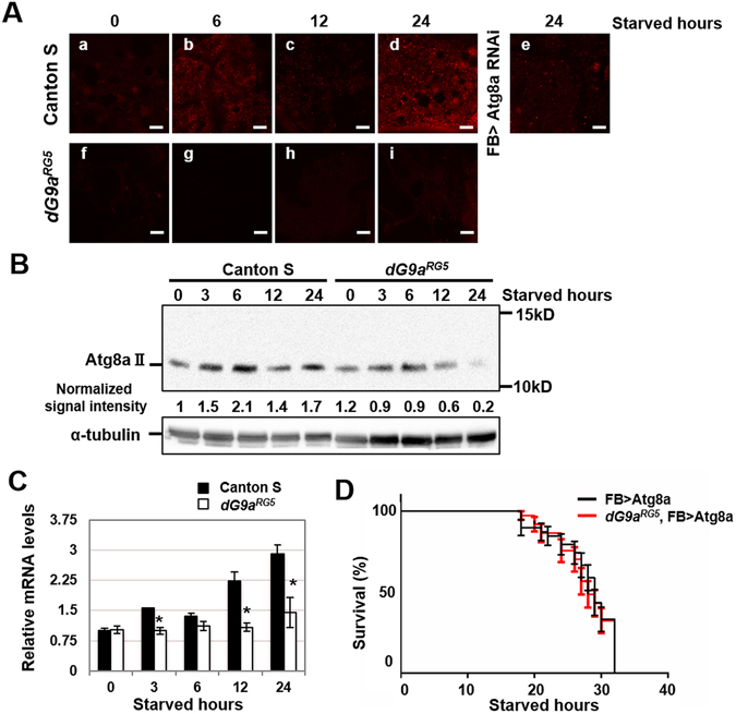
Epigenetics is now emerging as a key regulation in response to various stresses. We herein identified the Drosophila histone methyltransferase G9a (dG9a) as a key factor to acquire tolerance to starvation stress. The depletion of dG9a led to high sensitivity to starvation stress in adult flies, while its overexpression induced starvation stress resistance. The catalytic domain of dG9a was not required for starvation stress resistance. dG9a plays no apparent role in tolerance to other stresses including heat and oxidative stresses. Metabolomic approaches were applied to investigate global changes in the metabolome due to the loss of dG9a during starvation stress. The results obtained indicated that dG9a plays an important role in maintaining energy reservoirs including amino acid, trehalose, glycogen, and triacylglycerol levels during starvation. Further investigations on the underlying mechanisms showed that the depletion of dG9a repressed starvation-induced autophagy by controlling the expression level of Atg8a, a critical gene for the progression of autophagy, in a different manner to that in cancer cells. These results indicate a positive role for dG9a in starvation-induced autophagy.


Conflict of interest statement

The authors declare that they have no competing interests.

Figures

Figure 1
Figure 1
dG9a is critical for survival under starvation stress. (A) The results of a viability assay under starvation conditions using females of the wild-type (Canton S) (n = 153 from 8 independent experiments) and dG9a null mutant (dG9aRG5) (n = 135 from 7 independent experiments) P < 0.0001 (B). The results of a viability assay under starvation conditions using males of the wild-type (n = 131 from 7 independent experiments) and dG9a null mutant (dG9aRG5) (n = 138 from 7 independent experiments) P < 0.0001. (C) The results of a viability assay under heat stress using males of the wild-type (n = 80 from 4 independent experiments) and dG9a null mutant (dG9aRG5) (n = 80 from 4 independent experiments) P = 0.41. (D) The results of a viability assay under oxidative stress using males of the wild-type (n = 100 from 5 independent experiments) and dG9a null mutant (dG9aRG5) (n = 97 from 5 independent experiments) P = 0.80. (E) The results of a viability assay under starvation conditions using males of the FB (+; FB-GAL4; +) (n = 57 from 3 independent experiments) and FB > dG9a IR (w; UAS-dG9a IR/FB-GAL4; +) (n = 34 from 2 independent experiments) P < 0.0001. (F) The results of a viability assay under starvation conditions using males of the FB > dG9a IR (w; UAS-dG9a IR/FB-GAL4; +) (n = 59 from 3 independent experiments), males of the dG9a IR (w; UAS-dG9a IR/+; +) (n = 92 from 5 independent experiments) and males of Canton S (n = 60 from 3 independent experiments). dG9a IR and Canton S: P > 0.05. (AF) Error bars represent standard errors (SE).
Figure 2
Figure 2
dG9a function in the fat body is critical for adult fly viability under starvation stress. (A) Transient induction of dG9a in the adult fat body under starvation conditions. Fat bodies of the wild-type and dG9aRG5 mutant were immunostained with an anti-dG9a antibody (red) and stained with DAPI (cyan). The dG9aRG5 mutant was used as a negative control. (B) Quantification of dG9a signals in nuclei of the wild-type fat body. Values were adjusted by subtracting background fluorescence n = 10. (C) Quantification of H3K9me2 signals in nuclei of the wild-type and dG9aRG5 mutant fat bodies. Values were adjusted by subtracting background fluorescence (n = 10). (D) H3K9me2 signal in the adult fat body under starvation conditions. Fat bodies of the wild-type and dG9aRG5 mutant were immunostained with an anti-H3K9me2 antibody (green) and stained with DAPI (cyan). (E) The experimental design of dG9a overexpression under starvation conditions. (F) Confirmation of dG9a and dG9aΔ1532–1538 overexpression by semi-quantitative RT-PCR after 12 h of starvation. The primer included the core motif of the SET domain of dG9a. dG9a was more strongly expressed in FB > dG9a (FB-GAL4/GAL80ts.αTub84B; UAS-dG9a/+) than in FB > GFP (a control: FB-GAL4/GAL80ts.αTub84B; UAS-GFP/+). The amplicon size is 142 bp in FB > dG9a and FB > GFP. The amplicon size of FB > dG9aΔ(FB-GAL4/GAL80ts.αTub84B; UAS-dG9aΔ1532–1538/+) is 21 bp shorter because of the lack of the core motif of the SET domain. The full-length gel image is shown in Fig. S6A. (G) The results of a viability assay under starvation conditions using males of the control (GAL80ts FB > GFP: FB-GAL4/+; GAL80ts.αTub84B/UAS-GFP) (n = 40 from 2 independent experiments), another control (GAL80ts FB: FB-GAL4/+; GAL80ts.αTub84B/+) (n = 39 from 2 independent experiments), a dG9a overexpression line (GAL80ts FB > dG9a: FB-GAL4/+; GAL80ts.αTub84B/UAS-dG9a) (n = 58 from 3 independent experiments), and dG9aΔ1532–1538 overexpression line (GAL80ts FB > dG9aΔ: FB-GAL4/+; GAL80ts.αTub84B/UAS-dG9aΔ1532–1538) (n = 40 from 2 independent experiments). Significant differences were observed between GAL80ts FB > GFP and GAL80ts FB > dG9a (P < 0.0001) and between GAL80ts FB > GFP and GAL80ts FB > dG9aΔ (P < 0.0001). Significant differences were also noted between GAL80ts FB and GAL80ts FB > dG9a (P < 0.0001) and between GAL80ts FB and GAL80ts FB > dG9aΔ (P < 0.0001). No significant differences were found between GAL80ts FB > dG9a and GAL80ts FB > dG9aΔ (P = 0.23). (B,C,G) Error bars represent SE.
Figure 3
Figure 3
Fasted dG9aRG5 mutant flies show a distinct metabolic profile. (A) HCA data showing changes in the cellular metabolites of wild-type and dG9aRG5 mutant flies under starvation; the color scale is plotted on the top of the figure. Before starvation, flies with both genotypes shared similar profiles and were grouped into one cluster (grey). During starvation, flies with each genotype showed different profiles and were discriminated hierarchically into two clusters, wild-type (blue) and dG9aRG5 mutant (red). A similar discrimination based on genotypes was observed on the score plot of the supervised analysis PLS-DA (B). In each genotype, samples collected before fasting were clearly separated from samples collected during fasting. (C) Statistical validation by the permutation test with 20 permutations of PLS-DA (R2Y-intercepts = (0,0.15); Q2-interceps = (0, −0.27)).
Figure 4
Figure 4
Amino acid metabolism. A general view of the metabolites detected belonging to amino acids including: (A) The OPLS-DA score plot showed a clear separation between two genotypes with p(CV-ANOVA) = 1.99925E-23. (B) A plot formed by VIP scores and p(corr) revealed using OPLS-DA. The criteria VIP > 1.0 and |p(corr)|>0.5 were used to select potential metabolites (red) showing marked changes due to the loss of dG9a. (C) Essential amino acids (D) Non-essential amino acids (E) Uric acid (F) Urea. 4–5 replications for each time point. *The metabolites were significantly different in “genotype” between the wild-type and dG9aRG5 mutant in a two-way ANOVA with P < 0.05.
Figure 5
Figure 5
The loss of dG9a affects energy homeostasis in Drosophila during fasting. (A) Relative levels of TAG in the wild-type and dG9aRG5 mutant under starvation stress n = 3. *P < 0.05. (B) Relative levels of glycogen in the wild-type and dG9aRG5 mutant under starvation stress n = 5. *P < 0.05. The levels of trehalose (C) and glucose (D) from the GC-MS analysis. Only the level of trehalose showed a significantly different “genotype” between the wild-type and dG9aRG5 mutant in a two-way ANOVA with P = 9.17E-03. (E) The results of a viability assay with glucose supply (n = 40). When dG9aRG5 mutant flies were fed 1% and 10% glucose in PBS, they recovered their sensitivity and had similar viability to the wild-type.
Figure 6
Figure 6
dG9a is responsible for the induction of autophagy under starvation stress. (A) Immunostaining of wild-type and dG9aRG5 mutant fat bodies under starvation conditions with an anti-Atg8a antibody. Strains: (a–d) Canton S (e) w; FB-GAL4/+; Atg8aHMS01328/+ (f–i) dG9aRG5. Starved hours: (a,f) 0 h (b,g) 6 h (c,h) 12 h (d,i) 24 h. Scale bars, 10 μm. (B) A western blot analysis of extracts from the starved wild-type and dG9aRG5 mutant. Blots were probed with anti-Atg8a and anti-α-tubulin antibodies. Signal intensity normalized with that of α-tubulin is shown. The full-length image of the blot is shown in Fig. S6B. (C) Quantification of mRNA levels by a RT-qPCR analysis of Atg8a in the starved wild-type and dG9aRG5 mutant. Results were normalized to α-tubulin and displayed as relative values to that of the 0-h starved wild-type n = 3. *P < 0.05. (D) The results of a viability assay under starvation conditions using the males of dG9aRG5, FB > Atg8a (dG9aRG5; FB-GAL4/Atg8aScer/UAS.P/T.T:Avic/GFP-EGFP,T:Disc/RFP-mCherry; +) (n = 39 from 2 independent experiments) and FB > Atg8a (+; FB-GAL4/Atg8aScer/UAS.P/T.T:Avic/GFP-EGFP,T:Disc/RFP-mCherry; +) (n = 38 from 2 independent experiments) strains P = 0.79. (C,D) Error bars represent SE.

Similar articles

Cited by

References

    1. Stankiewicz AM, Swiergiel AH, Lisowski P. Epigenetics of stress adaptations in the brain. Brain Res Bull. 2013;98:76–92. doi: 10.1016/j.brainresbull.2013.07.003. - DOI - PubMed
    1. Skinner MK, Manikkam M, Guerrero-Bosagna C. Epigenetic transgenerational actions of environmental factors in disease etiology. Trends Endocrinol Metab. 2010;21:214–222. doi: 10.1016/j.tem.2009.12.007. - DOI - PMC - PubMed
    1. Heijmans BT, et al. Persistent epigenetic differences associated with prenatal exposure to famine in humans. Proc Natl Acad Sci USA. 2008;105:17046–17049. doi: 10.1073/pnas.0806560105. - DOI - PMC - PubMed
    1. Slade JD, Staveley BE. Extended longevity and survivorship during amino-acid starvation in a Drosophila Sir2 mutant heterozygote. Genome. 2016;59:311–318. doi: 10.1139/gen-2015-0213. - DOI - PubMed
    1. Artal-Martinez de Narvajas A, et al. Epigenetic regulation of autophagy by the methyltransferase G9a. Mol Cell Biol. 2013;33:3983–3993. doi: 10.1128/MCB.00813-13. - DOI - PMC - PubMed

Publication types

Substances

LinkOut - more resources