

Shape dependent cytotoxicity of PLGA-PEG nanoparticles on human cells
- PMID: 28779154
- PMCID: PMC5544670
- DOI: 10.1038/s41598-017-07588-9
Shape dependent cytotoxicity of PLGA-PEG nanoparticles on human cells
Abstract
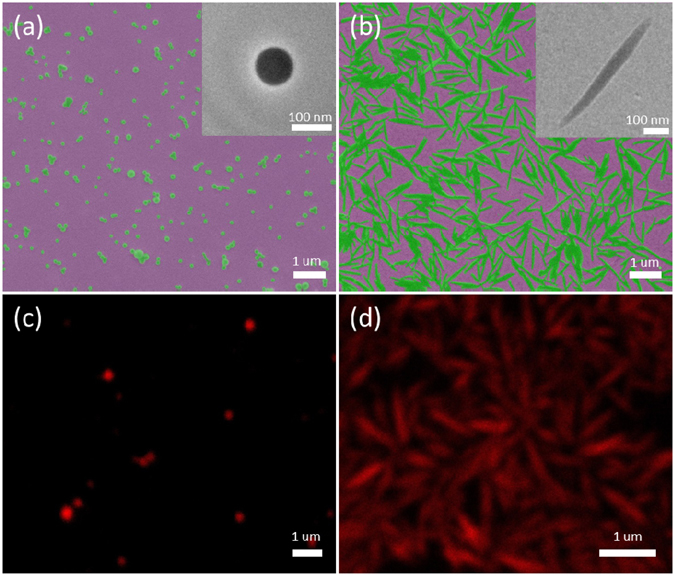
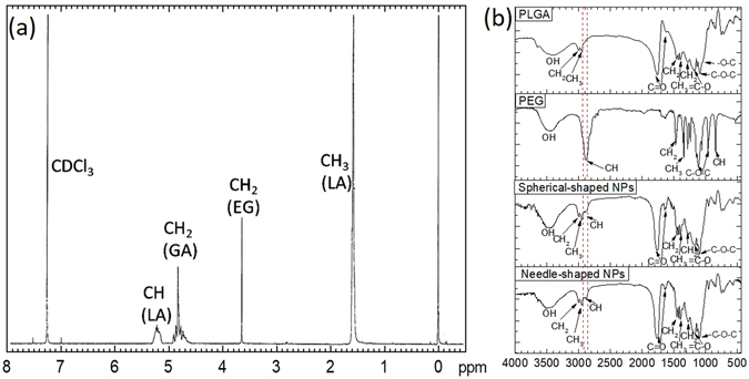
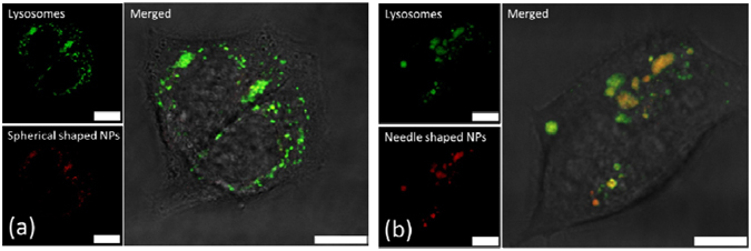
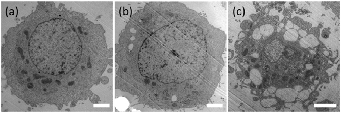
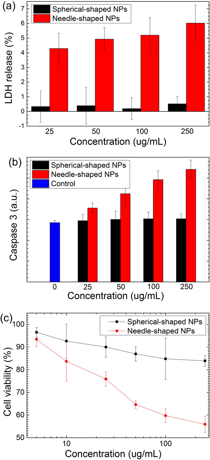
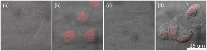
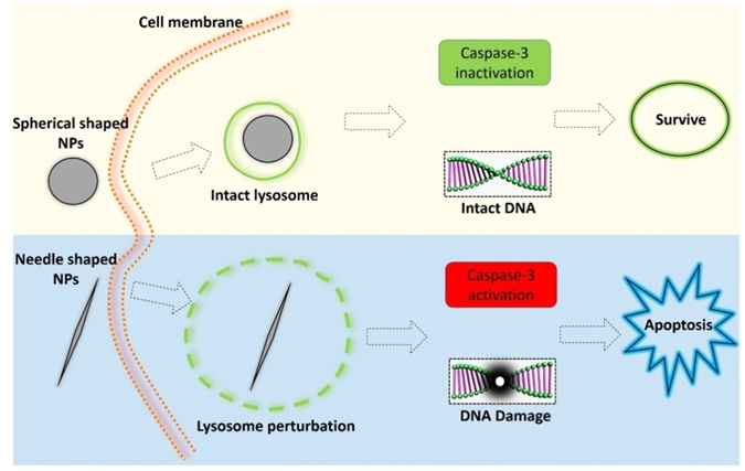
We investigated the influence of nanoparticles' shape on the physiological responses of cells, when they were fed with spherical and needle-shaped PLGA-PEG nanoparticles (the volume of the nanoparticles had been chosen as the fixed parameter). We found that both types of NPs entered cells via endocytosis and upon internalization they stayed in membrane bounded vesicles. Needle-shaped, but not the spherical-shaped NPs were found to induce significant cytotoxicity in the cell lines tested. Our study evidenced that the cytotoxicity of needle-shaped NPs was induced through the lysosome disruption. Lysosome damage activated the signaling pathways for cell apoptosis, and eventually caused DNA fragmentation and cell death. The present work showed that physiological response of the cells can be very different when the shape of the fed nanoparticles changed from spherical to needle-like. The finding suggests that the toxicity of nanomaterials also depends on their shape.
Conflict of interest statement
The authors declare that they have no competing interests.
Figures
Similar articles
-
Development and characterization of hyaluronic acid modified PLGA based nanoparticles for improved efficacy of cisplatin in solid tumor.Biomed Pharmacother. 2017 Nov;95:856-864. doi: 10.1016/j.biopha.2017.08.108. Epub 2017 Sep 10. Biomed Pharmacother. 2017. PMID: 28903181
-
Biocompatibility Assessment of Polyethylene Glycol-Poly L-Lysine-Poly Lactic-Co-Glycolic Acid Nanoparticles In Vitro and In Vivo.J Nanosci Nanotechnol. 2015 May;15(5):3710-9. doi: 10.1166/jnn.2015.9509. J Nanosci Nanotechnol. 2015. PMID: 26504996
-
Enhanced Antitumor Efficacy and Reduced Toxicity of Docetaxel Loaded Estradiol Functionalized Stealth Polymeric Nanoparticles.Mol Pharm. 2015 Nov 2;12(11):3871-84. doi: 10.1021/acs.molpharmaceut.5b00281. Epub 2015 Oct 1. Mol Pharm. 2015. PMID: 26375023
-
Enhanced anti-proliferative and pro-apoptotic effects of metformin encapsulated PLGA-PEG nanoparticles on SKOV3 human ovarian carcinoma cells.Artif Cells Nanomed Biotechnol. 2019 Dec;47(1):737-746. doi: 10.1080/21691401.2019.1573737. Artif Cells Nanomed Biotechnol. 2019. PMID: 30892093
-
Docetaxel-loaded nanoparticles based on star-shaped mannitol-core PLGA-TPGS diblock copolymer for breast cancer therapy.Acta Biomater. 2013 Nov;9(11):8910-20. doi: 10.1016/j.actbio.2013.06.034. Epub 2013 Jun 28. Acta Biomater. 2013. PMID: 23816645
Cited by
-
Poly(lactic-co-glycolic acid) nanoparticle fabrication, functionalization, and biological considerations for drug delivery.Biomicrofluidics. 2024 Sep 17;18(5):051503. doi: 10.1063/5.0201465. eCollection 2024 Sep. Biomicrofluidics. 2024. PMID: 39296325 Free PMC article. Review.
-
Size, shape, and flexibility influence nanoparticle transport across brain endothelium under flow.Bioeng Transl Med. 2019 Dec 26;5(2):e10153. doi: 10.1002/btm2.10153. eCollection 2020 May. Bioeng Transl Med. 2019. PMID: 32440560 Free PMC article.
-
Magnesium-ion-doped silica nanosheets as degradable drug carriers with enhanced antibacterial activity and cellular uptake.RSC Adv. 2025 Jan 31;15(5):3183-3191. doi: 10.1039/d4ra07626e. eCollection 2025 Jan 29. RSC Adv. 2025. PMID: 39896430 Free PMC article.
-
Targeting Glucose Metabolism in Cancer Cells as an Approach to Overcoming Drug Resistance.Pharmaceutics. 2023 Nov 10;15(11):2610. doi: 10.3390/pharmaceutics15112610. Pharmaceutics. 2023. PMID: 38004589 Free PMC article. Review.
-
Degradable Polymeric Bio(nano)materials and Their Biomedical Applications: A Comprehensive Overview and Recent Updates.Polymers (Basel). 2024 Jan 10;16(2):206. doi: 10.3390/polym16020206. Polymers (Basel). 2024. PMID: 38257005 Free PMC article. Review.
References
Publication types
MeSH terms
Substances
LinkOut - more resources
Full Text Sources
Other Literature Sources
