. 2017 Aug 4;7(1):7295.
doi: 10.1038/s41598-017-07821-5.

Dysfunctional high-density lipoproteins have distinct composition, diminished anti-inflammatory potential and discriminate acute coronary syndrome from stable coronary artery disease patients

Affiliations

Dysfunctional high-density lipoproteins have distinct composition, diminished anti-inflammatory potential and discriminate acute coronary syndrome from stable coronary artery disease patients

Mihaela G Carnuta et al. Sci Rep. .

Abstract

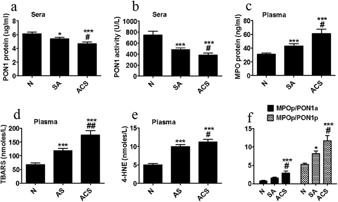
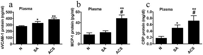
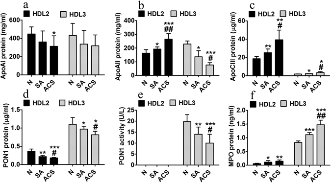
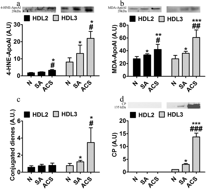
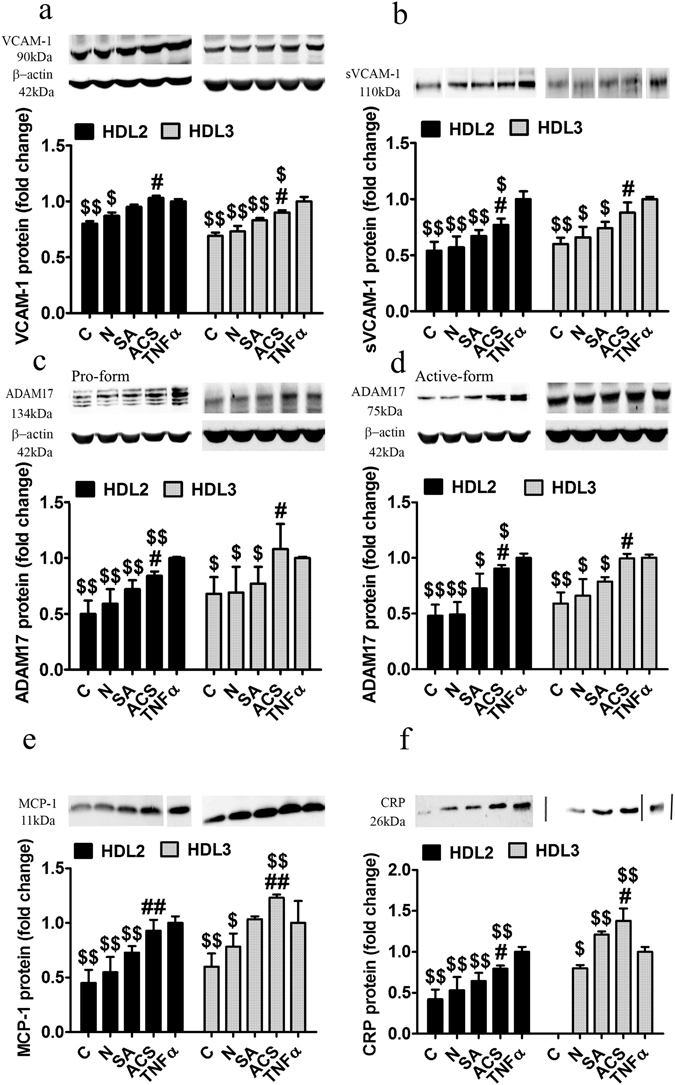
There is a stringent need to find means for risk stratification of coronary artery diseases (CAD) patients. We aimed at identifying alterations of plasma high-density lipoproteins (HDL) components and their validation as dysfunctional HDL that could discriminate between acute coronary syndrome (ACS) and stable angina (SA) patients. HDL2 and HDL3 were isolated from CAD patients' plasma and healthy subjects. ApolipoproteinAI (apoAI), apoAII, apoCIII, malondialdehyde (MDA), myeloperoxidase (MPO), ceruloplasmin and paraoxonase1 (PON1) were assessed. The anti-inflammatory potential of HDL subfractions was tested by evaluating the secreted inflammatory molecules of tumor necrosis factor α-activated endothelial cells (EC) upon co-incubation with HDL2 or HDL3. We found in ACS versus SA patients: 40% increased MPO, MDA, apoCIII in HDL2 and HDL3, 35% augmented apoAII in HDL2, and in HDL3 increased ceruloplasmin, decreased apoAII (40%) and PON1 protein and activity (15% and 25%). Co-incubation of activated EC with HDL2 or HDL3 from CAD patients induced significantly increased levels of secreted inflammatory molecules, 15-20% more for ACS versus SA. In conclusion, the assessed panel of markers correlates with the reduced anti-inflammatory potential of HDL subfractions isolated from ACS and SA patients (mostly for HDL3 from ACS) and can discriminate between these two groups of CAD patients.


Conflict of interest statement

The authors declare that they have no competing interests.

Figures

Figure 1
Figure 1
Biochemical parameters measured in sera/plasma of healthy subjects (N), stable angina patients (SA) and acute coronary syndrome patients (ACS). Levels of serum paraoxonase 1 (PON1) protein (a) and paraoxonase activity (b), plasma myeloperoxidase (MPO) protein (c), thiobarbituric reactive substances (TBARS) (d), free 4-hydroxy-2-nonenal (4-HNE) (e), and MPO protein/PON1 activity (MPOp/PON1a) and MPO protein/PON1 protein (MPOp/PON1p) ratios (f). Data are expressed as means ± SEM and analyzed with two-tailed Oneway ANOVA with Least Significant Difference (LSD) Post-hoc test; *p < 0.05, ***p < 0.001 vs. N; #p < 0.05, ##p < 0.01 vs SA.
Figure 2
Figure 2
Inflammatory parameters measured in plasma of healthy subjects (N), stable angina patients (SA) and acute coronary syndrome patients (ACS). Levels of soluble vascular cell adhesion molecule 1 (sVCAM-1) (a), monocyte chemoattractant protein 1 (MCP-1) (b) and C reactive protein (CRP) (c). Data are expressed as means ± SEM and analyzed with two-tailed Oneway ANOVA with Least Significant Difference (LSD) Post-hoc test; *p < 0.05, **p < 0.01 vs. N; #p < 0.05 vs. SA.
Figure 3
Figure 3
Characterization of HDL2 and HDL3 subfractions isolated by density gradient ultracentrifugation from seven independent pools of sera collected from healthy subjects (N), stable angina patients (SA) and acute coronary syndrome (ACS) patients. Levels of apolipoprotein AI (ApoAI) (a), ApoAII (b), ApoCIII (c), paraoxonase 1 (PON1) protein (d) and paraoxonase activity (e), and myeloperoxidase (MPO) protein (f). Data are expressed as means ± SD and analyzed with Independent Student’s T-test; *p < 0.05, **p < 0.01, ***p < 0.001 vs. N, #p < 0.05, ##p < 0.01 vs. SA.
Figure 4
Figure 4
Characterization of oxidative markers in HDL2 and HDL3 subfractions isolated by density gradient ultracentrifugation from seven independent pools of sera from healthy subjects (N), stable angina patients (SA) and acute coronary syndrome (ACS) patients. Levels of 4-hydroxy-2-nonenal associated to apolipoprotein AI (4-HNE-apoAI) (a), malondialdehyde associated to apolipoprotein AI (MDA-apoAI) (b), conjugated dienes (c) and ceruloplasmin (CP) (d). The western blot images were processed by cropping the corresponding parts from the blots. The full-length blots are supplied in the Supplementary information. Data are expressed as means ± SD and analyzed with Independent Student’s T-test; *p < 0.05, **p < 0.01, ***p < 0.001 vs. N, #p < 0.05, ##p < 0.01 vs. SA.
Figure 5
Figure 5
HDL2 and HDL3 functionality evaluated by their anti-inflammatory effects on endothelial cells (EC) activated with tumor necrosis factor α (TNFα). The vascular cell adhesion molecule 1 (VCAM-1) protein relative to β-actin (a) and soluble VCAM-1 (sVCAM-1) in the cultured media relative to cellular protein (b); protein expression of disintegrin and metalloproteinase domain-containing protein 17 (ADAM17) pro-form (c) and active form (d) relative to β-actin. Levels of monocyte chemoattractant protein 1 (MCP-1) (e) and C reactive protein (CRP) (f) secreted in the culture medium relative to cellular protein. All data come from four different experiments and are expressed as fold change versus TNFα-activated EC. C - cells unstimulated with TNFα, N - healthy subjects, SA - stable angina patients, ACS - acute coronary syndrome patients, TNFα - tumor necrosis factor α. The western blot (wb) images for (b), (e) and (f) were processed by cropping the lanes from the same blot and marked by dividing white lines where the order of the samples on wb was different from the one in the graph. The full-length blots are supplied in the Supplementary information. Data are presented as means ± SD and analyzed with Independent Student’s T-test. #p < 0.05, ##p < 0.01 vs. SA, $p < 0.05, $$p < 0.01 vs. TNFα.

Similar articles

Cited by

References

    1. Wang JC, Bennett M. Aging and atherosclerosis: mechanisms, functional consequences, and potential therapeutics for cellular senescence. Circulation research. 2012;111:245–259. doi: 10.1161/CIRCRESAHA.111.261388. - DOI - PubMed
    1. Simionescu M. Cellular dysfunction in inflammatory-related vascular disorders’ review series. The inflammatory process: a new dimension of a 19 century old story. Journal of cellular and molecular medicine. 2009;13:4291–4292. - PMC - PubMed
    1. Hafiane A, Genest J. HDL, Atherosclerosis, and Emerging Therapies. Cholesterol. 2013;2013:891403. doi: 10.1155/2013/891403. - DOI - PMC - PubMed
    1. Lim S, Park YM, Sakuma I, Koh KK. How to control residual cardiovascular risk despite statin treatment: focusing on HDL-cholesterol. International journal of cardiology. 2013;166:8–14. doi: 10.1016/j.ijcard.2012.03.127. - DOI - PubMed
    1. Camont L, Chapman MJ, Kontush A. Biological activities of HDL subpopulations and their relevance to cardiovascular disease. Trends in molecular medicine. 2011;17:594–603. doi: 10.1016/j.molmed.2011.05.013. - DOI - PubMed

Publication types