. 2017 Aug 4;7(1):7294.
doi: 10.1038/s41598-017-07876-4.

LIN-32/Atonal Controls Oxygen Sensing Neuron Development in Caenorhabditis elegans

Affiliations

LIN-32/Atonal Controls Oxygen Sensing Neuron Development in Caenorhabditis elegans

Teresa Rojo Romanos et al. Sci Rep. .

Abstract

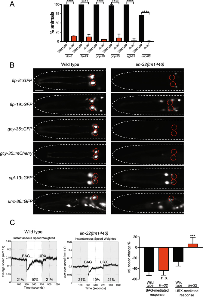
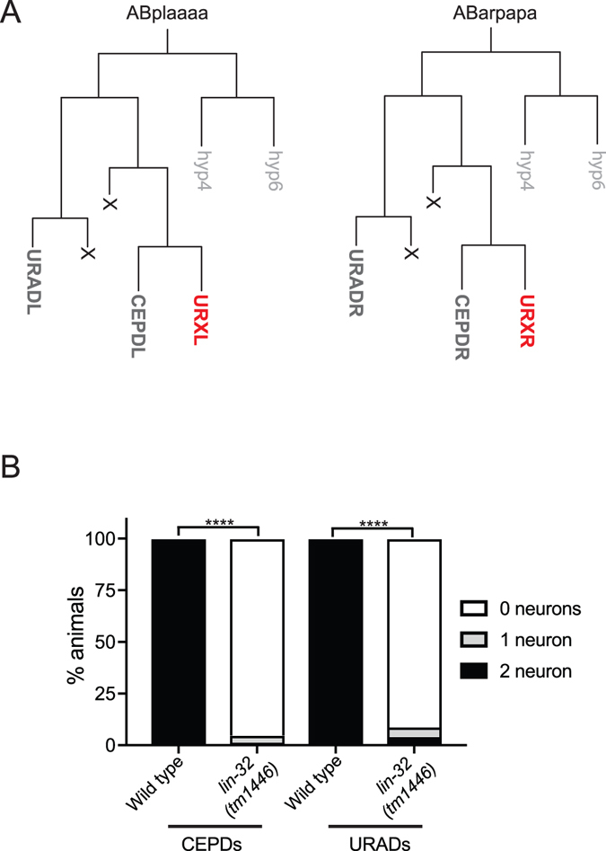
Development of complex nervous systems requires precisely controlled neurogenesis. The generation and specification of neurons occur through the transcriptional and post-transcriptional control of complex regulatory networks. In vertebrates and invertebrates, the proneural basic-helix-loop-helix (bHLH) family of transcription factors has multiple functions in neurogenesis. Here, we identified the LIN-32/Atonal bHLH transcription factor as a key regulator of URXL/R oxygen-sensing neuron development in Caenorhabditis elegans. When LIN-32/Atonal expression is lost, the expression of URX specification and terminal differentiation genes is abrogated. As such, lin-32 mutant animals are unable to respond to increases in environmental oxygen. The URX neurons are generated from a branch of the cell lineage that also produces the CEPDL/R and URADL/R neurons. We found development of these neurons is also defective, suggesting that LIN-32/Atonal regulates neuronal development of the entire lineage. Finally, our results show that aspects of URX neuronal fate are partially restored in lin-32 mutant animals when the apoptosis pathway is inhibited. This suggests that, as in other organisms, LIN-32/Atonal regulates neuronal apoptosis.


Conflict of interest statement

The authors declare that they have no competing interests.

Figures

Figure 1
Figure 1
rp1 is a lesion in lin-32 and causes URX developmental defects. (A) Quantification of rp1-induced URX defects using the flp-8::GFP reporter of URX fate. flp-8::GFP is expressed in the URX, AUA and PVM neurons. In rp1 animals, expression of flp-8::GFP is almost abolished in the URX and PVM neurons while expression in the AUA neurons is unaffected. 1URX-like and 2URX-like indicate that the neuronal morphology is similar to URX but the neuron is positioned incorrectly. N > 60. Statistical significance between wild-type and lin-32(rp1) animals was evaluated using a t-test. ****P < 0.0001. (B) Molecular identity of the rp1 allele and the previously described lin-32(tm1446) deletion allele. The rp1 lesion is a C to T transition that converts a glutamine (Q) to a premature amber stop codon. (C) Quantification of flp-8::GFP expression in the URX neurons in wild type, lin-32(tm1446) mutant and lin-32(tm1446); ezIs10(lin-32::GFP + unc-119(+) rescued animals. N > 60. Statistical significance between wild-type and lin-32(tm1446) animals was evaluated using one-way ANOVA analysis. ****P < 0.0001. Note the high number of 1URX-like and 2URX-like animals in rescued animals. These are neurons that exhibit a URX morphology but are mispositioned. We found that the lin-32::GFP rescuing transgene can cause URX mispositioning defects in wild-type animals (not shown) suggesting that dosage of LIN-32 is important for neuron position.
Figure 2
Figure 2
LIN-32 is required for URX specification and function. (A) Quantification of lin-32(tm1446)-induced URX defects in reporters for URX neuronal fate. Loss of lin-32 severely affects the expression of all URX reporters tested: flp-8, flp-19, gcy-36, gcy-35, egl-13 and unc-86. The black (wild type) and red (lin-32) bars represent the percentage of worms that show expression in either 1 or 2 URX neurons. 1-like and 2-like indicate that the neurons have a URX morphology but are mispositioned. N > 60. Statistical significance between wild-type and lin-32(tm1446) animals was evaluated using a t-test. ****P < 0.0001. (B) Micrographs of representative animals expressing fluorescent markers for the URX neurons (flp-8, flp-19, gcy-36, gcy-35, egl-13 and unc-86) in wild type and lin-32(tm1446) mutant animals. URX neuron positions are marked with red dashed circles. Anterior to the left. Ventral views except for unc-86::GFP which is a lateral view. Scale bar 20 μm. (C) lin-32 oxygen-sensing behavior analysis. Locomotion speed of wild type (left) and lin-32(tm1446) mutant animals (center) during O2 concentration shifts between 21% and 10%. The data represent averages of multiple assays. The right graph shows the quantification of changes in relative speed in response to changes in O2 concentration. lin-32(tm1446) mutants fail to respond to O2 upshifts (URX-mediated) but exhibit a similar response to wild type animals to O2 downshifts (BAG-mediated). Statistical significance between wild-type and lin-32(tm1446) animals was evaluated using one-way ANOVA analysis. ***P < 0.001; n.s., not significantly different from wild type controls. Assays were repeated at least four times using 80–120 animals per assay.
Figure 3
Figure 3
Mutations in lin-32 disturbs neuronal development in the ABplaaaa and ABarpapa lineages. (A) Lineage diagrams of ABplaaaa and ABarpapa from which the URXL and URXR neurons are generated. ABplaaaa and ABarpapa emanate two sublineages - a posterior lineage that generates two hypodermal cells (hyp4 and hyp6) and an anterior lineage that generates the URX, CEPD and URAD neurons plus two apoptotic deaths (marked with an X). (B) Quantification of dat-1::GFP (CEPD neurons) and flp-21::GFP (URAD neurons) expression in wild type and lin-32(tm1446) mutant animals. N > 65. Statistical significance between wild-type and lin-32(tm1446) animals was evaluated using a t-test. ****P < 0.0001.
Figure 4
Figure 4
The role of HAM-1, PIG-1 and HLH-2 in URX specification. (A) HAM-1 regulates URX differentiation. Quantification of gcy-36::GFP (left) and flp-8::GFP (right) expression in wild type, lin-32(tm1446), ham-1(n1438) and lin-32(tm1446); ham-1(n1438) mutant animals. N > 70. (B) PIG-1 is not required for URX specification. Quantification of gcy-36::GFP (left) and flp-8::GFP (right) expression in wild type, lin-32(tm1446), pig-1(gm344) and lin-32(tm1446); pig-1(gm344) mutant animals. N > 70. (C) HLH-2 is not required for URX specification. Quantification of gcy-36::GFP (left) and flp-8::GFP (right) expression in wild type, lin-32(tm1446), hlh-2(tm1768) and lin-32(tm1446); hlh-2(tm1768) mutant animals. N = 90. Statistical significance between wild-type and mutant strains was evaluated using one-way ANOVA analysis. ****P < 0.0001; *P < 0.01; n.s. not significantly different from control.
Figure 5
Figure 5
URX cell fate is restored by inhibiting apoptosis. (A) Quantification of flp-8::GFP expression in wild type, lin-32(tm1446), ced-3(n717), ced-3(n2452), lin-32(tm1446); ced-3(n717), lin-32(tm1446); ced-3(n2452) and ahr-1(ia3); ced-3(n717); egl-13(ku194) mutant animals. The lin-32(tm1446); ced-3 double mutants partially restore flp-8::GFP expression in the URX neurons. In contrast, in a URX specification factor mutant background the expression of flp-8::GFP cannot be restored by loss of ced-3. N > 85. Note that compound loss of ahr-1(ia3) and egl-13(ku194) causes ~95% loss of flp-8::GFP expression. (B) Quantification of gcy-35::mCherry expression in wild type, lin-32(tm1446), ced-3(n717), ced-3(n2452), lin-32(tm1446); ced-3(n717) and lin-32(tm1446); ced-3(n2452) mutant animals. N > 80. (C) Quantification of gcy-36::GFP expression in wild type, lin-32(tm1446), ced-3(n717), ced-3(n2452), lin-32(tm1446); ced-3(n717) and lin-32(tm1446); ced-3(n2452) mutant animals. N > 90. Statistical significance between wild-type and mutant strains was evaluated using one-way ANOVA analysis. ****P < 0.0001; ***P < 0.0005; **P < 0.005; n.s. not significantly different from control.

Similar articles

Cited by

References

    1. Johnston RJ, Hobert O. A microRNA controlling left/right neuronal asymmetry in Caenorhabditis elegans. Nature. 2003;426:845–849. doi: 10.1038/nature02255. - DOI - PubMed
    1. Hobert O. Regulation of terminal differentiation programs in the nervous system. Annu Rev Cell Dev Biol. 2011;27:681–696. doi: 10.1146/annurev-cellbio-092910-154226. - DOI - PubMed
    1. Powell LM, Jarman AP. Context dependence of proneural bHLH proteins. Curr Opin Genet Dev. 2008;18:411–417. doi: 10.1016/j.gde.2008.07.012. - DOI - PMC - PubMed
    1. Romani S, Campuzano S, Modolell J. The achaete-scute complex is expressed in neurogenic regions of Drosophila embryos. EMBO J. 1987;6:2085–2092. - PMC - PubMed
    1. Cabrera CV, Martinez-Arias A, Bate M. The expression of three members of the achaete-scute gene complex correlates with neuroblast segregation in Drosophila. Cell. 1987;50:425–433. doi: 10.1016/0092-8674(87)90496-X. - DOI - PubMed

Publication types