

Calcium depletion destabilises kinetochore fibres by the removal of CENP-F from the kinetochore
- PMID: 28779172
- PMCID: PMC5544769
- DOI: 10.1038/s41598-017-07777-6
Calcium depletion destabilises kinetochore fibres by the removal of CENP-F from the kinetochore
Abstract
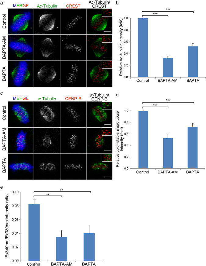
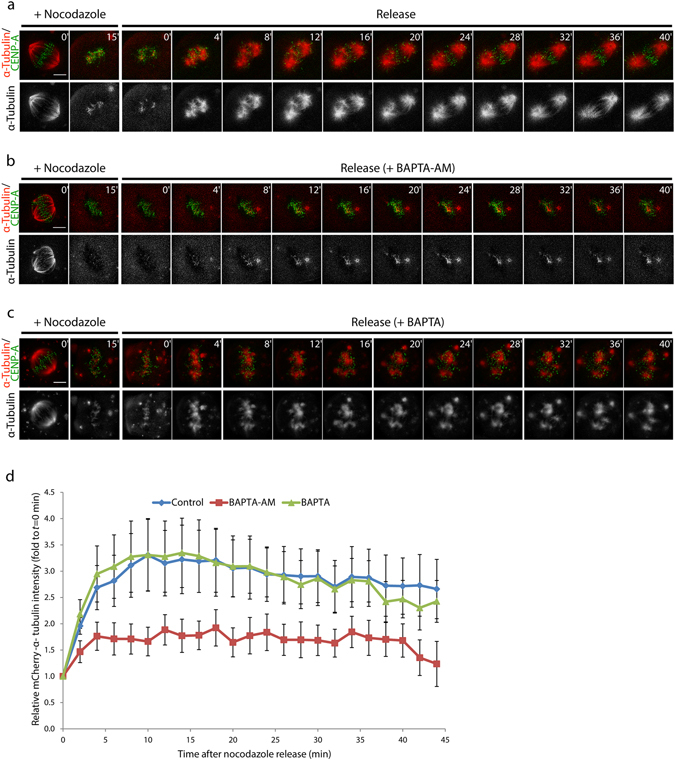
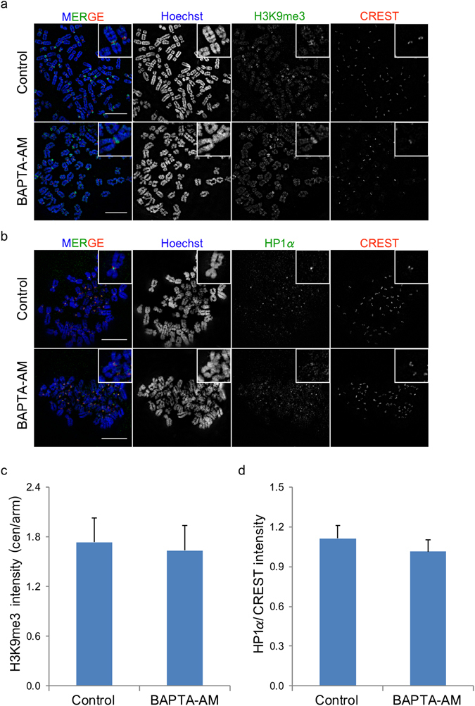
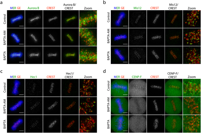
The attachment of spindle fibres to the kinetochore is an important process that ensures successful completion of the cell division. The Ca2+ concentration increases during the mitotic phase and contributes microtubule stability. However, its role in the spindle organisation in mitotic cells remains controversial. Here, we investigated the role of Ca2+ on kinetochore fibres in living cells. We found that depletion of Ca2+ during mitosis reduced kinetochore fibre stability. Reduction of kinetochore fibre stability was not due to direct inhibition of microtubule polymerisation by Ca2+-depletion but due to elimination of one dynamic component of kinetochore, CENP-F from the kinetochore. This compromised the attachment of kinetochore fibres to the kinetochore which possibly causes mitotic defects induced by the depletion of Ca2+.
Conflict of interest statement
The authors declare that they have no competing interests.
Figures
Similar articles
-
hNuf2 inhibition blocks stable kinetochore-microtubule attachment and induces mitotic cell death in HeLa cells.J Cell Biol. 2002 Nov 25;159(4):549-55. doi: 10.1083/jcb.200208159. Epub 2002 Nov 18. J Cell Biol. 2002. PMID: 12438418 Free PMC article.
-
Centromere Protein (CENP)-W Interacts with Heterogeneous Nuclear Ribonucleoprotein (hnRNP) U and May Contribute to Kinetochore-Microtubule Attachment in Mitotic Cells.PLoS One. 2016 Feb 16;11(2):e0149127. doi: 10.1371/journal.pone.0149127. eCollection 2016. PLoS One. 2016. PMID: 26881882 Free PMC article.
-
CENP-F is a novel microtubule-binding protein that is essential for kinetochore attachments and affects the duration of the mitotic checkpoint delay.Chromosoma. 2006 Aug;115(4):320-9. doi: 10.1007/s00412-006-0049-5. Epub 2006 Apr 7. Chromosoma. 2006. PMID: 16601978
-
Microtubule capture: a concerted effort.Cell. 2006 Dec 15;127(6):1105-8. doi: 10.1016/j.cell.2006.11.032. Cell. 2006. PMID: 17174890 Review.
-
A guide to classifying mitotic stages and mitotic defects in fixed cells.Chromosoma. 2018 Jun;127(2):215-227. doi: 10.1007/s00412-018-0660-2. Epub 2018 Feb 6. Chromosoma. 2018. PMID: 29411093 Review.
Cited by
-
Centromere Protein F in Tumor Biology: Cancer's Achilles Heel.Cancer Med. 2025 May;14(10):e70949. doi: 10.1002/cam4.70949. Cancer Med. 2025. PMID: 40387105 Free PMC article. Review.
-
Dissecting the role of the tubulin code in mitosis.Methods Cell Biol. 2018;144:33-74. doi: 10.1016/bs.mcb.2018.03.040. Methods Cell Biol. 2018. PMID: 29804676 Free PMC article.
References
Publication types
MeSH terms
Substances
LinkOut - more resources
Full Text Sources
Other Literature Sources
Miscellaneous
