. 2018 Feb;32(2):779-789.
doi: 10.1007/s00464-017-5740-5. Epub 2017 Aug 4.

Advanced Roux-en-Y hepaticojejunostomy with magnetic compressive anastomats in obstructive jaundice dog models

Affiliations

Advanced Roux-en-Y hepaticojejunostomy with magnetic compressive anastomats in obstructive jaundice dog models

Chao Fan et al. Surg Endosc. 2018 Feb.

Abstract

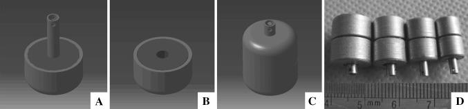
Background: Although commonly used procedure, Roux-en-Y hepaticojejunostomy (RYHJ) remains to be complicated, time consuming, and has a relatively poor prognosis. We designed the magnetic compressive anastomats (MCAs) to perform RYHJ more efficiently and safely.

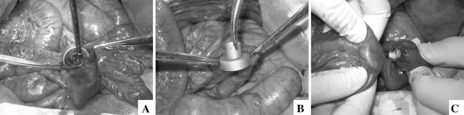
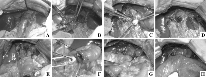
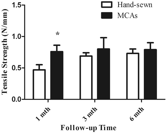
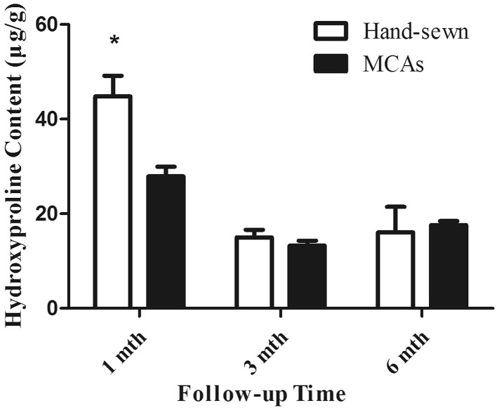
Materials and methods: 36 dogs were divided into two groups randomly. After obstructive jaundice model construction, RYHJ was performed with MCAs in study group or by hand-sewn in control group. Both groups were followed for 1, 3, and 6 months after RYHJ. The liver function and postoperative complications were recorded throughout the follow-up. At the end of each time point, dogs were sent for magnetic resonance imaging (MRI) and sacrificed. Anastomotic samples were taken for anastomotic narrowing rate calculation, histological analyses, tensile strength testing, and hydroxyproline content testing.

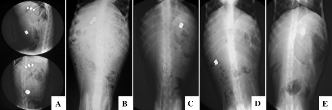
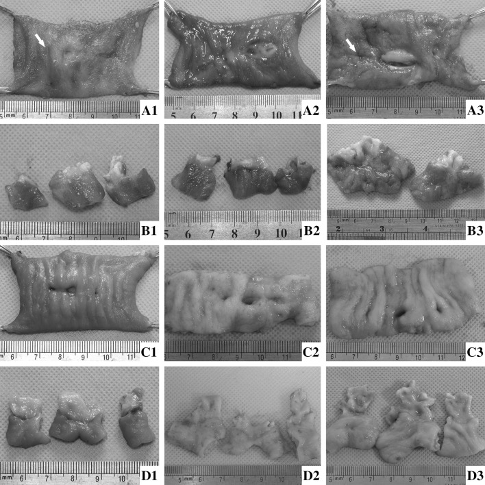
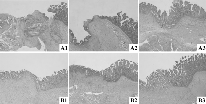
Results: The anastomotic construction times were 44.20 ± 23.02 min in study group, compared of 60.53 ± 11.89 min in control group (p < 0.05). The liver function recovered gradually after RYHJ in both groups (p > 0.05). All anastomats were expelled out of the body in 8.81 ± 2.01 days. The gross incidence of morbidity and mortality was 33.3% (6/18) and 16.7% (3/18) in study group compared with 38.9% (7/18) and 22.2% (4/18) in control group (p > 0.05), and there is no single case of anastomotic-specific complications happened in study group. The narrowing rates of anastomosis were 14.6, 18.5, and 18.7% in study group compared with 35.4, 36.9, and 34% in control group at 1st, 3rd, and 6th month after RYHJ (p < 0.05). In study group, preciser alignment of tissue layers and milder inflammatory reaction contributed to the fast and better wound healing process.

Conclusion: Perform RYHJ with MCAs is safer, more efficient than by hand-sewn method in obstructive jaundice dog models.

Keywords: Anastomat; Bilioenteric anastomosis; Hepaticojejunostomy; Magnetic compression anastomosis; Obstructive jaundice; Sutureless; Wound healing.


Conflict of interest statement

During the conduct of this work, Chao Fan, Hongke Zhang, Xiao-peng Yan, Jia Ma, ChunBao Wang, and Yi Lv have no conflicts of interest to disclose.

Figures

Fig. 1
Fig. 1
The design drawings and device of the BE-MCAs. A Mother part of BE-MCAs. B Daughter part of BE-MCAs. C Coupled BE-MCAs. D Real anastomats in different sizes
Fig. 2
Fig. 2
Enteroenterostomy with MCAs. A The daughter part of EE-MCAs was put in from the opening of distal jejunum. B The mother part of EE-MCAs was fixed at proximal end of jejunum. C Two parts of EE-MCAs were coupled together
Fig. 3
Fig. 3
Hepaticojejunostomy with MCAs. A Extrahepatic bile ducts dilated obviously after BDL. B Purse-string suturing at the end of biliary stumps. C The mother parts of BE-MCAs were inserted into the bile duct and fixed at the end of stumps successively. D Three mother parts of MCAs were fixed at hepatic duct stumps. E All guide strings passed through the jejunal wall. F Corresponding daughter part approximated to the porta hepatis through guidance. G The right and middle biliary-enteric anastomoses were constructed successfully. H Finished three anastomoses with MCAs
Fig. 4
Fig. 4
The expelling process of MCAs was monitored by X-ray. A The instant fluoroscopy showed all anastomats coupled well and retained at the right place (upper lateral view; lower anteroposterior view). B At 4th day after RYHJ, EE-MCA and the first BE-MCA were expelled out of the body, and two BE-MCAs remained in situ. C At 8th day after RYHJ, the second BE-MCA was missing, and the third one already dropped in small intestine. D At 9th day after RYHJ, the third BE-MCA was expelled at hepatic flexure of ascending colon. E At 10th day, all anastomats were expelled out of the body
Fig. 5
Fig. 5
The gross appearance of anastomoses in two groups at 1, 3, and 6th month after RYHJ. A1A3 The anastomoses were exposed from the jejunal cavity at 1, 3, and 6th month after RYHJ in control group. The white arrows in A1 and A3 were sites where anastomotic stricture happened, and the right anastomosis was totally blocked at A3. B1B3 The anastomoses were incised along the long axis, the mucosal side of anastomoses was shown at 1, 3, and 6th month after RYHJ in control group. The view of right anastomosis was absent in B3 because the lumen was blocked totally. C1C3 The anastomoses were exposed from the jejunal cavity at 1, 3, and 6th month after RYHJ in study group. D1D3 All the anastomoses were incised along the long axis, the mucosal side of anastomoses was shown at 1, 3, and 6th month after RYHJ in study group
Fig. 6
Fig. 6
Histologic section of the anastomotic sites between two groups (HE-stain, ×40). A1A3 were images of the control group at 1, 3, and 6th month after RYHJ. B1B3 were images of study group at 1, 3, and 6th month after RYHJ
Fig. 7
Fig. 7
Bar graph (mean, SD) of anastomotic tensile strength testing in two groups (Asterisk statistically significance)
Fig. 8
Fig. 8
Bar graph (mean, SD) of anastomotic hydroxyproline content testing in two groups (Asterisk statistically significance)

Similar articles

Cited by

References

    1. Laukkarinen J, Chow P, Sand J, Karkkainen P, Yu S, Somanesan S, Kee I, Song IC, Ng TH, Nordback I. Long-term changes in hepatobiliary physiology after Roux-en-Y hepaticojejunostomy. J Surg Res. 2007;143:270–275. doi: 10.1016/j.jss.2007.02.034. - DOI - PubMed
    1. Laukkarinen J, Sand J, Leppiniemi J, Kellomaki M, Nordback I. A novel technique for hepaticojejunostomy for nondilated bile ducts: a purse-string anastomosis with an intra-anastomotic biodegradable biliary stent. Am J Surg. 2010;200:124–130. doi: 10.1016/j.amjsurg.2009.05.012. - DOI - PubMed
    1. Antolovic D, Koch M, Galindo L, Wolff S, Music E, Kienle P, Schemmer P, Friess H, Schmidt J, Buchler MW, Weitz J. Hepaticojejunostomy—analysis of risk factors for postoperative bile leaks and surgical complications. J Gastrointest Surg. 2007;11:555–561. doi: 10.1007/s11605-007-0166-3. - DOI - PubMed
    1. Tanaka H, Fukuda A, Shigeta T, Kuroda T, Kimura T, Sakamoto S, Kasahara M. Biliary reconstruction in pediatric live donor liver transplantation: duct-to-duct or Roux-en-Y hepaticojejunostomy. J Pediatr Surg. 2010;45:1668–1675. doi: 10.1016/j.jpedsurg.2010.03.010. - DOI - PubMed
    1. Obora Y, Tamaki N, Matsumoto S. Non-suture micro-vascular anastomosis using magnet rings—preliminary-report. Surg Neurol. 1978;9:117–120. - PubMed

Publication types

MeSH terms

LinkOut - more resources