Review
. 2017 Aug;9(4):309-320.
doi: 10.1007/s12551-017-0279-1. Epub 2017 Aug 4.

Room-temperature ionic liquids meet bio-membranes: the state-of-the-art

Affiliations
Review

Room-temperature ionic liquids meet bio-membranes: the state-of-the-art

Antonio Benedetto. Biophys Rev. 2017 Aug.

Abstract

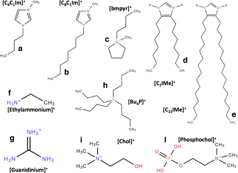
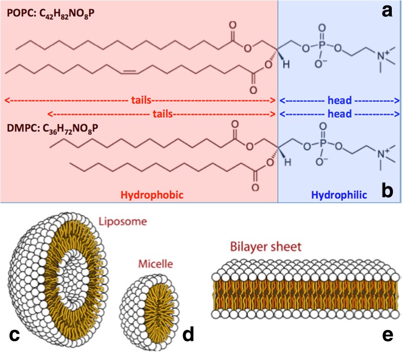
Room-temperature ionic liquids (RTIL) are a new class of organic salts whose melting temperature falls below the conventional limit of 100 °C. Their low vapor pressure, moreover, has made these ionic compounds the solvents of choice of the so-called green chemistry. For these and other peculiar characteristics, they are increasingly used in industrial applications. However, studies of their interaction with living organisms have highlighted mild to severe health hazards. Since their cytotoxicity shows a positive correlation with their lipophilicity, several chemical-physical studies of their interactions with biomembranes have been carried out in the last few years, aiming to identify the molecular mechanisms behind their toxicity. Cation chain length and anion nature of RTILs have seemed to affect lipophilicity and, in turn, their toxicity. However, the emerging picture raises new questions, points to the need to assess toxicity on a case-by-case basis, but also suggests a potential positive role of RTILs in pharmacology, bio-medicine and bio-nanotechnology. Here, we review this new subject of research, and comment on the future and the potential importance of this emerging field of study.

Keywords: Biomedicine; Biomembranes; Ionic liquids; Nanotechnology; Phospholipid bilayers; Toxicity.


Conflict of interest statement

Conflict of interest

Antonio Benedetto declares that he has no conflicts of interest.

Ethical approval

This article does not contain any studies with human participants or animals performed by any of the authors.

Figures

Fig. 1
Fig. 1
Chemical sketches of some selected RTILs cations: (a),(b),(c) the most common imidazolium and pyrridinium RTIL cations; (d),(e) the double-tail lipid-mimic imidazolium-based RTILs (Wang et al. 2015a); (f),(g) the ethylammonium and guanidinium RTIL cations that help and contrast protein amyloidogenesis, respectively (Byrne et al. , Byrne and Angell 2008, 2009); (h) a phosphonium-based RTIL cation; and (i),(l) the choline and phosphocholine cations also used in RTILs made of amino acids (Benedetto et al. 2014a)
Fig. 2
Fig. 2
Two of the most common phospholipids: a 1-palmitoyl-2-oleoyl-sn-glycero-3-phosphocholine (POPC) and b 1,2-Dimyristoyl-sn-glycero-3-phosphorylcholine (DMPC). They differentiate for the length of their (hydrophobic) hydrocarbon tails, whereas they share exactly the same (hydrophilic) head. When dispersed in aqueous environments, the hydrophobic-hydrophilic competition generates supramolecular structures such as uni-lamellar c liposomes, d micelles, and e bilayer sheets. Multi-lamellar structures can also be formed
Fig. 3
Fig. 3
Density distribution profiles as a function of height z from the surface of the substrate obtained by fitting the neutron reflectivity data taken from (Benedetto et al. 2014b). Neutron reflectometry has allowed us to model each single supported phospholipid bilayers with four different density distributions accounting for: (i) the inner lipid heads layer (cyan), (ii) the inner lipid tail layer (blue), (iii) the outer lipid tail layer (blue), (iv) the outer lipid heads layer (cyan); and also (v) the density distribution of the cations (red), whereas the anion (Cl) is almost invisible to neutrons. Three cases are here reported where two different phospholipid bilayers interact with aqueous solutions of two different RTILs at 0.5 M: a POPC and [Chol][Cl], b POPC and [bmim][Cl], and c DMPC and [bmim][Cl]. RTIL cations absorption accounts for 8%, 6.5%, and 11% of the lipid bilayer volume respectively. In c, the diffusion of the cations into the inner leaflet is apparent. In all the cases, phospholipid bilayers are in the liquid phase
Fig. 4
Fig. 4
Density distribution profiles as a function of height z obtained from our full-atom classical MD trajectories (Benedetto et al. 2015) for neat POPC bilayers (a) and bilayers doped with two RTILs (b). The computed profiles agreed with those measured by neutron reflectivity reported in Fig. 3: RTIL cations are absorbed in the lipid region, whereas the anions remain in the water in contact with the bilayers
Fig. 5
Fig. 5
Schematic view of one of the sample configurations used in our MD simulations (Benedetto et al. 2015). POPC domains in gray, water layers in red, [C4mim]+ in blue, and [PF6] in green. Inset (a): Representative configuration of POPC and [C4mim]+. Inset (b): water density profiles: the difference (area in red) points to a water excess in the POPC doped with [C4mim]+
Fig. 6
Fig. 6
Inhibition (%) of E. coli versus the concentration of the RTIL [BMIM][BF4] taken from (Bhattacharya et al. 2017). Figure reproduced with permission from the publisher
Fig. 7
Fig. 7
SAXS pattern of multilamellar POPC vesicles in interaction with RTILs from (Kontro et al. 2016). Reference MLV (light gray), MLV treated with [P4441][OAc] (black), and MLV treated with [emim][OAc] (dark gray). Figure reproduced with permission from the publisher
Fig. 8
Fig. 8
Phase diagram of [Cnmim][Cl] ionic liquid induced morphological changes to a supported α-PC bilayer taken from ref. (Yoo et al. 2016a). The EC50 toxicity line (in magenta) for IPC-cell shows a negative correlation between the toxic concentration and the RTIL cation chain length (Ranke et al. 2007a). The blue and gray lines are the predicted EC50 lines for wild-type (with cell wall) and mutant (without cell wall) strains of Chlamydomoas reinhardtii, respectively. The green line corresponds to the RTIL critical micelle concentration (CMC) of (Blesic et al. 2007). The symbols correspond to the specific morphologies as in the right image. Black square: neat bilayer; red circle: multilayer; blue triangle: multilayer and fiber/tube; pink diamond: multilayer, fiber/tube and vesicle; green hexagon: vesicle; navy star: disrupted bilayer. The solid black and red lines correspond to the onset of supported lipid bilayer disruption and the total disruption of the supported lipid bilayer respectively. Figure reproduced with permission from the publisher
Fig. 9
Fig. 9
Coarse-grained MD simulations of Ref. (Yoo et al. 2016b) suggest that it is the inability of some RTILs to diffuse from the outer leaflet to the inner leaflet of the phospholipid bilayer which is at the origin of the bilayer disruption. Figure reproduced with permission from the publisher
Fig. 10
Fig. 10
Epifluorescence images of mixed monolayers of DPPC with double-tail imidazolium-based RTILs of different chain-length at different molar fractions at the air–water interface at room temperature taken from Wang et al. . The membrane activity shows different behaviors depending on the chain length of the RTILs: the bilayers became more rigid for n = 15 (a), get inhibited for n = 11 (b), but almost unaffected for n = 7 (c). On the contrary, the biological activity of these RTILs goes the other way around, since the shortest one is the more toxic and the longest one stabilize the bilayer phase. Figure reproduced with permission from the publisher
Fig. 11
Fig. 11
A model for membrane interaction and structure formation of double-tail imidazolium-based RTILs taken from Drücker et al. . Liposomes (blue) are tethered via biotin linkers (green) and streptavidin (purple) on a self-assembled monolayer (brown), which itself is chemisorbed on a gold-coated sensor surface (orange). a C15-IMe·HI is able to form vesicles in solution that can then associate, fuse, and intercalate into bilayer membranes. b C11-IMe·HI is able to form both vesicles and micelles while in water, which can then intercalate and lyse bilayer membranes. Bilayer disintegration is accompanied by the formation of micelles and mixed micelles. c C7-IMe·HI dissolves to micelles and single molecules and can pass though the membrane without disintegration. Figure reproduced with permission from the publisher
Fig. 12
Fig. 12
Bilayer domain fluidization of small bulged domains to flat large domains with enhanced dye specificity in the presence of 10% the double-tail imidazolium-based RTIL C15IMe·HI from Drücker et al. (2017). Giant uni-lamellar vesicles of a DOPC/SSM/Chol (33:33:33), and b DOPC/SSM/Chol/ C15IMe·HI (33:23:33:10) at 38 °C, scale 20 μm. Figure reproduced with permission from the publisher
Fig. 13
Fig. 13
Effect of RTILs on gramicidin A ion channel from Ryu et al. (2015). a Neat system. b System doped with RTIL. The RTIL cation C10min (i) stabilizes the membrane–channel interaction by reducing the bilayer thickness and, in turn, its curvature closer to the channel location, and (ii) reduce the channel activity by electronic repulsion as sketched in a. The function of the channel seems also affected by the amount of the inorganic salt NaCl in the solution: the higher the amount, the higher the ion permeability. Figure reproduced with permission from the publisher

References

    1. Aroti A, Leontidis E, Dubois M, Zemb T. Effects of monovalent anions of the Hofmeister series on DPPC lipid bilayers part II: modeling the perpendicular and lateral equation-of-state. Biophys J. 2007;93:1580–1590. doi: 10.1529/biophysj.106.094482. - DOI - PMC - PubMed
    1. Bee M. Principles and applications in solid state chemistry, biology and materials science. Bristol PA: Adam Hilger; 1988.
    1. Benedetto A, Ballone P. Room temperature ionic liquids meet bio-molecules: a microscopic view of structure and dynamics. ACS Sustain Chem Eng. 2016;4:392–412. doi: 10.1021/acssuschemeng.5b01385. - DOI
    1. Benedetto A, Ballone P. Room temperature ionic liquids meet bio-molecules: a microscopic view of structure and dynamics. Phi Mag. 2016;96:870–894. doi: 10.1080/14786435.2015.1119323. - DOI
    1. Benedetto A, Kearley G. Elastic scattering spectroscopy (ESS): An instrument-concept for dynamics of complex (bio-)systems from elastic neutron scattering. Sci Rep. 2016;6:34266. doi: 10.1038/srep34266. - DOI - PMC - PubMed

LinkOut - more resources