. 2017 Oct;16(5):1146-1154.
doi: 10.1111/acel.12648. Epub 2017 Aug 4.

Dyrk1 inhibition improves Alzheimer's disease-like pathology

Affiliations

Dyrk1 inhibition improves Alzheimer's disease-like pathology

Caterina Branca et al. Aging Cell. 2017 Oct.

Abstract

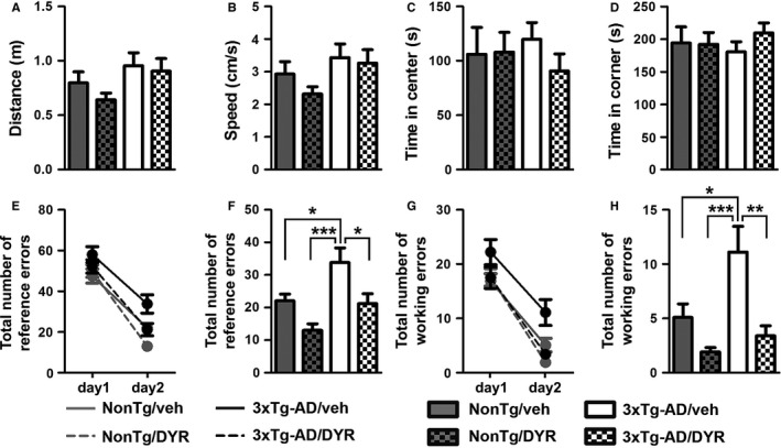
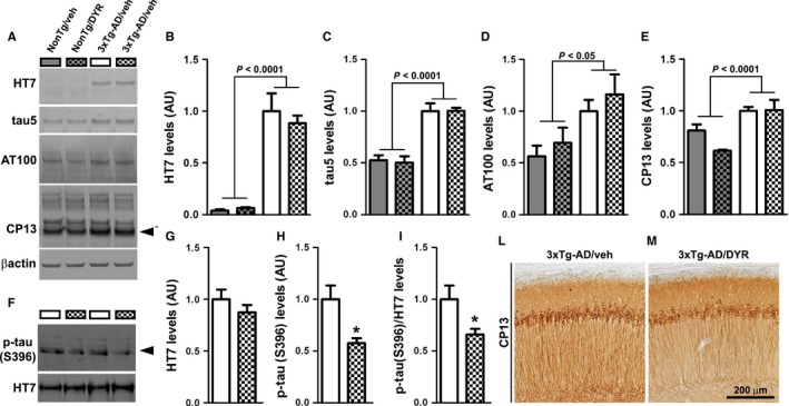
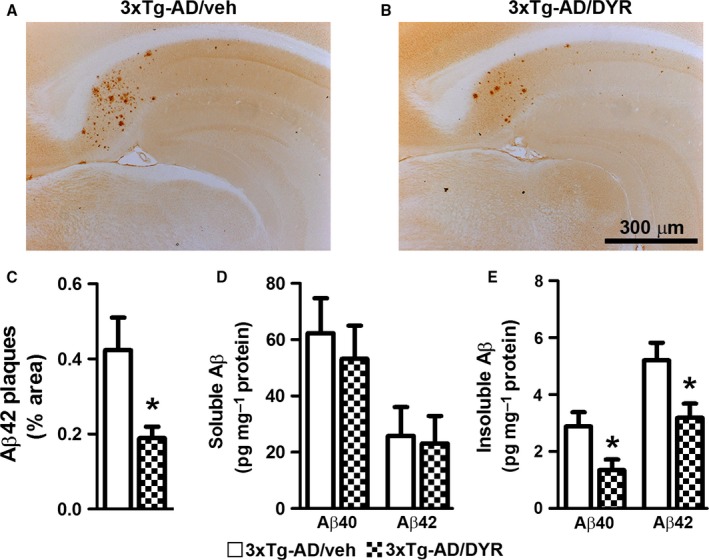
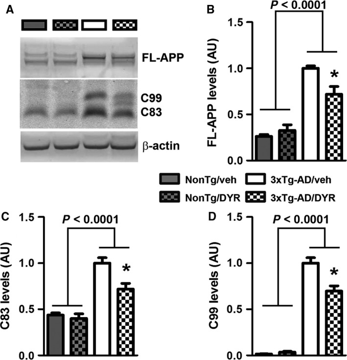
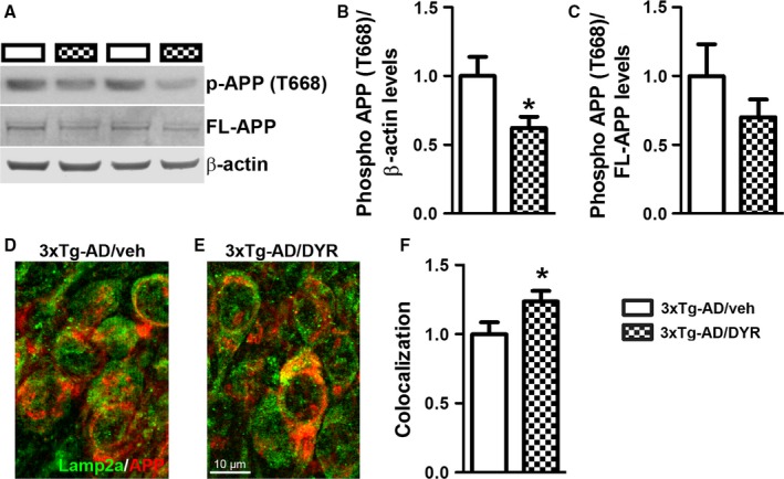
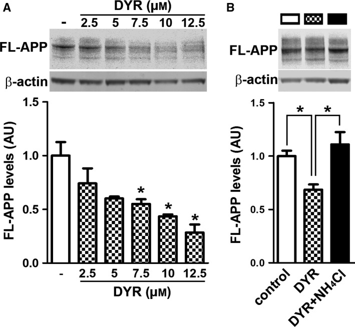
There is an urgent need for the development of new therapeutic strategies for Alzheimer's disease (AD). The dual-specificity tyrosine phosphorylation-regulated kinase-1A (Dyrk1a) is a protein kinase that phosphorylates the amyloid precursor protein (APP) and tau and thus represents a link between two key proteins involved in AD pathogenesis. Furthermore, Dyrk1a is upregulated in postmortem human brains, and high levels of Dyrk1a are associated with mental retardation. Here, we sought to determine the effects of Dyrk1 inhibition on AD-like pathology developed by 3xTg-AD mice, a widely used animal model of AD. We dosed 10-month-old 3xTg-AD and nontransgenic (NonTg) mice with a Dyrk1 inhibitor (Dyrk1-inh) or vehicle for eight weeks. During the last three weeks of treatment, we tested the mice in a battery of behavioral tests. The brains were then analyzed for the pathological markers of AD. We found that chronic Dyrk1 inhibition reversed cognitive deficits in 3xTg-AD mice. These effects were associated with a reduction in amyloid-β (Aβ) and tau pathology. Mechanistically, Dyrk1 inhibition reduced APP and insoluble tau phosphorylation. The reduction in APP phosphorylation increased its turnover and decreased Aβ levels. These results suggest that targeting Dyrk1 could represent a new viable therapeutic approach for AD.

Keywords: AD; 3xTg-AD; Alzheimer's disease; amyloid beta; plaques; tangles.


Figures

Figure 1
Figure 1
Chronic Dyrk1 inhibition improves learning and memory in 3xTg‐AD mice. (A, B) The graphs show total distance traveled and speed during the open‐field test. The data were not statistically significant among the four groups (= 0.0957 and 0.1016, respectively). (C, D) The graphs show the time spent in the center and periphery of the arena during open‐field testing. The data were not statistically significant among the four groups (= 0.7828 and 0.7827, respectively). (E‐H) We then evaluated the performance of the mice in the radial arm water maze (RAWM) by scoring spatial and working memory errors. The graphs show the average of the total errors that each mouse made across the 15 trials/day. (E) All groups show a decrease in total spatial errors at day 2, indicating learning (time effect, < 0.0001; group effect, = 0.0002; interaction effect, = 0.1841). (F) When we analyzed the total number of spatial errors at day 2, we found significant differences between groups (= 0.0001). As indicated in the figure, post hoc analyses showed that 3xTg‐AD/veh mice performed worse than all the other groups. Notably, 3xTg‐AD/DYR mice performed better than 3xTg‐AD/veh mice (< 0.05) and as well as NonTg mice (> 0.05). (G, H) Working memory errors: All the groups learned the task (time effect, < 0.0001; group effect, = 0.0001 interaction effect, = 0.4716). However, on day 2, we found significant differences between groups (< 0.0001). Post hoc analyses indicated that 3xTg‐AD/DYR mice performed significantly better than 3xTg‐AD/veh mice (< 0.01) and as well as NonTg mice (> 0.05). Both for working and reference errors, no differences were detected between NonTg/veh and NonTg/DYR mice (> 0.05). Data are presented as means ± SEM. Data in panels A‐D, F, and H were analyzed by one‐way ANOVA followed by Tukey's post hoc analyses. Data in panels E and G were analyzed by two‐way ANOVA. *P < 0.05; **P < 0.01; ***P < 0.001.
Figure 2
Figure 2
Chronic Dyrk1 inhibition reduces tau pathology. (A) Western blot on brain lysates probed with the indicated antibodies (= 4 mice/group for NonTg and = 5 mice/group for 3xTg‐AD). (B) Quantitative analysis of the HT7 blot showed a genotype effect (< 0.0001), as expected, but neither a treatment nor an interaction effect (= 0.5452 and 0.4102, respectively). (C) Quantitative analysis of the tau5 blot showed a genotype effect (< 0.0001), but neither a treatment nor an interaction effect (= 0.9146 and 0.8895, respectively). (D) Quantitative analysis of the AT100 blot showed a genotype effect (P = 0.0117), but neither a treatment nor an interaction effect (P = 0.4482 and 0.7140, respectively). (E) Quantitative analysis of the steady‐state levels of ~50 kDa band of CP13 (black arrowhead) showed a genotype effect (< 0.0001), but neither a treatment nor an interaction effect (= 0.2223 and 0.0662, respectively). (F–I) Western blot and quantitative analyses on insoluble brain fraction probed for total and p‐tau (S396). Quantitative analyses of the blots showed that total tau levels were similar between the two groups (= 0.2890). In contrast, the steady‐state levels of ~50 kDa band (black arrowhead) normalized to protein concentration or to total tau levels were significantly decreased in the 3xTg‐AD/DYR compared to 3xTg‐AD/veh (= 0.005 and 0.0211, respectively). (L‐M) Representative photomicrographs of treated and untreated 3xTg‐AD brain slices probed with the CP13 antibody (= 6 mice/group). Data in panels B‐E were normalized to β‐actin, presented as means ± SEM, and analyzed by two‐way ANOVA (genotype/treatment). Data in panels G‐H were normalized to protein concentration, presented as means ± SEM, and analyzed by Student's t‐test. *P < 0.05.
Figure 3
Figure 3
Dyrk1 inhibition reduces amyloid‐β (Aβ) pathology. (A‐B) Representative photomicrographs of 3xTg‐AD mice treated with Dyrk1‐inh or vehicle (= 6 mice/group). Sections were immunostained with an Aβ42‐specific antibody from Millipore. (C) The graph shows a significant decrease in the average area occupied in the hippocampus by plaques in treated vs. untreated 3xTg‐AD mice (= 0.0218). (D) Enzyme‐linked immunosorbent assay measurements from brain lysates (= 11 mice/group) revealed no difference in both Aβ40 and Aβ42 levels in the soluble fraction (= 0.6010 and P = 0.8539, respectively). (E) In contrast, insoluble Aβ40 and Aβ42 levels were significantly reduced in the brains of 3xTg‐AD/DYR mice compared to 3xTg‐AD/veh (= 0.0215 and = 0.0239, respectively). Data are presented as means ± SEM and were analyzed by Student's t‐test. *P < 0.05.
Figure 4
Figure 4
Dyrk1 inhibition alters APP processing. (A) Representative Western blots of proteins extracted from the brains of treated and untreated NonTg (= 4 mice/group) and 3xTg‐AD (= 5 mice/group) mice. Blots were probed with the indicated antibodies. (B) Quantitative analysis of the full‐length APP blot showed a genotype effect (< 0.0001), and a genotype‐treatment interaction effect (= 0.0074). Moreover, Bonferroni's post hoc analysis showed a significant reduction of full‐length APP for 3xTg‐AD/DYR (P < 0.01). (C–D) Quantitative analysis of the C83 and C99 blots showed a genotype effect (< 0.0001). Moreover, C99 and C83 levels were significantly decreased by treatment (< 0.01). Data were normalized to β‐actin, presented as means ± SEM, and analyzed by two‐way ANOVA (genotype/treatment) followed by post hoc Bonferroni's comparison. *P < 0.05.
Figure 5
Figure 5
Dyrk1 inhibition reduces APP phosphorylation, thereby modifying APP turnover. (A) Representative Western blots of proteins extracted from the hippocampi of treated and untreated 3xTg‐AD mice (= 9 mice/group). Blots were probed with the indicated antibodies. (B‐C) Quantitative analysis of the blots showed that while Dyrk1 inhibition did not change the ratio of phosphorylated over total APP, it significantly reduced the overall steady‐state levels of phosphorylation of APP at Thr668 (= 0.0480). However, the ratio pAPP over total APP was not significantly different between the two groups. (D–E) Representative microphotographs of hippocampal sections immunostained with the indicated antibodies (= 30 pictures from 6 mice/group). (F) Semiquantitative analysis showed that the number of yellow pixels (indicating a colocalization between APP and the lysosomal protein Lamp2A, analyzed as Pearson's correlation coefficient) was significantly higher in 3xTg‐AD/DYR mice compared with 3xTg‐AD/veh mice (P = 0.0436). Data are presented as means ± SEM and were analyzed by Student's t‐test. *P < 0.05.
Figure 6
Figure 6
Dyrk1 inhibition reduces APP levels by a lysosomal‐dependent mechanism. (A) Immunoblot analysis (anti‐APP antibody clone 22C11) of total extracts from HT22 cells treated for 24 hours with different concentrations of Dyrk1‐inh. One‐way ANOVA analysis showed a significant effect (P = 0.0011). Post hoc analyses with Tukey's correction showed that the reduction in APP levels was significantly different starting at 7.5 μm Dyrk1‐inh. (B) Immunoblot analysis (anti‐APP antibody clone 22C11) of total extracts from HT22 cells treated for 24 h with 7.5 μm Dyrk1‐inh in the presence or absence of the lysosomal inhibitor ammonium chloride (2 mm). One‐way ANOVA analysis showed a significant effect (P = 0.0069). Post hoc analyses with Tukey's correction showed that the reduction in APP levels elicited by Dyrk1‐inh treatment is reversed by lysosomal inhibition. Data were generated by normalizing the levels of the protein of interest to β‐actin used as loading control. Results presented as means ± SEM of three independent experiments. *P < 0.05.

Similar articles

Cited by

References

    1. Adayev T, Wegiel J, Hwang YW (2011) Harmine is an ATP‐competitive inhibitor for dual‐specificity tyrosine phosphorylation‐regulated kinase 1A (Dyrk1A). Arch. Biochem. Biophys. 507, 212–218. - PMC - PubMed
    1. Alzheimer's A (2015) 2015 Alzheimer's disease facts and figures. Alzheimer's Dement. 11, 332–384. - PubMed
    1. Azorsa DO, Robeson RH, Frost D, Meec hoovet B, Brautigam GR, Dickey C, Beaudry C, Basu GD, Holz DR, Hernandez JA, Bisanz KM, Gwinn L, Grover A, Rogers J, Reiman EM, Hutton M, Stephan DA, Mousses S, Dunckley T (2010) High‐content siRNA screening of the kinome identifies kinases involved in Alzheimer's disease‐related tau hyperphosphorylation. BMC Genom. 11, 25. - PMC - PubMed
    1. Branca C, Wisely EV, Hartman LK, Caccamo A, Oddo S (2014) Administration of a selective beta2 adrenergic receptor antagonist exacerbates neuropathology and cognitive deficits in a mouse model of Alzheimer's disease. Neurobiol. Aging 35, 2726–2735. - PMC - PubMed
    1. Caccamo A, Majumder S, Oddo S (2012) Cognitive decline typical of frontotemporal lobar degeneration in transgenic mice expressing the 25‐kDa C‐terminal fragment of TDP‐43. Am. J. Pathol. 180, 293–302. - PMC - PubMed

MeSH terms