. 2017 Oct 4;25(10):2372-2382.
doi: 10.1016/j.ymthe.2017.07.002. Epub 2017 Jul 8.

Microfluidic Transduction Harnesses Mass Transport Principles to Enhance Gene Transfer Efficiency

Affiliations

Microfluidic Transduction Harnesses Mass Transport Principles to Enhance Gene Transfer Efficiency

Reginald Tran et al. Mol Ther. .

Abstract

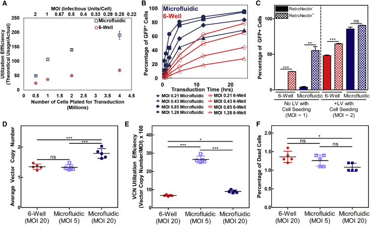
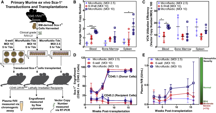
Ex vivo gene therapy using lentiviral vectors (LVs) is a proven approach to treat and potentially cure many hematologic disorders and malignancies but remains stymied by cumbersome, cost-prohibitive, and scale-limited production processes that cannot meet the demands of current clinical protocols for widespread clinical utilization. However, limitations in LV manufacture coupled with inefficient transduction protocols requiring significant excess amounts of vector currently limit widespread implementation. Herein, we describe a microfluidic, mass transport-based approach that overcomes the diffusion limitations of current transduction platforms to enhance LV gene transfer kinetics and efficiency. This novel ex vivo LV transduction platform is flexible in design, easy to use, scalable, and compatible with standard cell transduction reagents and LV preparations. Using hematopoietic cell lines, primary human T cells, primary hematopoietic stem and progenitor cells (HSPCs) of both murine (Sca-1+) and human (CD34+) origin, microfluidic transduction using clinically processed LVs occurs up to 5-fold faster and requires as little as one-twentieth of LV. As an in vivo validation of the microfluidic-based transduction technology, HSPC gene therapy was performed in hemophilia A mice using limiting amounts of LV. Compared to the standard static well-based transduction protocols, only animals transplanted with microfluidic-transduced cells displayed clotting levels restored to normal.

Keywords: CAR-T cells; cell manufacturing; cell therapy; gene therapy; hematopoietic stem cells; hemophilia; lentivirus; microfluidics.


Figures

None
Graphical abstract
Figure 1
Figure 1
Microfluidics Are an Enabling Platform for More Efficient LV Transduction (A) Microfluidics bring LV particles within closer proximity to target cells without requiring large quantities that would otherwise be wasted due to their short half-life. (B) Example of microfluidic devices that accommodate 106 (top) and 105 (bottom) cells with a surface area equivalent to a 6-well plate and a 96-well plate, respectively. The scale bar represents 1 cm. (C) Transductions using the same amount of GFP-LV and Jurkat cells (constant MOI) for various volumes/fluid heights reveal a height range (<100 μm) in which diffusion no longer limits vector integration as indicated by the plateau (n = 2–4). Data represent means ± SD. (D) Jurkat transductions comparing utilization efficiencies for a constant GFP-LV (v/v)% concentration at various transduction volumes demonstrates that minimizing total volume more efficiently utilizes available LV (n = 2–4). Data represent means ± SD. (E) A constant ratio of cells and virus (MOI of 1) loaded into 106 and 107 scale microfluidics for 5 hr produces nearly identical transduction of Jurkat cells with a GFP-LV. Data represent mean ± SD. Insets are photos of different versions of microfluidics used for each condition. The scale bar represents 3 cm.
Figure 2
Figure 2
Factors That Readily Increase Transduction Are Amplified in Microfluidic Systems and Are Optimized for Primary Human T Cell Transductions (A) Greater utilization of GFP-LV can be achieved in microfluidics by increasing the number of Jurkat cells targeted because virus-cell interactions are enhanced (n = 3). Data represent mean ± SD. (B) Assessment of Jurkat transduction kinetics between six-well plates and microfluidic systems at increasing MOIs using a GFP-LV. Both time and LV savings are possible using microfluidics. (C) RN coating can efficiently immobilize infectious particles in microfluidics compared to six-well plates. While RN significantly improves transduction in six-well plates, microfluidics marginally benefit from RN coating due to the high efficiency inherent in microfluidics (n = 3). Data represent mean ± SD. **p < 0.01, ***p < 0.001 (unpaired Student’s t test with Welch’s correction for unequal variances). (D and E) Microfluidic systems enable vector savings or increased gene transfer using a clinically processed fVIII-LV in primary human T cells as shown by VCN analysis (D) and VCN utilization efficiency (E). (F) Cell viability is not negatively affected by microfluidic transductions. Transduction time = 12 hr. (n = 5). Data represent means ± SD. *p < 0.05, ***p < 0.001 (one-way ANOVA). ns, not significant.
Figure 3
Figure 3
CD34+ Cells Are More Efficiently Transduced in Microfluidic Systems (A) VCN analysis demonstrates that transduction times and fVIII-LV usage are reduced by at least 50% using microfluidics. (B) Microfluidic systems enable greater VCN utilization efficiency compared to 24-well transductions. (C) Cell viability as measured by the trypan exclusion assay is not reduced from microfluidic systems. (D) Stem cell properties are not negatively impacted by microfluidic systems, as demonstrated by the human colony-forming assay (n = 3). Data represent means ± SD. *p < 0.05 (one-way ANOVA). Tdx, transduced; ns, not significant.
Figure 4
Figure 4
Transplantation of Microfluidic-Transduced Cells Results in Greater Rates of Gene Transfer and Higher fVIII Production in a Hemophilia A Animal Model (A) Pre-clinical simulation of murine gene therapy for hemophilia A using a clinically processed fVIII-LV. Microfluidic transductions are compared to an equivalent standard protocol using a six-well plate. (B and C) Microfluidic systems improve gene transfer efficiency, as indicated by VCN analysis (B) as well as VCN utilization efficiency (C) across various mouse tissues (n = 5 mice, MOI of 10, microfluidic; n = 4 mice, MOI of 10, six-well plate; and n = 4 mice, MOI of 2.5, microfluidic). Data represent means ± SD. *p < 0.05 and ***p < 0.001 (one-way ANOVA of individual tissue groups). (D) High levels of donor (CD45.1+) cell engraftment were maintained across all conditions. (E) Only mice transplanted with microfluidic-transduced Sca-1+ cells at an MOI of 10 were able to restore plasma fVIII levels to normal. Time course of averaged plasma fVIII levels for each group of mice. Tdx, transduced. Data represent mean ± SD.

References

    1. Aiuti A., Biasco L., Scaramuzza S., Ferrua F., Cicalese M.P., Baricordi C., Dionisio F., Calabria A., Giannelli S., Castiello M.C. Lentiviral hematopoietic stem cell gene therapy in patients with Wiskott-Aldrich syndrome. Science. 2013;341:1233151. - PMC - PubMed
    1. Cartier N., Hacein-Bey-Abina S., Bartholomae C.C., Veres G., Schmidt M., Kutschera I., Vidaud M., Abel U., Dal-Cortivo L., Caccavelli L. Hematopoietic stem cell gene therapy with a lentiviral vector in X-linked adrenoleukodystrophy. Science. 2009;326:818–823. - PubMed
    1. Cavazzana-Calvo M., Payen E., Negre O., Wang G., Hehir K., Fusil F., Down J., Denaro M., Brady T., Westerman K. Transfusion independence and HMGA2 activation after gene therapy of human β-thalassaemia. Nature. 2010;467:318–322. - PMC - PubMed
    1. Maude S.L., Frey N., Shaw P.A., Aplenc R., Barrett D.M., Bunin N.J., Chew A., Gonzalez V.E., Zheng Z., Lacey S.F. Chimeric antigen receptor T cells for sustained remissions in leukemia. N. Engl. J. Med. 2014;371:1507–1517. - PMC - PubMed
    1. Zufferey R., Dull T., Mandel R.J., Bukovsky A., Quiroz D., Naldini L., Trono D. Self-inactivating lentivirus vector for safe and efficient in vivo gene delivery. J. Virol. 1998;72:9873–9880. - PMC - PubMed

Publication types