Review
. 2018 Jan;43(1):218-230.
doi: 10.1007/s00261-017-1281-6.

Locoregional therapies for hepatocellular carcinoma and the new LI-RADS treatment response algorithm

Affiliations
Review

Locoregional therapies for hepatocellular carcinoma and the new LI-RADS treatment response algorithm

Ania Kielar et al. Abdom Radiol (NY). 2018 Jan.

Abstract

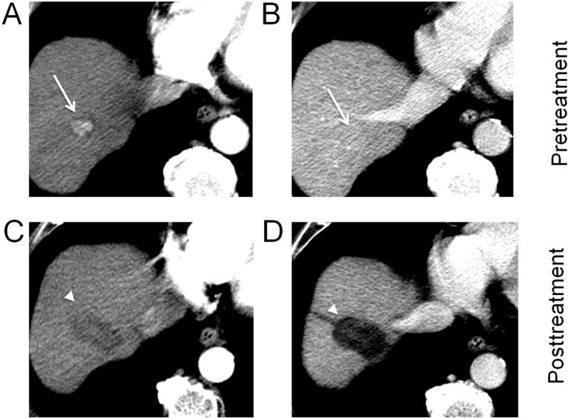
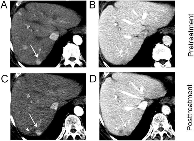
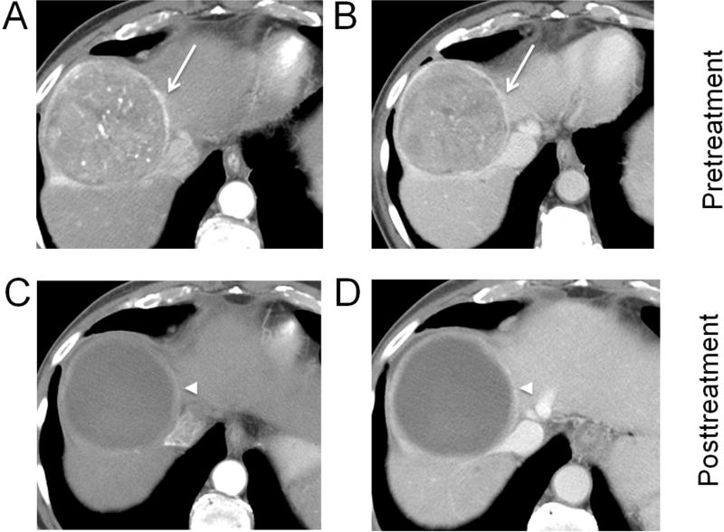
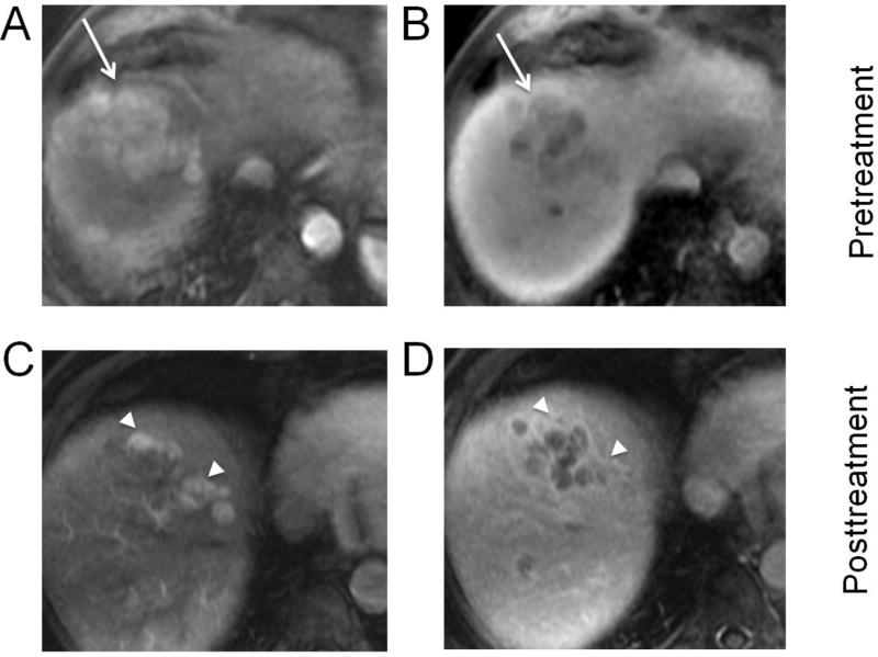
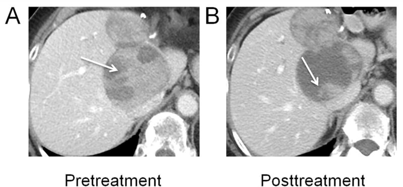
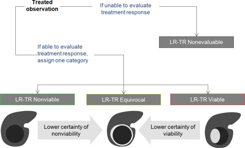
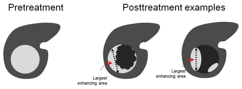
Radiologists play a central role in the assessment of patient response to locoregional therapies for hepatocellular carcinoma (HCC). The identification of viable tumor following treatment guides further management and potentially affects transplantation eligibility. Liver Imaging Reporting and Data Systems (LI-RADS) first introduced the concept of LR-treated in 2014, and a new treatment response algorithm is included in the 2017 update to assist radiologists in image interpretation of HCC after locoregional therapy. In addition to offering imaging criteria for viable and nonviable HCC, new concepts of nonevaluable tumors as well as tumors with equivocal viability are introduced. Existing guidelines provided by response evaluation criteria in solid tumors (RECIST) and modified RECIST address patient-level assessments and are routinely used in clinical trials but do not address the variable appearances following different locoregional therapies. The new LI-RADS treatment response algorithm addresses this gap and offers a comprehensive approach to assess treatment response for individual lesions after a variety of locoregional therapies, using either contrast-enhanced CT or MRI.

Keywords: Hepatocellular carcinoma; LI-RADS; Locoregional therapy; RECIST; Response.


Conflict of interest statement

Conflict of Interest:

The authors declare that they have no conflict of interest.

Figures

Fig. 1
Fig. 1. Hepatocellular carcinoma (HCC) treated with radiofrequency ablation
A biopsy proven HCC in the right hepatic lobe (arrows) shows arterial phase hyperenhancement (A) and no washout on portal venous phase (B). After radiofrequency ablation, an ablation zone (arrowheads) is now visible as a larger defect than the original tumor, with a linear ablation tract along the course of the electrode used (C, D). No residual enhancement is seen, and the lesion should be reported as LR-TR Nonviable.
Fig. 2
Fig. 2. Treatment with percutaneous ethanol ablation
A lesion in segment 7 (arrows) showing arterial phase hyperenhancement (A) and washout on portal venous phase (B) measures 1.3 cm (LR-4). After ethanol ablation (PEA), a nonenhancing ablation zone (arrowheads) is present, but more medially, a nodular area (arrows) of arterial phase hyperenhancement (C) and washout (D), measures 1.5 cm and is consistent with viable tumor (LR-TR Viable 1.5 cm).
Fig. 3
Fig. 3. Hepatocellular carcinoma (HCC) treated with drug-eluting bead transarterial chemoembolization (DEB-TACE)
Biopsy-proven HCC (arrows) in segment 8 showing arterial phase hyperenhancement (A), washout and capsule on portal venous phase (B). After DEB-TACE, no residual enhancement except for a thin rim of enhancement (arrowheads) is seen on arterial (C) and portal venous phases (D) (LR-TR Nonviable).
Fig. 4
Fig. 4. Hepatocellular carcinoma (HCC) treated with transarterial chemoembolization (TACE)
A lesion in segment 8 showing arterial phase hyperenhancement (A) and washout on portal venous phase (B) (arrows), measuring 8.0 cm (LR-5). After TACE, multiple nodular areas (arrowheads) of arterial phase hyperenhancement (C) and washout (D) anteriorly and medially are consistent with viable tumor. The largest nodular area of enhancement without intervening necrosis measures 2.5 cm and the lesion should be reported as: LR-TR Viable 2.5 cm (previously LR-5, 8.0 cm).
Fig. 5
Fig. 5. Hepatocellular carcinoma 2 days after treatment with transarterial embolization (TAE)
Foci of gas (arrowheads) are visible on portal venous phase CT within the embolized tumor. This is an early posttreatment expected finding that resolves over a few weeks and should not be confused with infection. Residual nodular enhancement measuring up to 10.1 cm along the right lateral margin (arrows) is consistent with viable tumor (LR-TR Viable 10.1 cm).
Fig. 6
Fig. 6. Hepatocellular carcinoma (HCC) treated with transarterial chemoembolization (TACE)
HCC in the right hepatic lobe on precontrast (A) and arterial phase (arrow) (B). After TACE, the presence of lipiodol appears hyperdense (arrowheads) on precontrast (C) and limits evaluation for enhancement on arterial phase imaging (D).
Fig. 7
Fig. 7. Hepatocellular carcinoma (HCC) with tumor in vein (arrows), treated with transarterial radioembolization (TARE)
Biopsy-proven HCC in the left hepatic lobe with tumor in vein on arterial phase (A) and portal venous phase (B). One month after TARE, the tumor in vein demonstrates decreased enhancement, but hyperenhancement in the surrounding treated left lateral segment (arrowheads) is visible on both arterial phase (C) and portal venous phase (D). Vague areas of enhancement within the tumor in vein are equivocal for tumor viability (LR-TR Equivocal 4.4 cm).
Fig. 8
Fig. 8. Hepatocellular carcinoma (HCC) treated with Stereotactic Body Radiotherapy (SBRT)
Multiphasic contrast-enhanced CT (A,B) demonstrating HCC in the hepatic dome (arrows), segment 4A, with arterial phase hyperenhancement (APHE), washout appearance, and threshold growth since 3 months prior, (LR-5, 1.7 cm). The 3-month post-SBRT follow-up CT (C,D) demonstrates unchanged size of the 1.7-cm tumor (arrowheads), with decreased prominence of APHE and washout appearance (LR-TR Equivocal 1.7 cm). The 6-month post-SBRT follow-up CT (E,F) demonstrates decreased tumor size, 0.5 cm, with persistent APHE and questionable washout appearance (LR-TR Viable 0.5 cm). HCCs after SBRT often demonstrate continued enhancement suggestive of tumor viability, but should be followed by serial imaging as long as the tumor size continues to regress. Note how the irradiated areas of penumbra (bounded by dashed line) shows slight differential enhancement compared to the rest of the liver parenchyma.
Fig. 9
Fig. 9. Multifocal hepatocellular carcinoma (HCC), treated with sorafenib
After prior left hepatic resection, two recurrent HCCs in the caudate lobe and along the resection margin are visible on portal venous phase (A). The larger tumor (arrows) shows decreased internal enhancement (B) during treatment with sorafenib, a multikinase inhibitor that targets tumor angiogenesis.
Fig. 10
Fig. 10. LI-RADS Treatment Response Algorithm with Tie-Breaking Rules
A treated observation is either nonevaluable (LR-TR Nonevaluable) or evaluable and assigned one of three categories: Nonviable, Equivocal, or Viable. When unsure about the category of response, the LR-TR Equivocal category should be selected.
Fig. 11
Fig. 11. Measurements of viable tumor after treatment
When reporting the size of the tumor after treatment, the viable tumor should be reported by measuring the single largest dimension covering the area of enhancement and avoiding intervening areas of nonenhancement. When multiple discrete areas of nodular enhancement are present, the single largest enhancing area is measured.

References

    1. Ahmed M, et al. Image-guided tumor ablation: standardization of terminology and reporting criteria--a 10-year update. Radiology. 2014;273(1):241–60. - PMC - PubMed
    1. Cheung W, et al. Irreversible electroporation for unresectable hepatocellular carcinoma: initial experience and review of safety and outcomes. Technol Cancer Res Treat. 2013;12(3):233–41. - PubMed
    1. Gaba RC, et al. Transcatheter Therapy for Hepatic Malignancy: Standardization of Terminology and Reporting Criteria. J Vasc Interv Radiol. 2016;27(4):457–73. - PubMed
    1. Benedict SH, et al. Stereotactic body radiation therapy: the report of AAPM Task Group 101. Med Phys. 2010;37(8):4078–101. - PubMed
    1. Halperin EC, et al. Perez and Brady's Principles and Practice of Radiation Obcology. 6. Lippincott Williams & Wilkins; 2013.

Publication types

MeSH terms