. 2017 Sep 5;25(9):1442-1448.e3.
doi: 10.1016/j.str.2017.07.003. Epub 2017 Aug 3.

KRAS G12C Drug Development: Discrimination between Switch II Pocket Configurations Using Hydrogen/Deuterium-Exchange Mass Spectrometry

Affiliations

KRAS G12C Drug Development: Discrimination between Switch II Pocket Configurations Using Hydrogen/Deuterium-Exchange Mass Spectrometry

Jia Lu et al. Structure. .

Abstract

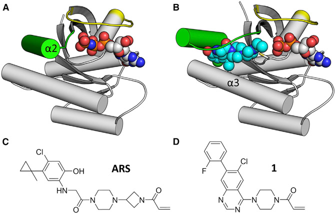
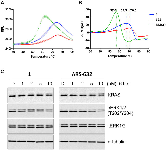
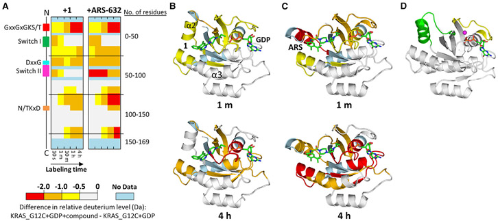
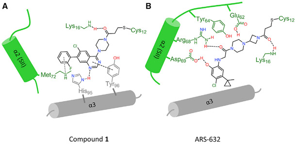
KRAS G12C, the most common RAS mutation found in non-small-cell lung cancer, has been the subject of multiple recent covalent small-molecule inhibitor campaigns including efforts directed at the guanine nucleotide pocket and separate work focused on an inducible pocket adjacent to the switch motifs. Multiple conformations of switch II have been observed, suggesting that switch II pocket (SIIP) binders may be capable of engaging a range of KRAS conformations. Here we report the use of hydrogen/deuterium-exchange mass spectrometry (HDX MS) to discriminate between conformations of switch II induced by two chemical classes of SIIP binders. We investigated the structural basis for differences in HDX MS using X-ray crystallography and discovered a new SIIP configuration in response to binding of a quinazoline chemotype. These results have implications for structure-guided drug design targeting the RAS SIIP.

Keywords: RAS; covalent inhibitor; drug discovery; protein dynamics.


Figures

Figure 1.
Figure 1.. Switch II Configuration Changes in Response to SIIP Inhibitors
(A) GTP-bound HRAS (PDB: 4L9W) shows that the helical portion of switch II (α2) engages the gamma phosphate of GTP, resulting in a closed conformation. Switch II in green, switch I in yellow, GTP in spheres. (B) ARS-853-bound KRAS G12C (PDB: 5F2E) shows an open switch II conformation. In addition to α2, ARS-853 (cyan spheres) also interacts with α3. (C and D) Chemical structures of ARS-853 and 1.
Figure 2.
Figure 2.. KRAS G12C-1 and KRAS G12C-ARS-632 Both Show Enhanced Thermal Stability and Similar in Potency for Inhibition of KRAS G12C-Depenent MAPK Signaling in H358 Cells
(A) Raw DSF measurements for KRAS G12C fully labeled with either ARS-632 or 1. (B) First derivative of (A) shows the shifts in Tm (dashed vertical lines) associated with protein labeling. (C) Impact of compound exposure on MAPK signaling. Levels of pERK were evaluated in the KRAS-G12C-containing cancer cell line H358 as a surrogate for RAS signaling through the MAPK pathway. Cells were treated with compound 1 or ARS-632 as indicated for 6 hr. Phosphorylation of ERK1/2 was determined by immunoblotting.
Figure 3.
Figure 3.. HDX MS Detects Changes in Switch II dynamics Induced by SIIP Binders
(A) Differences in HDX are represented by relative deuterium levels of inactive protein covalently bound to compound minus that of inactive GDP-bound protein alone, scale shown at top. Two-dimensional representations of KRAS G12C are given in linear fashion from N terminus (top) to C terminus (bottom), and the locations of key structural elements are shown on the left. All deuterium-labeling time points are shown, increasing from left to right. (B and C) Crystal structures of 1 (B) or ARS-853 (C) bound to KRAS G12C with HDX MS differences at labeling time point 1 min (upper panel) and 4 hr (lower panel) annotated using the color scheme from subpanel (A). (D) KRAS G12C bound to GDP alone (PDB: 4LDJ) provided for comparison. In (D), switch I is shown in yellow and switch II in green; magnesium is represented as a magenta sphere.
Figure 4.
Figure 4.. Schematic Representation of Key Interactions between Switch II Compounds and Adjacent α Helices
(A and B) Different interaction patterns are observed for 1 versus ARS-853 in the switch II binding pocket: (A) 1 is stabilized by pi-pi interaction with helix α3 residues His95 and Tyr96. (B) Arg68 and Asp69 in helix α2 (switch II) make hydrogen bonds to stabilize ARS-853.

Comment in

  • Remodeling KRAS.
    Deredge DJ, Wintrode PL. Deredge DJ, et al. Structure. 2017 Sep 5;25(9):1323-1324. doi: 10.1016/j.str.2017.08.012. Structure. 2017. PMID: 28877504

References

    1. Adams PD, Afonine PV, Bunkoczi G, Chen VB, Davis IW, Echols N, Headd JJ, Hung LW, Kapral GJ, Grosse-Kunstleve RW, et al. (2010). PHENIX: a comprehensive Python-based system for macromolecular structure solution. Acta Crystallogr. D Biol. Crystallogr 66, 213–221. - PMC - PubMed
    1. Campbell PM, and Der CJ (2004). Oncogenic Ras and its role in tumor cell invasion and metastasis. Semin. Cancer Biol 14, 105–114. - PubMed
    1. Emsley P, Lohkamp B, Scott WG, and Cowtan K (2010). Features and development of coot. Acta Crystallogr. D Biol. Crystallogr 66, 486–501. - PMC - PubMed
    1. Engen JR (2003). Analysis of protein complexes with hydrogen exchange and mass spectrometry. Analyst 128, 623–628. - PubMed
    1. Fasano O, Aldrich T, Tamanoi F, Taparowsky E, Furth M, and Wigler M (1984). Analysis of the transforming potential of the human H-ras gene by random mutagenesis. Proc. Natl. Acad. Sci. USA 81, 4008–4012. - PMC - PubMed

Publication types