. 2017 Aug 10;170(4):760-773.e15.
doi: 10.1016/j.cell.2017.06.052. Epub 2017 Aug 3.

Multi-invasions Are Recombination Byproducts that Induce Chromosomal Rearrangements

Affiliations

Multi-invasions Are Recombination Byproducts that Induce Chromosomal Rearrangements

Aurèle Piazza et al. Cell. .

Abstract

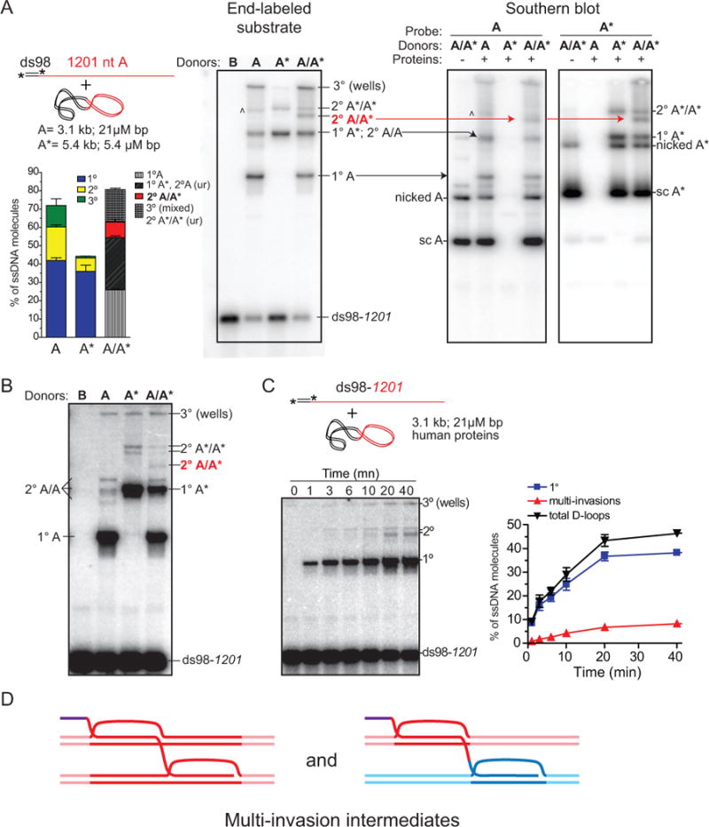
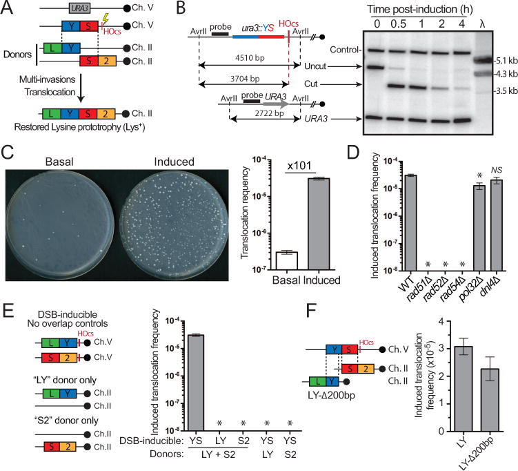
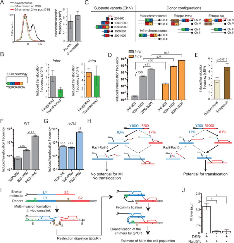
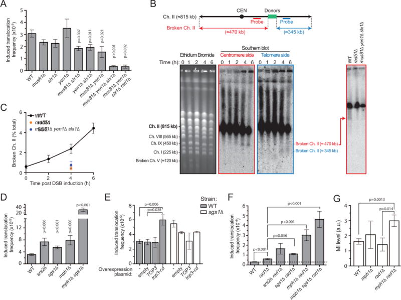
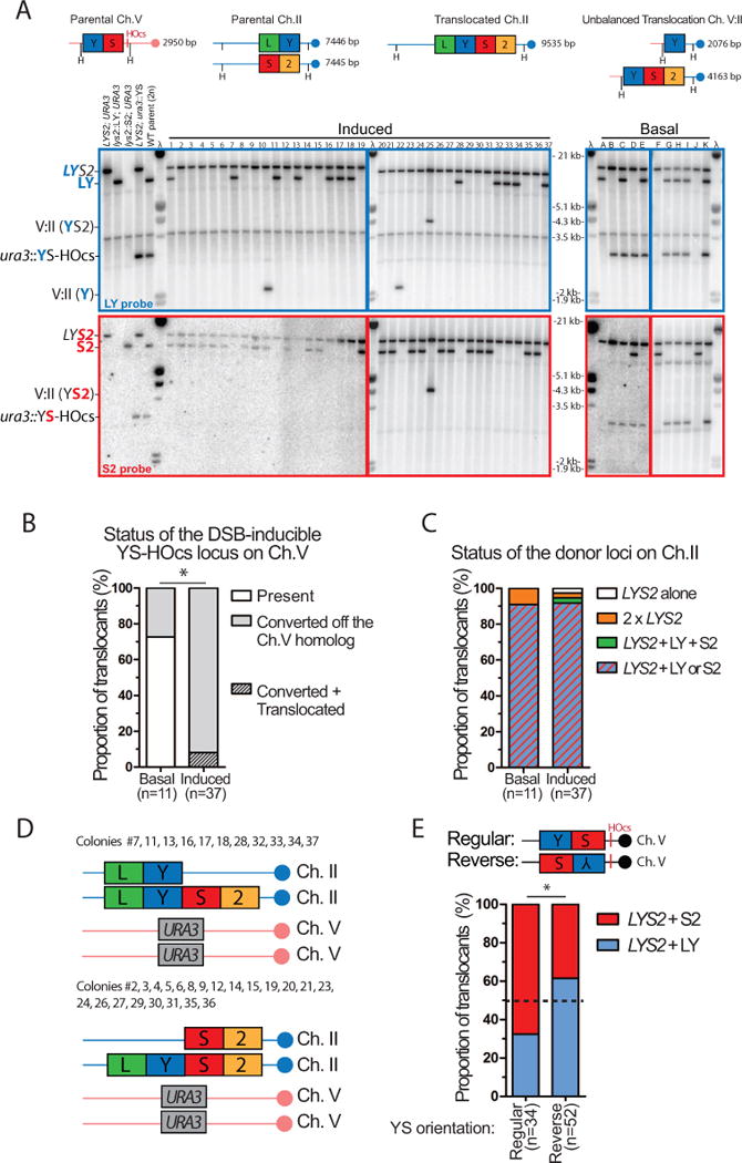
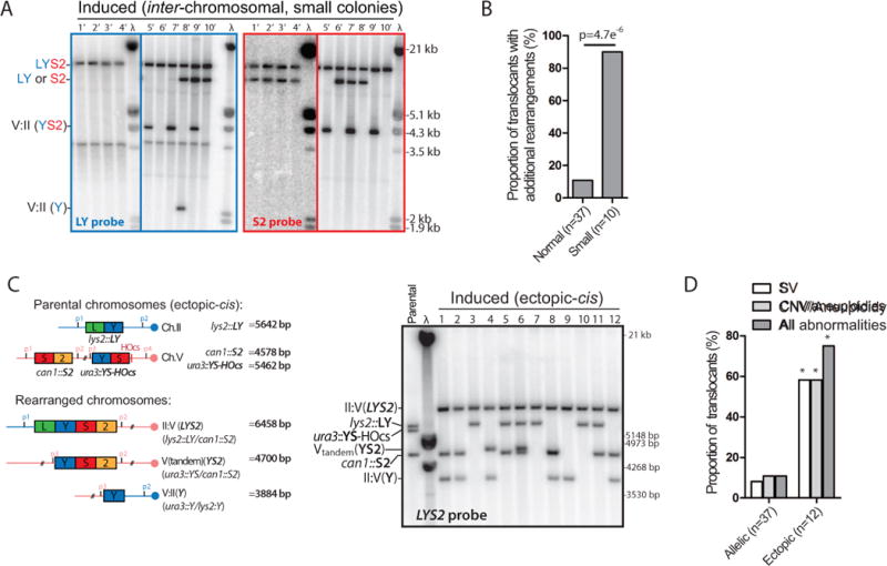
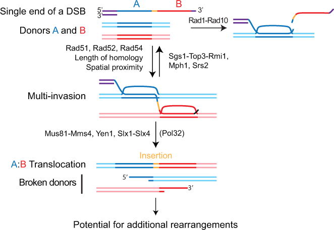
Inaccurate repair of broken chromosomes generates structural variants that can fuel evolution and inflict pathology. We describe a novel rearrangement mechanism in which translocation between intact chromosomes is induced by a lesion on a third chromosome. This multi-invasion-induced rearrangement (MIR) stems from a homologous recombination byproduct, where a broken DNA end simultaneously invades two intact donors. No homology is required between the donors, and the intervening sequence from the invading molecule is inserted at the translocation site. MIR is stimulated by increasing homology length and spatial proximity of the donors and depends on the overlapping activities of the structure-selective endonucleases Mus81-Mms4, Slx1-Slx4, and Yen1. Conversely, the 3'-flap nuclease Rad1-Rad10 and enzymes known to disrupt recombination intermediates (Sgs1-Top3-Rmi1, Srs2, and Mph1) inhibit MIR. Resolution of MIR intermediates propagates secondary chromosome breaks that frequently cause additional rearrangements. MIR features have implications for the formation of simple and complex rearrangements underlying human pathologies.

Keywords: D-loop; chromothripsis; endonuclease; genomic instability; helicase; homologous recombination; homology search; multi-invasion; translocation.


Conflict of interest statement

AUTHORS CONTRIBUTIONS

AP and WDH conceived the project and wrote the manuscript, with input and editing from WW. AP designed, performed and analyzed the in vivo and some in vitro experiments. WW designed, performed and analyzed most of the in vitro experiments.

Figures

Figure 1
Figure 1. Multi-invasions form in reconstituted D-loop reactions with yeast and human proteins
(A) D-loop reactions with yeast proteins. A ds98-1201 (1.3 μM nt/bp; 1 nM molecules) substrate was paired with two different sized dsDNA plasmids to distinguish multi-invasion (MI) products. Left panel: ds98-1201 end-labeled reaction. The main product band is a single D-loop (1°), the next band is 2° (MI: 2 dsDNAs), and the species in the well (3°) likely contains three dsDNAs. A minor product band (^) is likely a 3° invasion species with less complex shape and thus able to enter the gel. Right panels: Southern blot of D-loop reactions using A or A* specific probes. Arrows indicate the corresponding species identified with labeled ssDNA (right) or probing for the plasmids donors (left). (B) D-loop reactions with human proteins. Reaction conditions (except buffer composition) and analysis as in (A). (C) Time course with ds98-1201 substrate using the single donor A. Quantifications in (A) and (C) show the mean ±SD of n=3. (D) Scheme of multi-invasion (MI) joint molecule.
Figure 2
Figure 2. Requirements for intact chromosomal regions translocation induced by a DSB on another molecule
(A) Reference tripartite recombination system in diploid yeast. The heterozygous DSB-inducible construct (YS-HOcs) replaces URA3 on Ch. V. The LY and S2 donors represent the two halves of the LYS2 gene and share no homology with one another. They are located in allelic configuration referred to as inter-chromosomal. Their blunt translocation restores a LYS2 gene. (B) Southern blot analysis of DSB kinetics upon HO induction. Predicted sizes (bp) for the uncut and cut locus as well as the other Ch. V homolog (URA3) upon AvrII digestion are shown on the left. Control: RAD54 locus on Ch. VII. λ: length marker. (C) DSB-induction causes a 101-fold increase in the translocation (Lys+) frequency in wild type. Representative SD-LYS plates with 5×10⁷ cells plated are shown. (D) Induced translocation frequencies in wild type, rad51Δ, rad52Δ, rad54Δ, pol32Δ and dnl4Δ. No Lys+ colonies are detected in the HR mutants at our detection limit (~10−8). (E) No Lys+ colonies were observed with strains bearing DSB-inducible constructs devoid of homology overlap to the LY and S2 donors or strains lacking the LY or S2 donors. (F) Induced translocation frequency in the reference wild type or a strain bearing a LY donor truncated for its last 200 bp (LY-Δ200bp). (C–F) Bars represent mean ±SEM. *p<0.05.
Figure 3
Figure 3. A single ssDNA molecule concomitantly invades two donors and cause their translocation
(A) FACS profile and induced translocation frequency in asynchronous or G1-arrested cells. (B) Translocation frequency induced with either an integrated or a transformed YS2000-2000 construct in strains bearing the donors in the inter-chromosomal or intra-chromosomal configuration (Figure 3C). (C) Scheme of substrate length variants and donor configurations. (D) Homology length and physical proximity of the donors stimulate translocation frequency in wild type. (E) Induced translocation frequency in the ectopic-trans and ectopic-cis donor configurations. (F, G) Induced translocation frequency in wild type (F) or rad1Δ (G) with asymmetric homology length variants. (G) Fold over wild type is indicated. (A, C–G) Bars represent mean ±SEM. (H) Model for the Rad1-dependent differential effect of the DSB-proximal and -distal length of homology on MIR. (I) Rationale of MI-Capture assay. (J) The MI signal is DSB- and Rad51-dependent. Bars represent mean ±SEM of qPCR signal normalized over a control (ARG4) on Ch. VIII in wild type either un-induced (n=9) or 3 hrs after DSB induction (n=7), or in rad51Δ 3 hrs after DSB induction (n=3). *p<0.05. Controls for HOcs cleavage and ligation efficiency are reported in Figure S2F, G.
Figure 4
Figure 4. Genetic controls of MIR
(A) Induced translocation frequencies in single or multiple mutants for MUS81, SLX1, and YEN1, as well as mus81Δ slx1Δ yen1Δ rad1Δ. (B) Physical evidence for Ch. II breakage at the donors following DSB induction on Ch. V (see Figure S3D) in a Rad51- and SSE-dependent fashion (right, 4 hrs post-DSB induction). (C) DSB quantification at donor site on Ch. II (n=2). (D) Induced translocation frequency in the srs2Δ, sgs1Δ, mph1Δ and mph1Δ sgs1Δ. (E) Induced translocation frequency in wild type or sgs1Δ containing an empty overexpression plasmid or transiently overexpressing the WT or catalytic-deficient Top3. (F) Genetic interactions between RAD1 and SGS1, MPH1, and SRS2. (G) MI levels 3 hrs post-DSB induction in WT (n=7), mph1Δ (n=3), rad1Δ (n=3), and mph1Δ rad1Δ (n=9). Controls for DSB induction by HO and ligation efficiency are in Figure S3E and F. (A, C–G) Bars represent mean ±SEM.
Figure 5
Figure 5. Physical analysis of MIR translocants
(A) Southern blot analysis of basal (n=11) or induced Lys+ cells (n=37, normal colony size) obtained with the wild type inter-chromosomal strain. The expected size of the parental and translocated molecules upon HindIII digestion is shown on the left panel. Blots were probed with either the LY (top, blue) or the S2 probe (bottom, red) and phage λ DNA (molecular ladder). (B) Status of the DSB-inducible YS-HOcs construct in basal and induced cells. Translocated refers to Ch. V:II translocations depicted in (A). (C) Summary of the donor segregation pattern together with LYS2. (D) Summary of the genetic content of normal-size translocants exhibiting no additional chromosomal abnormalities (33/37). (E) The translocated LYS2 gene segregates preferentially with the donor corresponding to the DSB-proximal homology. Southern blot analysis of translocants induced with a DSB-inducible construct bearing the YS sequence in reverse orientation is shown Figure S4C. (B, E) *p<0.05, Fisher’s exact test.
Figure 6
Figure 6. MIR is frequently associated with additional chromosomal abnormalities
(A) Southern blot analysis of induced Lys+ cells (n=10, small colony size) obtained with the reference inter-chromosomal strain. Other legends as in Figure 5A. (B) Summary of additional chromosomal abnormalities in normal and small induced Lys+ colonies. (C) Southern blot analysis of induced translocants (n=12, normal colonies) obtained with the ectopic-cis strain. The expected size of the parental and translocated molecules upon PstI digestion is shown on the left panel. Blots were hybridized with the LYS2 probe and phage λ DNA (molecular ladder). (D) Summary of the SV detected by Southern blot and the CNV/aneuploidies determined by qPCR in normal-size translocants obtained in the inter-chromosomal and the ectopic-cis donor configurations (Figure S5C and F). *p<0.05, Fisher’s exact test.
Figure 7
Figure 7. Model for MIR
One ssDNA molecule with homology to two donors A and B, without the need for homology to one another, can form a multi-invasion intermediate. Formation of MI is stimulated by homology length and physical proximity of the donors. Cleavage of the invading strand by Rad1-Rad10 upon internal invasion irreversibly prevents MI formation and protects against MIR. The Srs2 and Mph1 (human FANCM) helicases and the Sgs1-Top3-Rmi1 (human BLM-TOPOnIα-RMI1-RMI2) helicase/topoisomerase complex disrupt MI and also inhibit MIR. Processing of MI by the overlapping activities of the Mus81-Mms4, Yen1, and Slx1-Slx4 SSE triggers MIR and transfers single-ended DSBs onto the donors. MIR occurs upon joining of the two opposite ends of the donors using the Rad51-ssDNA from the invading molecule as a synthesis template, which inserts sequence at the translocation junction. The two single-ended DSBs generated on the donors have potential to undergo secondary rearrangements. Detailed MIR mechanisms are proposed Figure S7.

References

    1. Anand RP, Tsaponina O, Greenwell PW, Lee CS, Du W, Petes TD, Haber JE. Chromosome rearrangements via template switching between diverged repeated sequences. Genes Dev. 2014;28:2394–2406. - PMC - PubMed
    1. Barlow JH, Lisby M, Rothstein R. Differential regulation of the cellular response to DNA double-strand breaks in G1. Mol Cell. 2008;30:73–85. - PMC - PubMed
    1. Bell JC, Kowalczykowski SC. RecA: regulation and mechanism of a molecular search engine. Trends Biochem Sci. 2016;41:646–646. - PMC - PubMed
    1. Binz SK, Dickson AM, Haring SJ, Wold MS. Functional assays for replication protein A (RPA) Methods Enzymol. 2006;409:11–38. - PubMed
    1. Bondurand N, Fouquet V, Baral V, Lecerf L, Loundon N, Goossens M, Duriez B, Labrune P, Pingault V. Alu-mediated deletion of SOX10 regulatory elements in Waardenburg syndrome type 4. Eur J Hum Genet. 2012;20:990–994. - PMC - PubMed