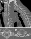
Cervical Flexion Myelopathy Eleven Years after a Cervical Spinal Cord Injury
- PMID: 28781306
- PMCID: PMC5596286
- DOI: 10.2169/internalmedicine.8322-16
Cervical Flexion Myelopathy Eleven Years after a Cervical Spinal Cord Injury
Abstract
We herein describe a 37-year-old man who developed cervical flexion myelopathy 11 years after suffering a cervical spinal cord injury. Cervical magnetic resonance imaging 11 years after the accident demonstrated atrophy and hyperintense lesions at the C6 and C7 levels in the cervical cord with an abnormal alignment of the vertebrae. In the neck flexion position, an anterior shift of the cervical cord was evident. Our patient's condition suggests that an abnormal alignment of the cervical spine and spinal cord injury due to a traumatic accident could be risk factors in the subsequent development of cervical flexion myelopathy.
Keywords: Hirayama disease; MRI; cervical flexion myelopathy; cervical spinal cord injury.
Figures


References
-
- Iwasaki Y, Tashiro K, Kikuchi S, Kitagawa M, Isu T, Abe H. Cervical flexion myelopathy: a “tight dural canal mechanism”. J Neurosurg 66: 935-937, 1987. - PubMed
-
- Kato Y, Imajo Y, Kanchiku T, Kojima T, Kataoka H, Taguchi T. Dynamic electrophysiological examination of cervical flexion myelopathy. J Neurosurg Spine 9: 180-185, 2008. - PubMed
-
- Tavee JO, Levin KH. Myelopathy due to degenerative and structural spine diseases. Continuum (Minneap Minn) 21: 52-66, 2015. - PubMed
-
- Watanabe K, Hasegawa K, Hirano T, Endo N, Yamazaki A, Homma T. Anterior spinal decompression and fusion for cervical flexion myelopathy in young patients. J Neurosurg Spine 3: 86-91, 2005. - PubMed
Publication types
MeSH terms
LinkOut - more resources
Full Text Sources
Other Literature Sources
Medical
Miscellaneous

