Case Reports
. 2017 Aug 15;56(16):2159-2164.
doi: 10.2169/internalmedicine.8323-16. Epub 2017 Aug 1.

Isolated Right Ventricular Stress (Takotsubo) Cardiomyopathy

Affiliations
Case Reports

Isolated Right Ventricular Stress (Takotsubo) Cardiomyopathy

Hitoshi Sumida et al. Intern Med. .

Abstract

A 79-year-old woman was admitted with a left femoral neck fracture and she immediately developed circulatory shock. Echocardiography showed a markedly enlarged right ventricle (RV) with systolic ballooning of the mid-ventricular wall and preserved contractility of the apex. The left ventricular (LV) motion was normal. Multi-detector-row computed tomography showed severe congestion of the contrast media in the right atrium with no forward flow to RV, but no pulmonary embolism. She was successfully treated with percutaneous veno-arterial extracorporeal membrane oxygenation. This case presented with acute, profound, but reversible RV dysfunction triggered by acute stress in a manner similar to that seen in LV stress cardiomyopathy.

Keywords: cardiac magnetic resonance; circulatory shock; echocardiography; mechanical support; right heart failure; stress (takotsubo) cardiomyopathy.


Figures

Figure 1.
Figure 1.
Electrocardiogram on admission showing sinus rhythm as well as S1 and T-wave inversion in leads V1-V4 (A). Electrocardiogram obtained after the detection of changes in the heart rate and a QRS complex pattern. The electrocardiograms displaying progressive changes in the heart rate followed by sinus arrest, complete right bundle-branch block pattern escape beats, T-wave inversion in leads III, V1-V3, and V4R, and no significant ST elevation, including inferior and V4R leads (B).
Figure 2.
Figure 2.
Echocardiography images obtained on admission. Apical four-chamber view of the heart during diastole and systole indicates right ventricular (RV) enlargement and free-wall systolic ballooning (yellow arrow) with apical motion sparing (red arrow). The left ventricle (LV) is small and no regional wall motion abnormalities are apparent (A). Parasternal short axis view of the heart during diastole and systole reveals RV enlargement. The interventricular septum (IVS) is flattened during diastole (white arrow), but not during systole, indicating a change in the volume without any superimposed pressure overload (B). LA: left atrium, LV: left ventricle, RA: right atrium, RV: right ventricle
Figure 3.
Figure 3.
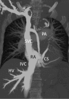
Maximum intensity projection (MIP) image of multi-detector row computed tomography shows excessive contrast in the systemic venous system, including the superior vena cava, inferior vena cava, hepatic vein, and coronary sinus, but faint contrast in the pulmonary circulation and no pulmonary embolism. These findings indicate severe right ventricular dysfunction; the majority of the contrast did not flow into the pulmonary circulation, but instead spilled over into the systemic venous system. CS: coronary sinus, HV: hepatic vein, IVC: inferior vena cava, PA: pulmonary artery, RA: right atrium, RV: right ventricle, SVC: superior vena cava
Figure 4.
Figure 4.
Cardiac magnetic resonance (CMR) imaging confirmed right ventricular (RV) enlargement and free wall systolic ballooning (yellow arrow) with apical motion sparing (red arrow). No regional left ventricular (LV) wall motion abnormalities are visible (A). CMR demonstrated no abnormalities of short-T1 inversion recovery (B), and late gadolinium enhancement (C). CMR angiography revealed that several non-obstructive RV branches diverged from the right coronary artery (RCA) perfused inferior LV wall (D).
Figure 5.
Figure 5.
Electrocardiography traces and echocardiography images obtained at 1 month post admission. A normal sinus rhythm without T-wave inversions is seen (A). Apical 4-chamber view of the heart in diastole and systole reveals right ventricle enlargement; free-wall systolic ballooning is not apparent (B). Parasternal short-axis view of the heart in diastole and systole reveals right ventricle enlargement; flattening of the interventricular septum in diastole is not apparent (C). IVS: interventricular septum, LA: left atrium, LV: left ventricle, RA: right atrium, RV: right ventricle

References

    1. Templin C, Ghadri JR, Diekmann J, et al. . Clinical features and outcomes of takotsubo (stress) cardiomyopathy. N Engl J Med 373: 929-938, 2015. - PubMed
    1. Eitel I, von Knobelsdorff-Brenkenhoff F, Bernhardt P, et al. . Clinical characteristics and cardiovascular magnetic resonance findings in stress (takotsubo) cardiomyopathy. JAMA 306: 277-286, 2011. - PubMed
    1. Medeiros K, O'Connor MJ, Baicu CF, et al. . Systolic and diastolic mechanics in stress cardiomyopathy. Circulation 129: 1659-1667, 2014. - PubMed
    1. Tsuchihashi K, Ueshima K, Uchida T, et al. . Transient left ventricular apical ballooning without coronary artery stenosis: a novel heart syndrome mimicking acute myocardial infarction. Angina Pectoris-Myocardial Infarction Investigations in Japan. J Am Coll Cardiol 38: 11-18, 2001. - PubMed
    1. Kato K, Kitahara H, Fujimoto Y, et al. . Prevalence and clinical features of focal takotsubo cardiomyopathy. Circ J 80: 1824-1829, 2016. - PubMed

Publication types

MeSH terms