

A Retrospective Study, an Initial Lesion of Primary Malignant Melanoma of the Esophagus Revealed by Endoscopy
- PMID: 28781311
- PMCID: PMC5596272
- DOI: 10.2169/internalmedicine.8378-16
A Retrospective Study, an Initial Lesion of Primary Malignant Melanoma of the Esophagus Revealed by Endoscopy
Abstract
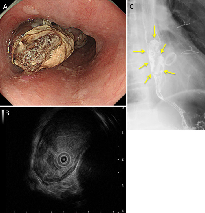
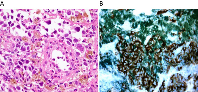
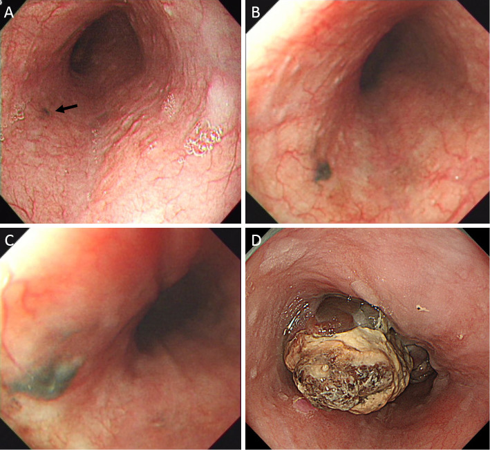
A 66-year-old man presented to his previous physician with epigastric discomfort in 2014. He was then referred to our hospital due to suspected primary malignant melanoma of the esophagus (PMME). A biopsy showed atypical cells containing melanin granules. A diagnosis of PMME was thus made. We investigated the endoscopic findings of the previous physician, which revealed a black point-like pigmentation at the same site since 2009. In 2010, black pigmentation was also observed at the same site. Although esophageal melanosis was suspected, no biopsy was performed. This case demonstrates the process by which esophageal melanomas develop into malignant melanomas.
Keywords: PMME; malignant melanoma; primary malignant melanoma of the esophagus.
Figures
Similar articles
-
A case of primary malignant melanoma of the esophagus with a widely expanded surface area.Clin J Gastroenterol. 2019 Oct;12(5):424-428. doi: 10.1007/s12328-019-00961-3. Epub 2019 Mar 18. Clin J Gastroenterol. 2019. PMID: 30887427
-
Superficial Primary Malignant Melanoma of the Esophagus Detected and Treated at Stage 0.Intern Med. 2024 Nov 15;63(22):3049-3053. doi: 10.2169/internalmedicine.2454-23. Epub 2024 Mar 25. Intern Med. 2024. PMID: 38522910 Free PMC article.
-
Primary Malignant Melanoma of the Esophagus With Unusual Endoscopic Findings: A Case Report and Literature Review.Medicine (Baltimore). 2016 Apr;95(17):e3479. doi: 10.1097/MD.0000000000003479. Medicine (Baltimore). 2016. PMID: 27124046 Free PMC article. Review.
-
Primary malignant melanoma of the esophagus.Virchows Arch A Pathol Anat Histol. 1979 Dec;385(1):49-59. doi: 10.1007/BF00433540. Virchows Arch A Pathol Anat Histol. 1979. PMID: 162100
-
Primary malignant melanoma of the esophagus with separate foci of melanoma in situ and atypical melanocytic hyperplasia in a patient positive for human immunodeficiency virus: a case report and review of the literature.Arch Pathol Lab Med. 2008 Oct;132(10):1675-8. doi: 10.5858/2008-132-1675-PMMOTE. Arch Pathol Lab Med. 2008. PMID: 18834229 Review.
Cited by
-
Primary malignant melanoma of the esophagus.Clin Case Rep. 2022 Apr 20;10(4):e05660. doi: 10.1002/ccr3.5660. eCollection 2022 Apr. Clin Case Rep. 2022. PMID: 35474989 Free PMC article.
-
Primary malignant melanoma of the esophagus arising from melanocytosis: a case report and literature review.Clin J Gastroenterol. 2025 Aug;18(4):545-551. doi: 10.1007/s12328-025-02129-8. Epub 2025 May 16. Clin J Gastroenterol. 2025. PMID: 40377881 Review.
-
A CT-Based Radiomics Nomogram Model for Differentiating Primary Malignant Melanoma of the Esophagus from Esophageal Squamous Cell Carcinoma.Biomed Res Int. 2023 Feb 20;2023:6057196. doi: 10.1155/2023/6057196. eCollection 2023. Biomed Res Int. 2023. PMID: 36860814 Free PMC article.
-
The Thousand Faces of Malignant Melanoma: A Systematic Review of the Primary Malignant Melanoma of the Esophagus.Cancers (Basel). 2022 Jul 30;14(15):3725. doi: 10.3390/cancers14153725. Cancers (Basel). 2022. PMID: 35954389 Free PMC article. Review.
References
-
- Bisceglio M, Perri F, Tucci A, et al. . Primary malignant melanoma of the esophagus: a clinicopathologic study of a case with comprehensive literature review. Adv Anat Pathol 18: 235-252, 2011. - PubMed
-
- Sabaanathan S, Eng J, Pradhan GN. Primary malignant melanoma of the esophagus. Am J Gastroenterol 84: 1475-1481, 1989. - PubMed
-
- Joob AW, Haines GK 3rd, Kies MS, et al. . Primary malignant melanoma of the esophagus. Ann Thorac Surg 60: 217-222, 1995. - PubMed
-
- Namiento T, Koito K, Ambo T, et al. . Primary malignant melanoma of the esophagus: diagnostic value of endoscopic ultrasonography. Am Surg 62: 716-718, 1996. - PubMed
-
- Dematos P. Primary malignant melanoma of the esophagus. J Surg Oncol 66: 201-206, 1997. - PubMed
Publication types
MeSH terms
LinkOut - more resources
Full Text Sources
Other Literature Sources
Medical