. 2017:2017:8791832.
doi: 10.1155/2017/8791832. Epub 2017 Jul 11.

Resveratrol Attenuates Microglial Activation via SIRT1-SOCS1 Pathway

Affiliations

Resveratrol Attenuates Microglial Activation via SIRT1-SOCS1 Pathway

Shuping Zhang et al. Evid Based Complement Alternat Med. 2017.

Abstract

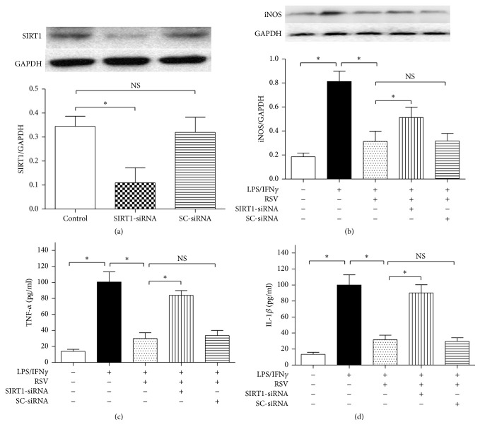
Microglial activation is involved in a variety of neurological disorders, and overactivated microglial cells can secrete large amount of proinflammatory factors and induce neuron death. Therefore, reducing microglial activation is believed to be useful in treating the disorders. In this study, we used 10 ng/ml lipopolysaccharide plus 10 U/ml interferon γ (LPS/IFNγ) to induce N9 microglial activation and explored resveratrol- (RSV-) induced effects on microglial activation and the underlying mechanism. We found that LPS/IFNγ exposure for 24 h increased inducible nitric oxide synthase (iNOS) and nuclear factor κB (NF-κB) p65 subunit expressions in the cells and enhanced tumor necrosis factor α (TNF-α) and interleukin 1β (IL-1β) releases from the cells. RSV of 25 μM reduced the iNOS and NF-κB p65 subunit expressions and the proinflammatory factors' releases; the knockdown of silent information regulator factor 2-related enzyme 1 (SIRT1) or suppressor of cytokine signaling 1 (SOCS1) by using the small interfering RNA, however, significantly abolished the RSV-induced effects on iNOS and NF-κB p65 subunit expressions and the proinflammatory factors' releases. These findings showed that microglial SIRT1-SOCS1 pathway may mediate the RSV-induced inhibition of microglial activation in the LPS/IFNγ-treated N9 microglia.


Figures

Figure 1
Figure 1
Resveratrol reduced iNOS expression and restored cell viability in the microglial cells exposed to LPS plus IFNγ. (a) The N9 microglial cells were divided into five groups, including 10 ng/ml LPS plus 10 U/ml IFNγ (LPS/IFNγ) exposure group, and four concentrations of resveratrol (RSV) treatment groups (5 μM, 10 μM, 25 μM, and 50 μM). After 24 h incubation, western blot analysis was taken to evaluate the iNOS protein expression in the cells (n = 4). (b) The cells were divided into six groups, including control, LPS/IFNγ, and four concentrations of resveratrol (RSV) treatment groups (5 μM, 10 μM, 25 μM, and 50 μM). After 24 h incubation, MTT assay was taken to evaluate the cell viability (n = 8). Results are expressed as means ± SD. P < 0.05; NS: no significance.
Figure 2
Figure 2
SIRT1-siRNA reversed resveratrol- (RSV-) induced effects on iNOS expression and proinflammatory factors' releases. (a) SIRT1-siRNA was effective in downregulating SIRT1 protein expression. The cells were divided into three groups as shown in the figure. After 6 h incubation, the SIRT1 protein expression was assessed by western blot analysis (n = 4). (b) Microglial iNOS protein expression (n = 4). (c) TNF-α concentration in the supernatants (n = 8). (d) IL-1β concentration in the supernatants (n = 8). The cells were divided into five groups and treated with different drugs as shown in the figure. After 24 h incubation, western blot and ELISA kits were used to assess iNOS expression and proinflammatory factors' releases, respectively. Results are expressed as means ± SD. P < 0.05; NS: no significance.
Figure 3
Figure 3
SIRT1-siRNA reversed resveratrol- (RSV-) induced effects on iNOS expression. The cells were divided into five groups and treated with different drugs as shown in the figure. After 24 h incubation, immunocytochemistry was used to observe the iNOS expression (red) in microglial cells, and the nuclei (blue) were counterstained with DAPI staining solution. Bar = 10 μm.
Figure 4
Figure 4
SIRT1-siRNA reversed resveratrol- (RSV-) induced effects on SOCS1 expression. The cells were divided into five groups and treated with different drugs as shown in the figure. After 24 h incubation, western blot was used to evaluate the SOCS1 protein expression in microglial cells (n = 4). Results are expressed as means ± SD. P < 0.05; NS: no significance.
Figure 5
Figure 5
SOCS1-siRNA reversed resveratrol- (RSV-) induced effects on iNOS expression and proinflammatory factors' releases. (a) SOCS1-siRNA was effective in reducing SOCS1 protein expression in N9 microglial cells. The N9 microglial cells were divided into three groups as shown in the figure. After 6 h incubation, the SOCS1 protein expression was assessed by western blot analysis (n = 4). (b) Microglial iNOS protein expression (n = 4). (c) TNF-α concentration in the supernatants (n = 8). (d) IL-1β concentration in the supernatants (n = 8). The cells were divided into five groups and treated with different drugs as shown in the figure. After 24 h incubation, western blot and ELISA kits were used to assess iNOS expression and proinflammatory factors' releases, respectively. Results are expressed as means ± SD. P < 0.05; NS: no significance.
Figure 6
Figure 6
SOCS1-siRNA reversed resveratrol- (RSV-) induced effects on NF-κB p65 subunit expression. The cells were divided into five groups and treated with the drugs as shown in the figure. After 24 h incubation, western blot was used to assess the protein expression of NF-κB p65 subunit (n = 4). Results are expressed as means ± SD. P < 0.05; NS: no significance.

References

    1. Shukla V., Shakya A. K., Perez-Pinzon M. A., Dave K. R. Cerebral ischemic damage in diabetes: an inflammatory perspective. Journal of Neuroinflammation. 2017;14(1) doi: 10.1186/s12974-016-0774-5. - DOI - PMC - PubMed
    1. Menzel L., Kleber L., Friedrich C., et al. Progranulin protects against exaggerated axonal injury and astrogliosis following traumatic brain injury. Glia. 2017;65(2):278–292. doi: 10.1002/glia.23091. - DOI - PubMed
    1. Carniglia L., Ramŕrez D., Durand D., et al. Neuropeptides and microglial activation in inflammation, pain, and neurodegenerative diseases. Mediators of Inflammation. 2017;2017:23. doi: 10.1155/2017/5048616.5048616 - DOI - PMC - PubMed
    1. Ding K., Wang H., Xu J., Lu X., Zhang L., Zhu L. Melatonin reduced microglial activation and alleviated neuroinflammation induced neuron degeneration in experimental traumatic brain injury: possible involvement of mTOR pathway. Neurochemistry International. 2014;76:23–31. doi: 10.1016/j.neuint.2014.06.015. - DOI - PubMed
    1. Bhoiwala D. L., Koleilat I., Qian J., et al. Overexpression of RCAN1 isoform 4 in mouse neurons leads to a moderate behavioral impairment. Neurological Research. 2013;35(1):79–89. doi: 10.1179/1743132812Y.0000000117. - DOI - PubMed

LinkOut - more resources