

Meloxicam decreases the migration and invasion of CF41.Mg canine mammary carcinoma cells
- PMID: 28781660
- PMCID: PMC5530185
- DOI: 10.3892/ol.2017.6400
Meloxicam decreases the migration and invasion of CF41.Mg canine mammary carcinoma cells
Abstract
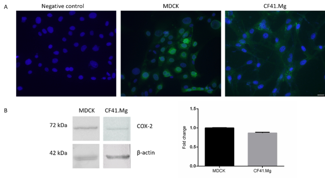
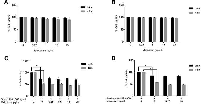
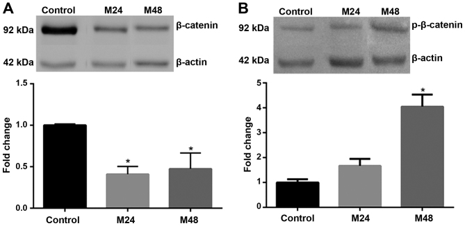
Cyclooxygenase (COX)-2 expression is positively correlated with malignant features in canine mammary carcinomas. Non-steroidal anti-inflammatory drugs (NSAIDs) inhibit COX activity and may therefore possess anticancer effects. Meloxicam is an NSAID that is widely used in human and veterinary medicine. High concentrations of meloxicam have been reported to be antitumorigenic in vitro; however, the effect of meloxicam at concentrations that are equivalent to those that can be obtained in vivo remains unknown. In the current study, the in vitro effects of low-dose meloxicam (0.25 µg/ml) on CF41.Mg canine mammary carcinoma cells were evaluated. The effects on cell proliferation, apoptosis, cell migration and invasion, in addition to the expression of different molecules associated with tumor invasiveness were analyzed. No effect on cell viability and apoptosis were observed. However, cell migration and invasion were significantly reduced following treatment with meloxicam. MMP-2 expression and activity were similarly reduced, explaining the impaired cell invasion. In addition, β-catenin expression was downregulated, while its phosphorylation increased. These results indicate that 0.25 µg/ml meloxicam reduces cell migration and invasion, in part through modulating MMP-2 and β-catenin expression. Additional studies are required to elucidate the mechanism associated with the anti-invasive effect of meloxicam on CF41.Mg cells. The results of the present study suggest that meloxicam has a potential adjunctive therapeutic application, which could be useful in controlling the invasion and metastasis of canine mammary carcinomas.
Keywords: canine mammary carcinoma; cell invasion; cyclooxygenase-2; meloxicam.
Figures


Similar articles
-
Melatonin decreases in vitro viability and migration of spheres derived from CF41.Mg canine mammary carcinoma cells.BMC Vet Res. 2019 Nov 4;15(1):390. doi: 10.1186/s12917-019-2142-z. BMC Vet Res. 2019. PMID: 31684950 Free PMC article.
-
Meloxicam suppresses hepatocellular carcinoma cell proliferation and migration by targeting COX-2/PGE2-regulated activation of the β-catenin signaling pathway.Oncol Rep. 2016 Jun;35(6):3614-22. doi: 10.3892/or.2016.4764. Epub 2016 Apr 20. Oncol Rep. 2016. PMID: 27109804
-
Meloxicam affects the inflammatory responses of bovine mammary epithelial cells.J Dairy Sci. 2019 Nov;102(11):10277-10290. doi: 10.3168/jds.2019-16630. Epub 2019 Aug 22. J Dairy Sci. 2019. PMID: 31447141
-
Pharmacology of meloxicam, a new non-steroidal anti-inflammatory drug with an improved safety profile through preferential inhibition of COX-2.Br J Rheumatol. 1996 Apr;35 Suppl 1:4-12. doi: 10.1093/rheumatology/35.suppl_1.4. Br J Rheumatol. 1996. PMID: 8630636 Review.
-
Meloxicam: a reappraisal of pharmacokinetics, efficacy and safety.Expert Opin Pharmacother. 2005 Oct;6(12):2117-40. doi: 10.1517/14656566.6.12.2117. Expert Opin Pharmacother. 2005. PMID: 16197363 Review.
Cited by
-
CircRNA Expression Profiles in Canine Mammary Tumours.Vet Sci. 2022 Apr 22;9(5):205. doi: 10.3390/vetsci9050205. Vet Sci. 2022. PMID: 35622733 Free PMC article.
-
Cyclooxygenase-2 as a Biomarker with Diagnostic, Therapeutic, Prognostic, and Predictive Relevance in Small Animal Oncology.J Vet Res. 2020 Mar 24;64(1):151-160. doi: 10.2478/jvetres-2020-0018. eCollection 2020 Mar. J Vet Res. 2020. PMID: 32258812 Free PMC article.
-
Formulation and Functional Characterization of a Cannabidiol-Loaded Nanoemulsion in Canine Mammary Carcinoma Cells.Pharmaceutics. 2025 Jul 26;17(8):970. doi: 10.3390/pharmaceutics17080970. Pharmaceutics. 2025. PMID: 40870993 Free PMC article.
-
Characterization of six canine prostate adenocarcinoma and three transitional cell carcinoma cell lines derived from primary tumor tissues as well as metastasis.PLoS One. 2020 Mar 13;15(3):e0230272. doi: 10.1371/journal.pone.0230272. eCollection 2020. PLoS One. 2020. PMID: 32168360 Free PMC article.
-
Contribution of non-steroidal anti-inflammatory drugs to breast cancer treatment: In vitro and in vivo studies.Vet World. 2024 May;17(5):1052-1072. doi: 10.14202/vetworld.2024.1052-1072. Epub 2024 May 15. Vet World. 2024. PMID: 38911075 Free PMC article. Review.
References
-
- Torres CG, Pino AM, Sierralta WD. A cyclized peptide derived from alpha fetoprotein inhibits the proliferation of ER-positive canine mammary cancer cells. Oncol Rep. 2009;21:1397–1404. - PubMed
LinkOut - more resources
Full Text Sources
Other Literature Sources
Research Materials
Miscellaneous