. 2016:1:16019.
doi: 10.1038/npjgenmed.2016.19. Epub 2016 Jul 20.

Development and validation of a whole-exome sequencing test for simultaneous detection of point mutations, indels and copy-number alterations for precision cancer care

Affiliations

Development and validation of a whole-exome sequencing test for simultaneous detection of point mutations, indels and copy-number alterations for precision cancer care

Hanna Rennert et al. NPJ Genom Med. 2016.

Abstract

We describe Exome Cancer Test v1.0 (EXaCT-1), the first New York State-Department of Health-approved whole-exome sequencing (WES)-based test for precision cancer care. EXaCT-1 uses HaloPlex (Agilent) target enrichment followed by next-generation sequencing (Illumina) of tumour and matched constitutional control DNA. We present a detailed clinical development and validation pipeline suitable for simultaneous detection of somatic point/indel mutations and copy-number alterations (CNAs). A computational framework for data analysis, reporting and sign-out is also presented. For the validation, we tested EXaCT-1 on 57 tumours covering five distinct clinically relevant mutations. Results demonstrated elevated and uniform coverage compatible with clinical testing as well as complete concordance in variant quality metrics between formalin-fixed paraffin embedded and fresh-frozen tumours. Extensive sensitivity studies identified limits of detection threshold for point/indel mutations and CNAs. Prospective analysis of 337 cancer cases revealed mutations in clinically relevant genes in 82% of tumours, demonstrating that EXaCT-1 is an accurate and sensitive method for identifying actionable mutations, with reasonable costs and time, greatly expanding its utility for advanced cancer care.


Conflict of interest statement

COMPETING INTERESTS The authors declare no conflict of interest.

Figures
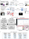
Figure 1
Figure 1
Schematic view of EXaCT-1 assay workflow. (a) (1) Slides are assessed by pathologist for neoplastic content, and tumour tissue marked and macrodissected. (2) DNA from fresh or FFPE tumour tissue and matched normal control specimen is extracted. (3) DNA is then enriched for exome sequences (357,999 exons corresponding to 21,522 genes) with HaloPlex technology described in this protocol in four major steps: fragmentation by restriction enzyme digestion, hybridisation to HaloPlex probes and introduction of Illumina sequence motifs, solid-phase capture and DNA ligation, and amplification of targeted fragments by PCR, followed by (4) sequencing on an Illumina HiSeq 2500 rapid mode, four samples per lane. (5) Paired-end reads are aligned to the human genome and (6) variant calls are made. (7) Variants are annotated and classified by our internally developed informatics pipeline, using publicly available and our own developed knowledge base. (8) The case is reviewed and results interpreted by a molecular pathologist who also signs it out in the LIS. (9) A report is then automatically generated and (10) dispensed to medical records. EXaCT-1 data analysis pipeline. (b) Schematic view of EXaCT-1 assay validation. (c) DNA derived from matched normal/tumour pairs from either fresh or FFPE specimens as well as standardised commercial DNA material were used to demonstrate the accuracy, sensitivity, specificity, reproducibility and precision of the assay using bioinformatics process for the detection of numerous types of variants (SNV, indel and CNV gain), according to NYS-DOH NGS guidelines for somatic genetic variant detection. IPM, Institute for Precision Medicine.
Figure 2
Figure 2
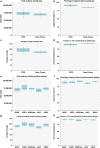
EXaCT-1 sequencing quality metrics. The EXaCT-1 was validated using 57 archival samples with known mutations comprising a diverse representation of solid tumours and haematological cancers. Total reads, percentage of captured reads, average coverage and fraction of >10× covered bases by sample type (ad) and mutation-positive sample (eh) in FFPE (n=41) and fresh (n=16) tumour specimens. Additional QC metrics for all cases are available in Supplementary Tables 2 and 3. No statistically significant difference between FFPE and non-FFPE tissue and mutation-harbouring sample was observed in these analyses.
Figure 3
Figure 3
EXaCT-1 reportable range. Number and percentage of common low coverage bases shared by 36 (80%) and all 45 (100%) control (germline) samples obtained from normal tissue. (a) The germline samples were expected to have low genomic complexity and therefore provide more accurate assessment of coverage and performance statistics. Over 95% of the HaloPlex exome bases were covered at >10×. (b) These guaranteed regions that have constant higher than 10× coverage is our reportable range, which is listed on our website (https://rubinlab.med.cornell.edu/IPMWES/Haloplex_Exome_Sequencing_reportable_region.xlsx). This poorly covered 4.29% genomic area of the HaloPlex exome will be excluded from our reportable range. The reportable range is listed on https://rubinlab.med.cornell.edu/IPMWES/HaloPlex_low_coverage_region.xlsx.
Figure 4
Figure 4
Coverage analysis for 49 Tier 1 clinically relevant genes. (a) The barplots show the mean bp coverage per gene across all validation samples (n=57). Each row is a gene and the barplot represents the distribution of the mean coverage across all specimens. This metric is measured by dividing the total coverage by the total number of coding bp. The dashed red line is the 30× threshold. (b) The heatmap shows percentage of coding bp (averaged across all samples of a group) of a gene (each row) below a specific coverage threshold (each column). The colours of the heatmap represent the percentage of the coding bp of a gene below a coverage threshold. The white colour shows that <10% of the coding bp of a gene are below a coverage threshold. For the majority of genes, only <10–20% of the coding regions are covered at <10×. (c) Fraction of coding bp covered by <30× reads across the validation sample cohort. On average, ~20% of the gene-coding regions were covered at <30×.
Figure 5
Figure 5
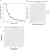
EXaCT-1 analytical sensitivity and power analysis. (a) Power analysis shows that at a VAF threshold of 10–12% VAF, at least 28× total coverage is needed (P⩽0.05) for avoiding false-negative results if no mutation is present (0 reads). (b) Analytical sensitivity for low VAF detection for selected mutations. Synthetic mixed samples were generated from mutation-positive cell lines diluted into HapMap DNA with mixed proportion as determined by Ion Torrent AmpliSeq Cancer Hot Spot assay (EGFR, KRAS, BRAF and JAK2) (a) and by digital droplet PCR (HER2 copy number). (c) With proportions ranging between 50 and 2%. Experiments were performed in triplicate. The threshold of EXaCT-1 for detecting variants at low allele frequency was established at 10%.
Figure 6
Figure 6
EXaCT-1 mutation correlation studies. (a) EXaCT-1 performance was validated using Reference FFPE DNA (Horizon sample) covering 11 onco-relevant variants at allele frequencies ranging from 2–25% across 6 genes commonly mutated in cancer. VAF results demonstrate a good agreement (Pearson correlation=0.76) between EXaCT-1 AF results and the expected reference DNA results. (b) Concordance between EXaCT-1 and Ion Torrent AmpliSeq assay across 9 genes (26 mutations) in 22 clinical cancer samples. The results demonstrate a good agreement between both tests over a wide range of allele frequencies with a Pearson correlation values of 0.92.
Figure 7
Figure 7
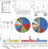
EXaCT-1 mutation testing statistics in IPM patient cohort. (a) Number of cases by cancer type, (b) mutation load by cancer diagnosis and (c) stage. (d) Number of gene variations by gene and by tier. (e) Commonly mutated Tier 1 and (f) Tier 2 genes (shown are 17 most commonly occurring genes out of 153 Tier 2 cancer genes). (g) Spectrum of unique mutations (N=40) in most commonly mutated Tier 1 cancer genes (N=15). IPM, Institute for Precision Medicine.

References

    1. Rabbani, B., Tekin, M. & Mahdieh, N. The promise of whole-exome sequencing in medical genetics. J. Hum. Genet. 59, 5–15 (2014). - PubMed
    1. Biesecker, L. G. Exome sequencing makes medical genomics a reality. Nat. Genet. 42, 13–14. - PubMed
    1. Majewski, J., Schwartzentruber, J., Lalonde, E., Montpetit, A. & Jabado, N. What can exome sequencing do for you? J. Med. Genet. 48, 580–589 (2011). - PubMed
    1. Schreiber, M., Dorschner, M. & Tsuang, D. Next-generation sequencing in schizophrenia and other neuropsychiatric disorders. Am. J. Med. Genet. B Neuropsychiatr. Genet. 162B, 671–678 (2013). - PubMed
    1. Dias-Santagata, D. et al. Rapid targeted mutational analysis of human tumours: a clinical platform to guide personalized cancer medicine. EMBO Mol. Med. 2, 146–158 (2010). - PMC - PubMed