Case Reports
. 2017 Jul 13:7:28.
doi: 10.4103/jcis.JCIS_28_17. eCollection 2017.

Mandibular Canal Enlargement: Clinical and Radiological Characteristics

Affiliations
Case Reports

Mandibular Canal Enlargement: Clinical and Radiological Characteristics

Chong Jun Ai et al. J Clin Imaging Sci. .

Abstract

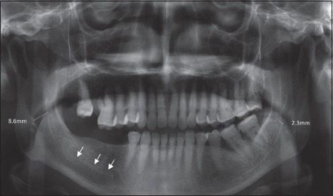
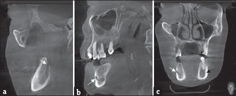
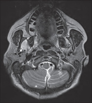
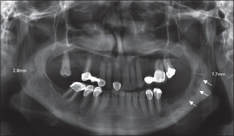
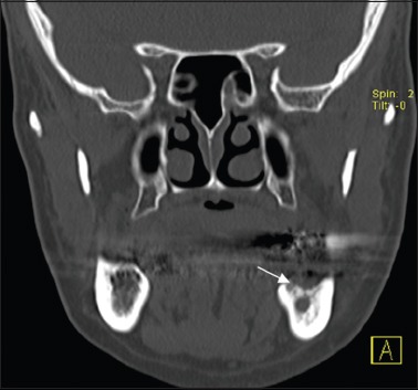
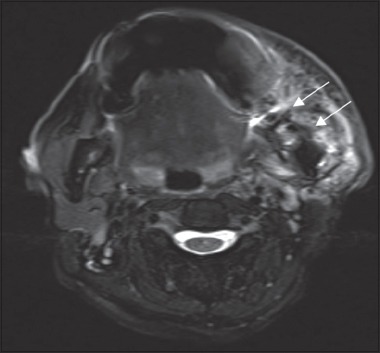
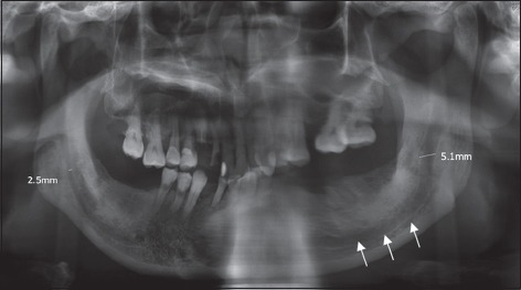
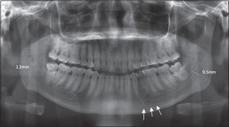
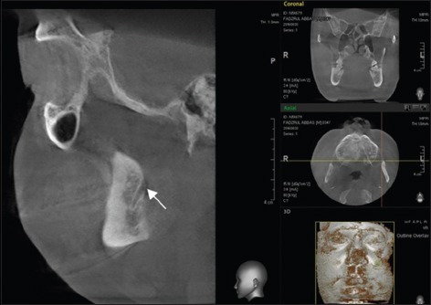
Enlargement of the mandibular canal is a rare radiological finding. Clinically, it may or may not be associated with sensory deficits. We report four cases of widening of the mandibular canal observed with various methods of imaging with different clinical characteristics. We describe this unique radiological finding and elaborate the importance of quality assessment of the imaging that is vital for accurate diagnosis and treatment planning. Clinicians should be mindful when assessing the imaging whenever the size of the mandibular canal is implicated. The case ranged from a benign tumor to malignancy, radiological errors, and artifacts. A more superior imaging or treatment modality was necessary to ascertain the diagnosis.

Keywords: Clinical characteristics; imaging characteristics; mandibular canal.


Conflict of interest statement

There are no conflicts of interest.

Figures

Figure 1
Figure 1
57-year-old female with cerebrovascular accident who presented with sudden onset of loss of taste and tongue deviation. Panoramic radiograph showing widening of the right mandibular canal (arrows).
Figure 2
Figure 2
57-year-old female with cerebrovascular accident who presented with sudden onset of loss of taste and tongue deviation. Cone-beam computed tomography image showing various tomographic views. (a and b) Sagittal view showing enlargement of the mandibular canal from the mandibular foramen (white arrow) toward the mental foramen (white arrow). (c) Coronal view comparing the size of the mandibular canal bilaterally (white arrow: Right canal, black arrow: Left canal).
Figure 3
Figure 3
57-year-old female with cerebrovascular accident who presented with sudden onset of loss of taste and tongue deviation. Magnetic resonance imaging showing well-defined T1-hypointense and T2-hyperintense lesion at the right masticator space invaginating into the right mandibular canal at axial section (white arrow).
Figure 4
Figure 4
60-year-old female with squamous cell carcinoma who presented with left lower lip hypoesthesia. Panoramic radiograph showing ill-defined widening of the left mandibular canal (white arrows).
Figure 5
Figure 5
60-year-old female with squamous cell carcinoma who presented with left lower lip hypoesthesia. Sagittal view of the computed tomography scan showing cortical irregularity with periosteal reaction of the medial aspect of the left mandible (white arrow).
Figure 6
Figure 6
60-year-old female with squamous cell carcinoma who presented with left lower lip hypoesthesia. Axial view of the magnetic resonance imaging showing the mass extending to the left angle of mandible and into the mandibular canal (white arrows).
Figure 7
Figure 7
86-year-old male with reduced taste sensation of the left lower lip and tongue. Panoramic radiograph showing widening of the left mandibular canal (white arrows).
Figure 8
Figure 8
34-year-old male with left recurrent pericoronitis. Panoramic radiograph showing “double mandibular canal” (white arrows).
Figure 9
Figure 9
34-year-old male with left recurrent pericoronitis. Sagittal section of the cone-beam computed tomography showing a single mandibular canal (white arrow).

References

    1. Vartiainen VM, Siponen M, Salo T, Rosberg J, Apaja-Sarkkinen M. Widening of the inferior alveolar canal: A case report with atypical lymphocytic infiltration of the nerve. Oral Surg Oral Med Oral Pathol Oral Radiol Endod. 2008;106:e35–9. - PubMed
    1. Srinivasan K, Seith A, Gadodia A, Sharma R, Kumar A, Roychoudhury A, et al. Evaluation of the inferior alveolar canal for cysts and tumors of the mandible-comparison of multidetector computed tomography and 3-dimensional volume interpolated breath-hold examination magnetic resonance sequence with curved multiplanar reformatted reconstructions. J Oral Maxillofac Surg. 2012;70:2327–32. - PubMed
    1. Juodzbalys G, Wang HL, Sabalys G. Anatomy of mandibular vital structures. Part I: Mandibular canal and inferior alveolar neurovascular bundle in relation with dental implantology. J Oral Maxillofac Res. 2010;1:e2. - PMC - PubMed
    1. Kalantar Motamedi MH, Navi F, Sarabi N. Bifid mandibular canals: Prevalence and implications. J Oral Maxillofac Surg. 2015;73:387–90. - PubMed
    1. Keat-Siong O, Hong-Sai L. Entrapment of the inferior dental nerve by a 3-rooted mandibular third molar - Case report. Asian J Oral Maxillofac Surg. 2002;14:105–8.

Publication types